D0366 Dental Code: The Complete Guide to Cone Beam Computed Tomography in Modern Dentistry
For over a century, dental diagnosis relied on a two-dimensional world. The familiar dental X-ray, whether a small bitewing or a panoramic film, provided a flat, often distorted, glimpse into the complex three-dimensional anatomy of the maxillofacial region. While invaluable, these 2D images forced clinicians to rely on experience, intuition, and sometimes guesswork to interpret depth, spatial relationships, and the true nature of pathological conditions. It was akin to navigating a vast city with only a simple, outdated map—functional for the main roads but perilous for the intricate alleyways and underground structures.
The dawn of the 21st century ushered in a paradigm shift with the adaptation of computed tomography (CT) technology for the dental office. However, it was the refinement into Cone Beam Computed Tomography (CBCT) that truly revolutionized the field. This technology collapsed the gap between 2D inference and 3D reality, offering dentists an unprecedented, crystal-clear window into the hidden architecture of their patients’ craniofacial structures. To standardize and track this advanced diagnostic service, the American Dental Association (ADA) introduced a specific code: D0366.
This code is more than just a billing number; it is the gateway to a higher standard of care, representing a commitment to precision, predictability, and patient safety. This exhaustive guide will delve into every facet of the D0366 dental code, exploring the transformative technology it represents, its vast clinical applications, and its critical role in shaping the future of evidence-based dentistry.
2. Demystifying the Code: What Exactly is D0366?
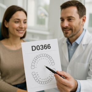
In the ADA’s Current Dental Terminology (CDT), D0366 is defined as: “Cone Beam Computed Tomography (CBCT) – image capture with limited field of view; less than one whole jaw.”
This precise definition contains several key components that must be understood:
-
Cone Beam Computed Tomography (CBCT): This specifies the exact technology used. It is distinct from a medical CT scan or a traditional dental X-ray.
-
Image Capture: This refers to the actual acquisition of the scan data.
-
Limited Field of View (FOV): This is the most critical differentiator. The FOV is the volume of anatomy captured during the scan, measured in centimeters. A “limited” or “focused” FOV means the scan is targeted to a specific area of interest—for example, a few teeth, a single jaw (maxilla or mandible), or the temporomandibular joints. This is in contrast to larger FOV scans that might capture the entire skull.
Why is FOV so important? The principle of ALARA (As Low As Reasonably Achievable) is a cornerstone of radiology, mandating that radiation exposure be minimized while still obtaining the necessary diagnostic information. By using a limited FOV, the clinician can precisely tailor the scan to the clinical question, significantly reducing unnecessary radiation exposure to areas not relevant to the diagnosis. D0366 is typically used for tasks like evaluating a single implant site, a problematic tooth root, or a small pathological lesion.
It is crucial to note that the CDT code set includes other CBCT codes:
-
D0367: CBCT – image capture with field of view of one full jaw or both jaws.
-
D0368: CBCT – image capture with field of view of both jaws including temporomandibular joints (TMJ).
-
D0369: CBCT – image capture with field of view of craniofacial structures.
D0366, with its limited scope, is often the most commonly used code for a wide array of specific diagnostic problems.
3. The Technology Behind the Code: How CBCT Works
Understanding the mechanics of CBCT illuminates why it is so superior to traditional radiography.
The Process:
-
Image Acquisition: The patient is positioned in the CBCT machine, which consists of a rotating gantry. On one arm of the gantry is an X-ray source, and directly opposite is a digital detector. Unlike a medical CT scanner where the X-ray source and detector rotate continuously in a tight circle around the patient, a CBCT machine rotates once, or sometimes twice, around the patient’s head in a complete 360-degree (or slightly less) arc.
-
The “Cone Beam”: As the machine rotates, the X-ray source emits a divergent, cone-shaped beam (as opposed to the fan-shaped beam of a traditional CT scanner) that passes through the patient’s tissues.
-
Data Capture: The digital detector on the opposite side captures between 150 and 600+ individual 2D projection images (called “basis” images) from different angles throughout the rotation. This process typically takes between 10 and 40 seconds, depending on the machine and settings.
-
Reconstruction: After the scan is complete, powerful proprietary software on a connected computer workstation takes these hundreds of 2D images and uses a sophisticated algorithm known as Feldkamp-Davis-Kress (FDK) reconstruction to create a perfect 3D volumetric data set. This “cube” of data contains all the information about the patient’s anatomy within the scanned field of view.
The Output: A Multiplanar Reconstructable Dataset
This is the heart of the D0366 value proposition. The raw data is not a single image but a digital volume that can be manipulated and sliced in any plane:
-
Axial Plane: Slices from feet to head (the base view).
-
Coronal Plane: Slices from front to back.
-
Sagittal Plane: Slices from side to side.
-
Oblique Planes: The software allows the clinician to reorient the dataset to create slices along the axis of a single tooth or any other structure of interest.
Furthermore, the software can generate 3D surface-rendered models that provide a photorealistic view of the bones and, with certain software, even the skin surface. This data can also be exported for use in CAD/CAM software for designing surgical guides, orthodontic appliances, and prosthetics.
4. A World of Difference: CBCT vs. Traditional Dental Imaging
To appreciate D0366, one must understand what it replaces and the limitations it overcomes.
| Feature | 2D Panoramic / Periapical X-Ray | 3D Cone Beam CT (D0366) |
|---|---|---|
| Dimensionality | Two-dimensional (flat) | Three-dimensional (volumetric) |
| Anatomy | Superimposed structures; no depth perception | Separate, distinct structures; precise spatial relationships |
| Distortion | Present, especially in panoramic radiographs | Minimal, geometrically accurate |
| Information Density | Limited grayscale contrast | Voxel-based data with high contrast resolution |
| Measurement | Inaccurate due to magnification and distortion | Highly accurate 1:1 measurements |
| Viewing | Static film or digital image | Interactive, multiplanar reconstructions |
| Radiation Dose | Very Low (Panoramic: ~5-25 µSv) | Low to Moderate (Limited FOV: ~11-100 µSv) |
Key Limitations of 2D Imaging that CBCT Solves:
-
Anatomic Superimposition: Critical structures like the mandibular canal, mental foramen, or maxillary sinus can be superimposed over the roots of teeth, making it impossible to determine their true relationship. This is a major risk factor in surgery.
-
Geometric Distortion: Panoramic images have a focal trough—a narrow zone where structures are in focus. Anything outside this zone is blurred and distorted, compromising diagnostic accuracy.
-
Lack of Bucco-Lingual Information: A periapical X-ray cannot show the width of the jawbone or the position of a root in the bone from cheek-side to tongue-side. This is critical for implant planning.
Comparative Overview of Dental Imaging Modalities
5. The Clinical Applications of D0366: A Revolution in Patient Care
The applications of a limited FOV CBCT scan (D0366) are vast and transformative across nearly every dental specialty.
Implantology: The Gold Standard for Precision
This is perhaps the most common indication for D0366. Precise implant planning requires knowing:
-
Bone Quality and Quantity: The exact height and width of the alveolar bone.
-
Anatomical Avoidance: The precise 3D location of the inferior alveolar nerve, mental foramen, maxillary sinus, and any concavities in the jawbone.
-
Prosthetic-Driven Planning: Determining the ideal implant position based on the planned final restoration, not just the available bone.
Using the D0366 dataset, surgeons can virtually place implants in the software, ensuring perfect positioning and allowing for the fabrication of computer-guided surgical stents that transfer the virtual plan to the operatory with sub-millimeter accuracy. This minimizes surgical risk, improves outcomes, and reduces patient recovery time.
Endodontics: Seeing the Unseeable
For root canal specialists, D0366 is a diagnostic powerhouse:
-
Diagnosing Complex Pain: Identifying cracks (vertical root fractures), missed canals, and periapical pathosis that are invisible on 2D films.
-
Managing Retreatments: Visualizing the true anatomy of previously treated teeth, including the position of separated instruments, perforations, and resorptive defects.
-
Surgical Planning: For apicoectomies, providing a detailed map of the root apex and its relationship to adjacent structures to ensure a safe and effective procedure.
Oral and Maxillofacial Surgery: Navigating Complex Anatomy
Beyond implants, surgeons use D0366 for:
-
Impacted Tooth localization: Precisely determining the position of impacted canines or third molars (wisdom teeth) in all three dimensions relative to nerves, adjacent roots, and the sinus.
-
Pathology Assessment: Evaluating the size, extent, and internal structure of cysts, tumors, and other lesions.
-
Trauma: Diagnosing and planning the repair of complex fractures of the jaws and midface.
-
TMJ Analysis: Detailed evaluation of bony changes in the temporomandibular joint.
Orthodontics: Beyond the Cephalogram
While lateral cephalograms are standard, D0366 offers orthodontists:
-
Accurate Impacted Canine Localization: A primary use of limited FOV CBCT in orthodontics.
-
Airway Analysis: Assessing the volume and patency of the nasal and pharyngeal airways, which can be a factor in sleep apnea and craniofacial development.
-
Root Resorption Monitoring: Detecting and monitoring external root resorption during treatment with far greater accuracy than 2D images.
-
Mini-Implant Placement: Planning the safe placement of temporary anchorage devices (TADs).
Periodontics: Assessing Bone Destruction in 3D
CBCT allows periodontists to visualize the morphology of bony defects (e.g., furcation involvements, intrabony defects) in three dimensions, which is crucial for diagnosing severity and planning regenerative surgical therapy.
Temporomandibular Joint (TMJ) Analysis
A limited FOV scan centered on the TMJs provides exquisite detail of the bony components of the joint, allowing for the diagnosis of degenerative changes (osteoarthritis), ankylosis, and other arthritic conditions.
Airway Analysis and Sleep Apnea
Dentists involved in oral appliance therapy for sleep apnea use CBCT to assess airway dimensions and visualize anatomic obstructions that contribute to sleep-disordered breathing.
Forensic and Identification Dentistry
The unique anatomy captured in a CBCT scan can serve as a powerful tool for human identification when other methods are not available.
6. The D0366 Procedure: From Consultation to Diagnosis
-
Clinical Indication: The process begins with a clinical examination where the dentist identifies a problem that cannot be fully diagnosed with 2D imaging alone (e.g., unexplained pain, a complex treatment plan).
-
Informed Consent: The dentist explains the need for the CBCT scan, its benefits, the alternatives, and the associated risks, including radiation exposure. Written consent is obtained.
-
Preparation: The patient removes any metal objects (eyeglasses, jewelry, hairpins, hearing aids) that could cause artifacts. A lead apron with a thyroid collar is often placed, though its necessity is a topic of modern debate as the primary beam is focused on the head.
-
Positioning: The patient is carefully positioned in the machine using laser lights or facial supports to ensure the correct FOV is captured. The technician selects the appropriate exposure parameters (kV, mA, scan time, FOV size) based on the diagnostic task.
-
Scan Acquisition: The patient must remain perfectly still during the brief rotation of the machine. Some units offer a seated position, while others require standing.
-
Reconstruction and Interpretation: The scan data is reconstructed and the dentist (or a specialized oral and maxillofacial radiologist) interprets the volumes, creating cross-sectional slices and 3D models as needed. A formal report is generated.
-
Diagnosis and Treatment Planning: The findings are integrated with the clinical examination to form a definitive diagnosis and inform the treatment plan.
7. Understanding the Benefits: Why D0366 is a Game-Changer
-
Enhanced Diagnostic Accuracy: Eliminates guesswork and provides a definitive view of the area of interest.
-
Improved Patient Safety: Reduces the risk of surgical complications (e.g., nerve damage, sinus perforation).
-
Precision Treatment Planning: Enables minimally invasive, flapless surgeries and predictable outcomes.
-
Efficient Treatment: Can reduce operative time and improve the efficiency of procedures.
-
Patient Education: 3D visualizations are powerful tools for helping patients understand their diagnosis and the necessity of proposed treatments.
-
Medicolegal Protection: Provides an objective, 3D record of the pre-treatment condition.
8. Weighing the Considerations: Safety, Limitations, and Risks
-
Radiation Exposure: This is the primary concern. While the effective dose from a limited FOV CBCT (D0366) is low (often comparable to a few days of natural background radiation or a cross-country flight) and significantly lower than a medical CT scan, it is higher than conventional dental X-rays. The ALARA principle must always be followed.
-
Artifacts: Metallic restorations (amalgam, implants) can cause streak artifacts that obscure adjacent anatomy. Patient movement can cause blurring.
-
Soft Tissue Visualization: Standard CBCT provides excellent detail of bony structures but poor soft tissue contrast compared to medical MRI or CT with contrast.
-
Cost and Access: The equipment is expensive, which can translate to a higher cost for the patient and may not be available in all dental practices.
-
Operator Dependency: The quality of the scan and the accuracy of interpretation depend heavily on the operator’s technical skill and the clinician’s training in 3D radiology.
9. The Cost and Insurance Landscape of D0366
The cost of a D0366 scan varies widely ($150 – $500+) based on geographic location, the practice’s overhead, and whether a radiologist’s interpretation fee is included. Dental insurance coverage is increasingly common but not universal. Many plans now cover CBCT when it is deemed medically necessary for specific procedures like implant planning or complex endodontic diagnosis. Pre-authorization is often required. Patients should always check with their insurance provider to understand their benefits.
10. The Future of D0366 and CBCT Technology
The technology behind D0366 is rapidly evolving. Future trends include:
-
Dose Reduction: Continued development of algorithms and detectors to further minimize radiation dose.
-
Enhanced Soft Tissue Imaging: New software and hardware techniques to improve the visualization of muscles, nerves, and blood vessels.
-
Artificial Intelligence (AI): AI algorithms are being integrated to automate tasks like nerve canal tracing, implant placement suggestions, and pathological lesion detection, increasing speed and consistency in interpretation.
-
Point-of-Care 3D Printing: The seamless integration of CBCT data with 3D printers to instantly create models and surgical guides in the dental office.
-
Functional Imaging: The potential fusion of CBCT data with other modalities like MRI or data from jaw motion trackers to create a dynamic 4D model of the masticatory system.
11. Conclusion
The D0366 dental code represents far more than a procedural billing entry; it symbolizes the culmination of dentistry’s journey into a new era of precision health. Cone Beam Computed Tomography has fundamentally transformed diagnostic capabilities, shifting practice from inference-based to evidence-based. By providing a meticulous, three-dimensional map of the maxillofacial region, it empowers clinicians across all specialties to deliver care with unprecedented accuracy, safety, and predictability. As technology continues to advance, making CBCT faster, safer, and more intelligent, its role as an indispensable tool in achieving optimal patient outcomes will only become more deeply entrenched, solidifying the legacy of D0366 as a cornerstone of modern dental practice.
12. Frequently Asked Questions (FAQs)
Q1: Is a CBCT scan (D0366) safe? How much radiation is involved?
A: Yes, when justified for a specific diagnostic purpose, it is considered safe. The radiation dose from a limited FOV CBCT scan is low, typically ranging from about 11 to 100 microsieverts (µSv). To put this in perspective, this is comparable to the natural background radiation everyone receives over a few days to a couple of weeks and is significantly lower than a medical CT scan of the head.
Q2: Why do I need a CBCT scan if I just had regular X-rays?
A: Regular X-rays are a great first-line tool. However, they are 2D and can superimpose structures, hiding important details. Your dentist may recommend a CBCT scan to see a specific area in 3D, much like a architect needing a 3D blueprint instead of a flat sketch to understand complex spatial relationships, especially before surgery or for diagnosing complex problems.
Q3: Will my insurance cover the cost of a D0366 procedure?
A: Coverage is increasingly common but not guaranteed. It is most likely to be covered for specific, pre-authorized procedures like dental implant planning, complex root canal diagnosis, or impacted tooth removal. You should always contact your dental insurance provider before the scan to verify your benefits and any pre-authorization requirements.
Q4: What is the difference between a medical CT scan and a dental CBCT scan?
A: While both produce 3D images, they use different technology. Medical CT uses a fan-shaped beam and collects data in a spiral (helical) pattern, providing excellent soft tissue detail but at a higher radiation dose. Dental CBCT uses a cone-shaped beam and a single rotation, providing superb bony detail with a lower radiation dose, making it ideal for dental and craniofacial applications.
Q5: Can I get a copy of my CBCT scan?
A: Absolutely. You have a right to your medical and dental records. You can request a copy, typically provided on a CD or DVD that contains the proprietary DICOM (Digital Imaging and Communications in Medicine) files. These can be viewed with free software available online.
13. Additional Resources
-
American Dental Association (ADA): Guidelines for the use of CBCT in dentistry. https://www.ada.org
-
American Academy of Oral and Maxillofacial Radiology (AAOMR): The leading professional organization for experts in dental radiology. Provides position statements and clinical recommendations. https://www.aaomr.org
-
Food and Drug Administration (FDA): Information on radiation-emitting products, including CBCT devices. https://www.fda.gov/radiation-emitting-products
-
RadiologyInfo.org: A patient-friendly resource from the American College of Radiology and the Radiological Society of North America explaining medical and dental imaging procedures. https://www.radiologyinfo.org/en/info/dentalconect