D0369 Dental Code: A Comprehensive Guide to Cone Beam Computed Tomography in Modern Dentistry
Imagine a surgeon embarking on a complex procedure with only a crude, two-dimensional map, lacking critical information about depth, hidden obstacles, and precise topography. Until the turn of the 21st century, this was the reality for many dental professionals. While traditional two-dimensional X-rays provided invaluable insight, they were fundamentally limited—a flat representation of a complex three-dimensional structure. They superimposed anatomy, distorted dimensions, and left critical diagnostic information hidden in the shadows. The introduction of Cone Beam Computed Tomography (CBCT) marked a seismic shift in dental diagnostics, a revolution as significant as the move from film to digital radiography. At the heart of this revolution in clinical practice and administrative billing lies a specific code: D0369.
This code is more than just a billing identifier; it is the gateway to a transformative level of patient care. D0369 represents a diagnostic paradigm that empowers clinicians to visualize the maxillofacial region with unprecedented clarity, accuracy, and detail. It allows for the exploration of bone structure, tooth orientation, nerve pathways, and soft tissues in three dimensions, enabling diagnoses and treatment plans that were previously based on estimation and experience alone. This article delves deep into the world of D0369 dental code, exploring its technological foundations, its vast array of clinical applications, the procedure itself, and its profound impact on shaping the future of precision, minimally invasive, and predictably successful dentistry. We will move beyond the code to understand the tool, its uses, and its indispensable role in modern patient care.

2. Demystifying D0369: Beyond the Alphanumeric Code
The American Dental Association (ADA) maintains the Current Dental Terminology (CDT), a standardized coding system used for reporting dental services and procedures. This system ensures uniformity in communication between dental offices and insurance companies. Code D0369 is explicitly defined within this system.
Official CDT Description for D0369: Cone Beam CT – craniofacial data capture.
This succinct description encompasses the capture of the three-dimensional volumetric data set using cone beam computed tomography technology. It is crucial to understand what this code includes and, just as importantly, what it does not. D0369 covers the acquisition of the scan itself. It involves the patient preparation, the positioning, the exposure, and the initial reconstruction of the raw data into a primary image set.
However, D0369 is distinct from the interpretation and report. The analysis of the captured data, the manipulation of the 3D volume, the taking of measurements, and the creation of a formal diagnostic report by a qualified professional are billed under a separate code: D0381. This separation is critical. It acknowledges that the act of taking the scan and the expertise required to interpret it are two distinct services, often performed by different individuals (e.g., a general dentist captures the image, but an oral radiologist or oral surgeon interprets it).
D0369 is categorized under “Imaging Laboratory and Physiological Data” in the CDT, highlighting its role as a sophisticated diagnostic data capture method rather than a simple radiographic image.
3. The Technology Behind the Code: How CBCT Works
To appreciate the value of D0369, one must understand the fundamental technology it represents. While both CBCT and medical CT (Computed Tomography) scanners produce cross-sectional images, they achieve this through different means, with CBCT being specifically optimized for the hard tissues of the maxillofacial region.
The Core Principle: A CBCT system consists of a rotating gantry. On one arm of this gantry is an X-ray source, and directly opposite is a digital detector. During a single scan, this apparatus rotates 180 to 360 degrees around the patient’s head. As it rotates, the X-ray source emits a cone-shaped beam (as opposed to the fan-shaped beam of a medical CT) that passes through the patient and is captured on the detector on the other side. From hundreds of different projection angles, a series of 2D images, known as “basis images” or “projection images,” are acquired.
Reconstruction: This collection of 2D basis images is then processed by sophisticated software using an algorithm called Feldkamp-Davis-Kress (FDK) reconstruction. This algorithm mathematically reconstructs the data into a three-dimensional volumetric data set, often called a “voxel map” (a 3D pixel). This voxel-based data set is the primary output of a D0369 procedure.
Key Technological Components:
-
X-ray Generator: Determines the energy (kVp) and current (mA) of the beam, which influence contrast and patient dose.
-
Detector: Typically an amorphous silicon flat-panel detector that captures the X-ray projections with high efficiency and speed.
-
Software: The true powerhouse. Reconstruction software creates the 3D volume, and visualization software allows the clinician to navigate through this volume in axial, coronal, sagittal, and panoramic planes. Advanced software enables tools like curved planar reformation, volume rendering, and tissue segmentation.
This entire process, from data capture to the creation of an explorable 3D volume, is what a patient and insurer are billed for when code D0369 is used.
4. A World of Difference: CBCT vs. Traditional Dental Imaging
The advent of CBCT did not render traditional dental imaging obsolete; rather, it provided a powerful complementary tool for specific diagnostic challenges. Understanding the differences is key to knowing when to employ D0369.
Traditional 2D Imaging (Periapicals, Panoramic, Cephalometric):
-
Nature: Provides a single, static, two-dimensional image.
-
Superimposition: All structures in the path of the X-ray beam are superimposed onto a single plane. This can hide pathologies, distort anatomy, and make precise localization impossible.
-
Distortion: Panoramic X-rays, in particular, have inherent magnification and distortion, making them unreliable for accurate measurement.
-
Limited Information: Provides excellent detail for caries detection and evaluating the periodontal bone crest but fails to show the buccal-lingual dimension.
Cone Beam CT (D0369):
-
Nature: Provides a dynamic, three-dimensional volumetric data set.
-
No Superimposition: Structures can be visualized in isolation from any angle. A nerve canal can be traced without being obscured by adjacent roots or bone.
-
Accuracy: Provides 1:1 life-size images with sub-millimeter resolution, allowing for precise linear, angular, and volumetric measurements.
-
Comprehensive Information: Reveals the intricate interplay between teeth, bone, nerves, sinuses, and airways in all three planes of space.
The following table summarizes the key distinctions:
CBCT (D0369) vs. Traditional Dental Imaging
| Feature | Traditional 2D Imaging (Panoramic/PA) | CBCT (D0369) |
|---|---|---|
| Dimensionality | 2D (Flat) | 3D (Volumetric) |
| Superimposition | Significant (anatomy overlaps) | None (anatomy can be isolated) |
| Geometric Accuracy | Distorted and magnified | Highly accurate, 1:1 life-size |
| Diagnostic Information | Limited to structures visible in 2D plane | Comprehensive, multi-planar |
| Measurement Capability | Inaccurate and unreliable | Highly precise and reproducible |
| Patient Dose | Low | Higher than a single PA, but often lower than a full series of PAs and significantly lower than medical CT |
| Primary Use | Initial screening, caries detection, periodontal evaluation | Complex treatment planning, precise localization, advanced diagnosis |
5. The Clinical Applications of D0369: Where Precision is Paramount
The utility of D0369 spans nearly every specialty in dentistry. Its ability to provide accurate 3D information has become the standard of care for numerous procedures.
Implantology: The Gold Standard for Planning
This is perhaps the most well-known application. CBCT allows for:
-
Bone Assessment: Precise measurement of bone height, width, density, and morphology at the proposed implant site.
-
Critical Structure Mapping: Accurate 3D localization of the inferior alveolar nerve, mental foramen, maxillary sinus, and nasal floor to avoid injury during surgery.
-
Virtual Implant Placement: Using the D0369 data set, software allows for the virtual placement of implants of specific sizes and brands within the 3D model of the jaw, ensuring ideal positioning for function and aesthetics.
-
Surgical Guide Fabrication: The virtual plan can be used to design and 3D-print a surgical guide that directs the drill during surgery, translating the virtual plan into reality with extreme precision, often enabling flapless surgery.
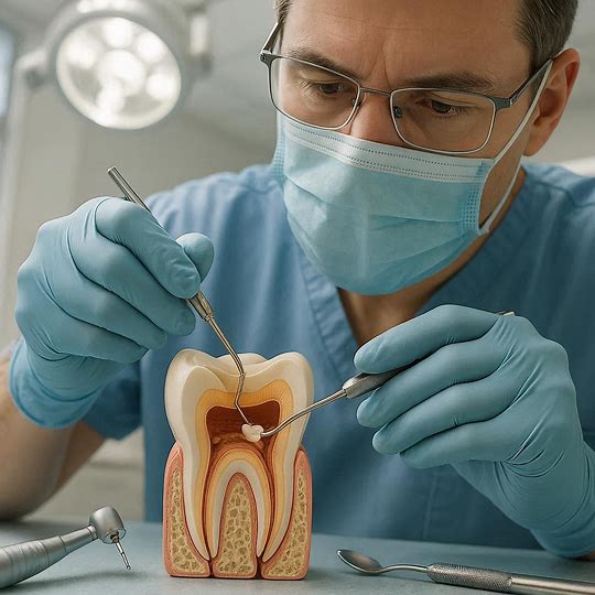
Endodontics: Illuminating the Root Canal System
For endodontists, D0369 is a game-changer:
-
Diagnosing Complex Pain: Identifying cracks (vertical root fractures), missed canals, and periapical pathosis that are invisible on 2D films.
-
Managing Retreatments: Visualizing the true anatomy of previously treated teeth, locating separated instruments, and assessing perforations.
-
Surgical Planning: For apicoectomies, providing a detailed map of the root apex and its relationship to adjacent structures to minimize surgical access and maximize success.
Oral and Maxillofacial Surgery: Navigating Complex Anatomy
Surgeons rely on D0369 for:
-
Impacted Tooth Removal: Especially for mandibular third molars, precisely determining the tooth’s position in relation to the inferior alveolar nerve, greatly reducing the risk of nerve paresthesia.
-
Pathology Assessment: Determining the exact size, extent, and nature of cysts, tumors, and other lesions within the jawbones.
-
Trauma Management: Evaluating complex fractures of the jaws, condyles, and midface in multiple planes, which is essential for effective reduction and fixation.
-
Orthognathic Surgery: Planning for corrective jaw surgery by creating a 3D virtual model of the skull for precise osteotomy planning and prediction of soft tissue changes.
Orthodontics: Beyond the Cephalometric Radiograph
Modern orthodontics uses D0369 for:
-
Airway Analysis: Assessing adenoid hypertrophy and nasopharyngeal airway volume, which can be a factor in sleep-disordered breathing and craniofacial development.
-
Impacted Tooth Localization: Precisely locating impacted canines and other teeth to plan effective and efficient traction pathways.
-
Root Resorption Monitoring: Accurately assessing the amount of root resorption during treatment, which is difficult to quantify with 2D images.
-
3D Cephalometrics: Moving beyond 2D tracing to perform analysis on a 3D reconstructed skull model, eliminating projection errors.
Periodontics: Assessing Bone Morphology and Defects
Periodontists use CBCT to:
-
Visualize Intrabony Defects: See the true morphology, depth, and number of walls of periodontal bone defects, which is critical for determining the prognosis and planning regenerative procedures.
-
Furcation Involvement: Accurately classify furcation involvements in maxillary molars.
-
Evaluate Root Anatomy: Assess the shape and concavities of roots adjacent to defects.
Temporomandibular Joint (TMJ) Analysis
CBCT provides exquisite detail of the bony components of the TMJ, allowing for the diagnosis of degenerative changes (osteoarthritis), ankylosis, and condylar fractures. While it does not image the soft tissue disc (for which MRI is used), it is the best modality for assessing bony pathology.
Airway Analysis and Sleep Apnea
Dentists involved in oral appliance therapy for sleep apnea use D0369 to visualize the entire airway in 3D, identifying the site and severity of obstruction, and to evaluate skeletal relationships that may contribute to the condition.
Diagnosis of Pathologies and Trauma
CBCT is an exceptional tool for identifying and characterizing a wide range of conditions, from periapical cemento-osseous dysplasia to sclerosing osteitis, and for providing a comprehensive view of traumatic injuries to the dentition and facial skeleton.
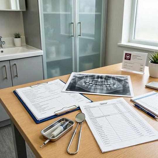
6. The D0369 Procedure: From Patient Preparation to Image Acquisition
The process of acquiring a CBCT scan is straightforward but requires careful attention to detail to ensure patient safety and diagnostic efficacy.
-
Clinical Justification (The “Why”): This is the most critical first step. Due to the higher radiation dose compared to 2D imaging, a CBCT scan must be justified. The dentist must determine that the diagnostic benefits outweigh the risks and that the same information cannot be obtained with a lower-dose 2D image. This decision is based on a clinical examination and patient history.
-
Patient Preparation: The patient is asked to remove any metal objects from the head and neck region (eyeglasses, earrings, hearing aids, dentures, etc.) as these can cause artifacts in the image. A lead apron with a thyroid collar is placed on the patient for protection, even though the primary beam is highly collimated.
-
Positioning: The patient is carefully positioned within the CBCT unit. This may involve using a chin rest, head supports, and laser positioning lights to align the patient’s head according to the desired field of view (FOV). Minimizing movement is crucial.
-
Parameter Selection: The operator selects the appropriate scan parameters based on the clinical question:
-
Field of View (FOV): This is the size of the anatomical area scanned. A small FOV (e.g., 5×5 cm) is used for a single implant site or tooth. A large FOV (e.g., 16×22 cm) captures the entire craniofacial skeleton for orthognathic or airway planning. Using the smallest FOV necessary is a key principle of the ALARA (As Low As Reasonably Achievable) radiation safety protocol.
-
Voxel Size: The resolution of the scan. Smaller voxel sizes (e.g., 0.08 mm) provide higher resolution but may require a higher dose and longer processing time. Larger voxel sizes (e.g., 0.4 mm) are sufficient for larger anatomical surveys.
-
kVp and mA: The energy and current settings that influence image contrast and dose.
-
-
Scan Acquisition: The machine rotates around the patient’s head, typically taking 10-40 seconds to complete the scan. The patient must remain perfectly still during this time. Some machines offer a “pediatric” or “low-dose” mode for specific applications.
-
Initial Reconstruction: Once the scan is complete, the primary reconstruction of the 3D volume happens almost instantly within the machine’s software. The raw data is saved, and the reconstructed volume is now ready for viewing and interpretation.
7. Interpreting the Results: The Art and Science of Reading a CBCT Scan
The acquisition of the data (D0369) is only half the story. The real value is unlocked in the interpretation (D0381). Reading a CBCT scan requires specialized training and a systematic approach.
-
Navigation: The interpreter uses software to scroll through the volumetric data set in the axial, coronal, and sagittal planes simultaneously. They can also create reconstructed panoramic and cross-sectional views.
-
Windowing: Adjusting the contrast and brightness (window/level settings) is essential to optimize the visualization of different tissues (bone, soft tissue, air).
-
Systematic Evaluation: A trained radiologist or clinician follows a specific protocol to examine all anatomy within the FOV:
-
Teeth and Supporting Structures: Periodontal bone levels, lamina dura, furcations.
-
Jawbones: Mandible and maxilla, looking for symmetry, cortication, and any radiolucencies/radiopacities.
-
Airways: Nasal cavity, nasopharynx, oropharynx.
-
Sinuses: Maxillary sinuses for mucosal thickening or pathology.
-
Tempromandibular Joints: Condylar head shape, cortex integrity.
-
Critical Anatomical Landmarks: Inferior alveolar canal, mental foramen, incisive canal, maxillary sinus floor, nasal floor.
-
-
Diagnostic Report: The findings are documented in a formal report that describes any normal variants, abnormalities, pathologies, and their precise locations. This report becomes a permanent part of the patient’s medical record.
8. Weighing the Considerations: Benefits, Risks, and Safety Protocols
Benefits:
-
Unmatched Diagnostic Information: Provides 3D data that is simply unattainable with 2D imaging.
-
Improved Treatment Outcomes: Leads to more accurate diagnoses and more predictable and successful treatments, especially in surgery and implantology.
-
Enhanced Patient Communication: The 3D models can be shown to patients, helping them understand their condition and the proposed treatment, leading to increased informed consent.
-
Minimally Invasive Procedures: Enables flapless surgery and smaller surgical approaches, reducing patient morbidity and healing time.
Risks and Limitations:
-
Ionizing Radiation: The primary risk is exposure to ionizing radiation. While a CBCT dose is significantly lower than a medical CT scan of the same area, it is higher than a single panoramic or periapical X-ray.
-
Artifacts: Metallic restorations (amalgam, implants) can cause streak artifacts that obscure adjacent anatomy. Patient movement can cause blurring.
-
Soft Tissue Resolution: CBCT is excellent for bone but has poor soft tissue contrast compared to MRI. It cannot reliably differentiate between different soft tissues.
-
Cost and Training: The equipment is expensive, and its operation and interpretation require significant training and expertise.
Safety Protocol: ALARA
The guiding principle for all radiographic imaging, especially CBCT, is ALARA (As Low As Reasonably Achievable). This means:
-
Justification: Never perform a “routine” CBCT scan. It must be clinically indicated.
-
Optimization: Use the smallest FOV, the lowest resolution (largest voxel size), and the lowest mA and kVp settings that will still answer the clinical question.
-
Limitation: Use lead shielding (apron, thyroid collar) and ensure equipment is regularly maintained and calibrated.
9. The Financial Dimension: Cost, Insurance, and Reimbursement for D0369
The cost of a CBCT scan varies widely based on geographic location, the dental practice, the size of the FOV, and whether interpretation by a radiologist is included. Costs can range from $200 to $800 per scan.
Insurance and Reimbursement: Dental insurance plans vary greatly in their coverage of D0369. Many plans now recognize its value for specific procedures like implant planning and impacted tooth removal and will provide partial coverage. However, coverage is rarely automatic. Pre-authorization is often required, and the dentist must submit strong clinical justification (e.g., narrative, clinical photos, 2D X-rays) to demonstrate the medical necessity of the 3D scan. It is often considered a “diagnostic” service subject to the patient’s deductible and coinsurance.
10. The Future of CBCT and 3D Imaging in Dentistry
The technology behind D0369 continues to evolve rapidly. Future trends include:
-
Ultra-Low Dose Protocols: Advances in detector technology and reconstruction algorithms will continue to drive patient dose down while maintaining image quality.
-
Artificial Intelligence (AI): AI algorithms are being integrated to automate tasks like nerve canal tracing, cephalometric analysis, caries and pathology detection, and implant planning, increasing speed and consistency.
-
Enhanced Soft Tissue Imaging: Developments in contrast agents and dual-energy CBCT may improve the ability to visualize soft tissues.
-
Point-of-Care 3D Printing: The seamless integration of CBCT data with 3D printers will allow for the immediate in-office fabrication of surgical guides, models, and custom implants.
-
Functional Imaging: The ability to not just see static anatomy but also to assess function, such as dynamic TMJ imaging or blood flow.
11. Conclusion: D0369 as a Cornerstone of Precision Dentistry
The D0369 dental code represents far more than a billing entry; it signifies a fundamental shift towards data-driven, precision-based dentistry. By providing a detailed three-dimensional map of the complex craniofacial region, CBCT technology has elevated diagnostic confidence, revolutionized treatment planning across all specialties, and improved patient safety and outcomes. While its use must always be prudently justified by the ALARA principle, its role as an indispensable tool in modern dental practice is firmly established, paving the way for a future where diagnosis and treatment are more accurate, predictable, and minimally invasive than ever before.
12. Frequently Asked Questions (FAQs)
Q1: Is a CBCT scan the same as a medical CT scan?
A: No. While both produce 3D images, they use different technologies. CBCT uses a cone-shaped beam that rotates once around the head, making it faster and emitting a significantly lower radiation dose for dental-specific applications. Medical CT uses a fan-shaped beam and a spiral acquisition pattern, which provides superior soft tissue detail but at a higher dose.
Q2: How much radiation am I exposed to during a D0369 scan?
A: The effective dose varies dramatically based on the machine settings and the size of the field of view. A small FOV scan can have an effective dose comparable to a few days of natural background radiation (similar to a panoramic X-ray or a cross-country flight), while a large FOV scan might be equivalent to a few months of background radiation. Your dentist will always use the lowest dose possible to get the necessary information.
Q3: Why would my general dentist refer me to a specialist for a CBCT scan?
A: Many general dentists have CBCT machines in their offices. However, some may refer patients to an oral surgeon, endodontist, or oral radiologist for the scan. This is often because the specialist has a machine with a specific field of view or resolution needed for the case, or because the specialist will be the one performing the advanced interpretation and treatment planning based on the scan.
Q4: Can I refuse a CBCT scan if my dentist recommends one?
A: Absolutely. All medical and dental procedures require your informed consent. You have the right to refuse. However, it is important to have a discussion with your dentist about why they are recommending the scan and what the potential diagnostic limitations and risks might be if you proceed with treatment without the 3D information.
Q5: How long does it take to get the results?
A: The 3D images are available immediately after the scan is completed. However, if a formal interpretation and report by a radiologist are required (coded as D0381), this may take anywhere from a few hours to a few days, depending on the service used.
13. Additional Resources
-
American Dental Association (ADA): The official source for the Current Dental Terminology (CDT) manual, which defines code D0369.
-
American Academy of Oral and Maxillofacial Radiology (AAOMR): The leading professional organization for experts in dental radiology. They publish clinical recommendations and guidelines for the use of CBCT.
-
International Association of Dentomaxillofacial Radiology (IADMFR): A global organization that provides resources and promotes research in the field.
-
Image Gently Alliance: A coalition dedicated to promoting safe and effective use of medical imaging in children. While focused on medicine, their principles align with the ALARA concept in dentistry.
-
Your Dentist or Specialist: The best resource for your specific situation is always the dental professional who knows your health history and treatment needs.