The Complete Guide to Tooth Extraction Surgery: From Anxiety to Aftercare
- On
- InDENTAL
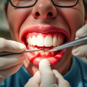
The phrase “tooth extraction” often conjures images of medieval pliers, brute force, and sheer agony. This antiquated perception, however, is a stark contrast to the reality of modern tooth extraction surgery—a highly sophisticated, predictable, and patient-centered procedure grounded in science, technology, and profound empathy. Today, exodontia (the technical term for tooth removal) is not merely an act of pulling a tooth; it is a deliberate surgical intervention designed to eliminate pathology, alleviate pain, and restore the foundation for oral health and well-being.
This comprehensive guide aims to demystify every facet of the tooth extraction process. Whether you are a patient facing an upcoming procedure, a student of dentistry, or simply someone seeking to understand this common surgical practice, this article will serve as an authoritative resource. We will journey from the initial decision-making process and the critical pre-operative planning stages through the intricacies of the procedure itself, and finally, into the detailed pathway of recovery and healing. Our goal is to replace anxiety with knowledge and fear with confidence, empowering you with the understanding that modern tooth extraction is a routine, safe, and manageable step on the path to optimal oral health.

Table of Contents
ToggleChapter 1: The Why – Indications for Tooth Extraction
A tooth is not removed on a whim. The decision is always a last resort, made after careful consideration of all possible alternatives to save the natural tooth. The indications for extraction are varied and multifaceted.
The Unsalvageable Tooth: Decay, Trauma, and Infection
The most common reason for extraction is severe tooth decay (dental caries) that has destroyed so much of the tooth structure that it cannot be restored with a filling or crown. When decay reaches the pulp chamber, it causes infection and inflammation (pulpitis), leading to a dental abscess. While root canal therapy can often save such a tooth, if the infection is too severe, has caused significant bone loss, or the tooth is too structurally compromised, extraction becomes the only viable option to prevent the spread of infection. Similarly, teeth fractured below the gumline or split longitudinally (vertical root fracture) are often impossible to save and require removal.
Orthodontic Motivations: Creating Space for a Perfect Smile
Orthodontists often recommend the extraction of one or more teeth to address crowding and create the space necessary to properly align the remaining teeth. This is a strategic decision aimed at achieving a stable, functional, and aesthetic bite. Typically, first premolars are chosen for extraction in these cases due to their position in the dental arch.
Periodontal Disease: When the Foundation Fails
Advanced periodontal (gum) disease destroys the bone and ligaments that support teeth, leading to significant mobility. When teeth become so loose that they affect function and comfort, and when the bone loss is too advanced for regenerative procedures to be successful, extraction is indicated. Removing these hopeless teeth eliminates a chronic source of infection and allows for treatment planning with prosthetic replacements like dentures or implants.
Strategic Extractions: Wisdom Teeth and Prosthetic Preparation
Third molars, or wisdom teeth, are frequently extracted prophylactically. They often erupt misaligned, become impacted (trapped in the jawbone), and can cause a host of problems, including pericoronitis (infection around the crown), cysts, damage to adjacent teeth, and crowding. Furthermore, for patients planning to get dentures, sometimes teeth are extracted to make room for the prosthetic appliance.
Medical Necessity: Oncology, Radiation, and Organ Transplantation
In some medical contexts, teeth may need to be extracted to mitigate risk. For example, before radiation therapy for head and neck cancer, teeth in the field of radiation with a poor prognosis are often removed to prevent a serious bone infection called osteoradionecrosis later on. Similarly, patients about to undergo organ transplantation or cardiac valve surgery may have compromised teeth extracted to eliminate potential sources of infection that could have devastating consequences under immunosuppression.
Chapter 2: The Pre-Operative Phase – Meticulous Planning for Optimal Outcomes
The success of an extraction is determined long before the first instrument touches the tooth. The pre-operative phase is a critical process of assessment, planning, and preparation.
The Comprehensive Consultation: More Than Just an X-Ray
The process begins with a thorough clinical examination. The surgeon will visually inspect the tooth, check its mobility, and assess the condition of the surrounding gums. They will also evaluate your bite and the relationship of the tooth to be extracted with its neighbors and opposing teeth.
Medical History: The Cornerstone of Safety
This is arguably the most important part of the consultation. You must disclose your complete medical history, including:
-
Medications: Especially blood thinners (anticoagulants like warfarin, clopidogrel, aspirin), bisphosphonates (for osteoporosis), and immunosuppressants.
-
Medical Conditions: Such as diabetes, hypertension, heart conditions, bleeding disorders, liver disease, or a history of bacterial endocarditis.
-
Allergies: To medications, latex, or other substances.
This information allows the surgeon to mitigate risks, consult with your physician if necessary, and choose the safest possible protocol for your procedure.
Imaging Revolution: The Critical Role of Panoramic X-Rays and 3D CBCT Scans
While a small periapical X-ray shows the tooth and its immediate root structure, modern extraction planning, especially for surgical cases, relies on advanced imaging.
-
Panoramic X-Ray (OPG): Provides a broad view of both jaws, all teeth, the sinuses, and nerve canals. It is essential for assessing wisdom teeth and planning multiple extractions.
-
Cone Beam Computed Tomography (CBCT): This 3D imaging technology is the gold standard for complex cases. It allows the surgeon to visualize the exact position of the tooth roots in relation to vital structures like the inferior alveolar nerve (which provides sensation to the lip and chin) and the maxillary sinus. This precise anatomical understanding is crucial for avoiding complications and planning an atraumatic surgery.
* Comparing Pre-Operative Imaging Modalities*
| Feature | Periapical X-Ray | Panoramic X-Ray (OPG) | Cone Beam CT (CBCT) |
|---|---|---|---|
| View | 2D, detailed view of 1-2 teeth | 2D, wide view of entire jaw | 3D, high-resolution view of specified area |
| Best For | Simple extractions, assessing decay | Initial assessment, wisdom teeth planning | Complex & surgical extractions, proximity to nerves/sinuses |
| Information | Root shape, number, periapical infection | Tooth angulation, gross relationship to structures | Precise 3D location, bone density, nerve canal location |
| Radiation Dose | Low | Low to Moderate | Moderate (higher than 2D, lower than medical CT) |
Informed Consent: Understanding Risks, Benefits, and Alternatives
Before the procedure, your surgeon will discuss the planned extraction with you in detail. This informed consent process includes:
-
The reason for the extraction.
-
The procedure itself and what to expect.
-
The benefits of removing the tooth.
-
Risks and potential complications, such as pain, swelling, bleeding, infection, dry socket, sinus communication (for upper teeth), temporary or permanent nerve damage (for lower teeth), and root fracture.
-
Alternatives to extraction (if any exist) and the risks of not proceeding.
You will have the opportunity to ask questions and must provide your voluntary consent before treatment begins.
Pre-Procedure Instructions: Fasting, Medications, and Logistics
Depending on the type of anesthesia planned, you may be instructed to fast (no food or drink) for 6-8 hours beforehand. You may be advised to take certain prescribed medications (e.g., antibiotics) ahead of time. It is also crucial to arrange for a responsible adult to drive you home if you will be receiving sedation, and to clear your schedule for adequate rest post-operatively.
Chapter 3: A World of Anesthesia – Ensuring a Pain-Free and Anxiety-Free Experience
Modern dentistry offers a spectrum of anesthesia options to ensure patient comfort, tailored to the complexity of the procedure and the patient’s anxiety level.
Local Anesthesia: Numbing the Specific Area
This is the foundation of all dental surgical procedures. A local anesthetic (like Lidocaine or Articaine) is injected near the tooth to block pain signals from the specific nerves serving that area. You will be awake and aware, but you should feel no pain, only pressure and movement. For most simple extractions, local anesthesia is sufficient.
Sedation Dentistry: From Minimal to Deep Sedation
For anxious patients or more complex surgeries, various levels of sedation can be used in conjunction with local anesthesia.
-
Nitrous Oxide (“Laughing Gas”): A minimal sedation technique. You inhale a mixture of nitrous oxide and oxygen through a nasal mask. It induces a state of relaxation and euphoria while keeping you fully conscious and able to follow instructions. The effects wear off quickly once the gas is turned off.
-
Oral Conscious Sedation: Involves taking a prescribed sedative pill (like Diazepam or Triazolam) about an hour before the procedure. You will remain conscious but in a deeply relaxed, drowsy state. Many patients have little memory of the procedure afterward. This requires a companion for transportation.
-
Intravenous (IV) Sedation: Administered directly into the bloodstream by the surgeon or an anesthesiologist, IV sedation allows for a deeper level of sedation where you are on the edge of consciousness. The level can be adjusted throughout the procedure, and it provides profound amnesia. This is common for complex wisdom tooth removals.
General Anesthesia: For Complex Cases
Under general anesthesia, you are completely unconscious and unaware. This is typically reserved for the most complex cases, such as the removal of multiple impacted teeth, or for patients with extreme anxiety or special needs. It is performed in a hospital or surgical center setting with a dedicated anesthesiologist monitoring your vital signs throughout.
Chapter 4: The Art and Science of the Procedure – Surgical vs. Simple Extraction
The surgical approach is determined by the visibility and accessibility of the tooth.
Simple Extraction: The Protocol for Visible Teeth
This is performed on teeth that are fully erupted and visible in the mouth.
-
Administration of Local Anesthetic: The area is thoroughly numbed.
-
Luxation: The surgeon uses an instrument called a luxator or periodontal elevator. This thin, flat instrument is carefully inserted into the periodontal ligament space—the tiny gap between the tooth root and the bone. The surgeon applies controlled pressure and leverages against the bone itself (not the adjacent tooth) to progressively widen this space and sever the ligament fibers that attach the tooth to the bone. This process loosens the tooth from its socket.
-
Delivery with Forceps: Once sufficiently luxated, dental forceps are applied to the crown of the tooth. The forceps are designed to adapt to the specific shape and root anatomy of different teeth (e.g., upper incisor forceps vs. lower molar forceps). The surgeon uses a combination of controlled rotational and rocking motions to further expand the bony socket and gently deliver the tooth out. The goal is to remove the tooth intact with minimal trauma to the surrounding tissues.
Surgical Extraction: The Protocol for Complex Cases
This is required for teeth that are not accessible, such as broken teeth at the gumline, severely decayed teeth, or teeth that are impacted (like most wisdom teeth).
-
Incision and Flap Reflection: After anesthesia, the surgeon makes a small incision in the gum tissue to create a “flap.” This flap is gently lifted away from the underlying bone, providing direct visual and physical access to the surgical site.
-
Bone Removal (Osteotomy): If the tooth is covered by bone, a high-speed surgical handpiece (drill) is used to remove a small amount of the overlying bone. Modern techniques often use Piezoelectric surgery devices, which use ultrasonic vibrations to cut mineralized tissue (bone) with incredible precision while sparing soft tissues like nerves and blood vessels, leading to less trauma and faster healing.
-
Tooth Sectioning (Odontosection): Often, to remove the tooth safely without removing excessive bone, the tooth is divided into sections. The crown may be separated from the roots, or a multi-rooted tooth may be split into individual roots. Each smaller piece is then removed individually through the created access.
-
Debridement and Irrigation: The empty socket is thoroughly cleaned of any debris, tooth fragments, or infected tissue. It is irrigated copiously with sterile saline to ensure it is clean.
-
Suturing: The gum flap is repositioned and closed with sutures (stitches). Sutures help control bleeding, protect the underlying blood clot, and promote primary healing. They may be resorbable (dissolve on their own in 7-10 days) or non-resorbable (require removal by the surgeon).
Chapter 5: The Immediate Aftermath – Navigating the First 24 Hours
Your actions in the first day post-surgery are paramount to a comfortable recovery and the prevention of complications.
The Blood Clot: Your Body’s Natural Bandage
The formation of a stable blood clot in the socket is the single most important event in the healing process. This clot acts as a protective layer over the underlying bone and nerve endings, and it serves as the foundation for new tissue growth. Protecting this clot is the primary goal of all post-operative instructions.
Post-Operative Instructions: The Holy Grail of Healing
-
Biting on Gauze: You will be asked to bite down on a piece of sterile gauze placed over the extraction site with firm, steady pressure for 30-60 minutes. This direct pressure helps the blood vessels constrict and facilitates clot formation. If oozing persists, replace with a fresh gauze for another 30 minutes.
-
The Ice Pack: Apply an ice pack to the outside of your cheek, in the area of the extraction, for 15-20 minutes on, then 15-20 minutes off, for the first 24-48 hours. This significantly reduces swelling and inflammation.
-
Rest and Activity: Plan to rest for the remainder of the day. Keep your head elevated with pillows, even when sleeping. Avoid all strenuous activity, bending over, and lifting for at least 48-72 hours, as this can increase blood pressure and lead to throbbing or bleeding.
Diet: The Transition from Liquids to Soft Foods
-
First few hours: Stick to cool liquids (water, apple juice). Avoid hot beverages.
-
First 24-48 hours: Consume a soft, cool, or lukewarm diet (yogurt, pudding, applesauce, mashed potatoes, smoothies, lukewarm soup). Do not use a straw. The sucking action creates negative pressure in your mouth that can dislodge the precious blood clot, leading to a dry socket.
-
Avoid: Spicy, crunchy, hard, or hot foods that can irritate the wound.
Pain Management: Pharmacological and Non-Pharmacological Approaches
Some discomfort is normal as the anesthesia wears off. Your surgeon will likely prescribe a pain medication or recommend an over-the-counter anti-inflammatory like ibuprofen (Advil, Motrin), which is excellent for reducing both pain and inflammation. Take the first dose before the numbness fully subsides. Applying ice is a key non-pharmacological method for pain control.
Chapter 6: The Healing Journey – From Socket to Solid Bone (Weeks 1-8+)
Healing is a biological process that occurs in sequential, overlapping stages.
Stages of Socket Healing:
-
Clot Formation (First 24 hours): The socket fills with blood, which coagulates.
-
Granulation Tissue (Days 3-7): The blood clot is infiltrated by fibroblasts and new blood vessels, forming granulation tissue. This is the framework for new tissue growth.
-
Soft Tissue Closure (Weeks 1-2): The gum tissue begins to close over the socket. The sharp edges of the bony socket begin to smooth out.
-
Bone Formation (Weeks 4-8+): Osteoblasts (bone-forming cells) slowly fill the socket with new bone. This process can take several months to complete fully.
Managing Common Symptoms:
-
Swelling: Usually peaks at 48-72 hours, then gradually subsides. Ice for the first 48 hours, then after 48 hours, switching to moist heat can help disperse lingering swelling.
-
Bruising: Discoloration of the cheek and neck is normal and will resolve on its own.
-
Trismus (Stiff Jaw): Difficulty opening your mouth wide is common due to inflammation of the jaw muscles. It resolves as healing progresses. Gentle stretching after a few days can help.
Oral Hygiene Post-Extraction:
-
Day of surgery: Avoid rinsing your mouth. Brush your teeth gently, carefully avoiding the surgical site.
-
Day 2 onward: Begin gentle rinsing with a warm saltwater solution (1/2 teaspoon salt in 8 oz of warm water) after meals and before bed. This keeps the area clean without aggressive swishing.
The Dreaded Dry Socket (Alveolar Osteitis):
This is the most common complication, occurring in about 2-5% of extractions (higher for mandibular wisdom teeth). It happens when the blood clot dislodges or dissolves prematurely, exposing the raw bone and nerves to air, food, and fluid. This causes a severe, throbbing pain that often radiates to the ear and is not well-controlled by pain medication. It typically starts 2-3 days after the extraction.
-
Prevention: Follow all post-op instructions meticulously: no straws, no smoking, no spitting, and avoid disturbing the area.
-
Treatment: There is no way to regenerate the clot. Treatment is palliative. The surgeon will gently irrigate the socket to remove debris and then pack it with a medicated sedative dressing (e.g., with eugenol) that soothes the pain and protects the nerve. This dressing may need to be changed every few days until pain subsides and granulation tissue begins to form.
Chapter 7: Advanced Techniques and Technologies in Modern Exodontia
The field is continuously evolving with innovations designed to improve precision, reduce trauma, and accelerate healing.
Piezoelectric Surgery
As mentioned, this technology uses ultrasonic frequencies to cut bone with micron-level precision. It is particularly invaluable when working near delicate structures like nerves and sinus membranes, as it is inherently selective for mineralized tissue and spares soft tissue.
Platelet-Rich Fibrin (PRF)
This is an advanced biotechnology derived from the patient’s own blood. A small sample of blood is drawn and centrifuged to concentrate the platelets, growth factors, and fibrin. This PRF membrane can be placed into the empty extraction socket before suturing. It dramatically accelerates soft tissue healing and bone regeneration, reduces post-operative pain and swelling, and significantly lowers the risk of dry socket.
Socket Preservation
When a tooth is extracted, the surrounding jawbone that supported it naturally begins to resorb and atrophy over time. This can create problems for future replacement with a dental implant, which requires adequate bone volume. Socket preservation, or alveolar ridge preservation, is a technique where a bone graft material is placed into the socket immediately after extraction. This graft acts as a scaffold that helps maintain the bone’s width and height, preserving the site for a future implant.
Robotic-Assisted and Guided Surgery
Using 3D CBCT scans and digital intraoral scans, surgeons can now plan the entire extraction procedure virtually on a computer. They can then 3D-print a surgical guide that fits over the teeth adjacent to the extraction site. This guide has precision channels that dictate the exact angle and depth for bone removal and tooth sectioning, making the procedure incredibly accurate, minimally invasive, and predictable.
Chapter 8: Special Considerations and Complex Cases
Wisdom Tooth Extractions
This is a category unto itself due to the complexity of impactions. Teeth can be soft tissue impacted, partially bony impacted, or fully bony impacted. They can be positioned vertically, horizontally, or even upside down (inverted). The proximity of the roots to the inferior alveolar nerve is a primary concern, meticulously assessed via CBCT to avoid paresthesia (numbness).
Extractions in the Esthetic Zone
Removing a front tooth requires extreme care to preserve the delicate gum tissue and underlying bone for a natural-looking future restoration, whether it’s a bridge or an implant. Techniques like “Socket Shield” involve leaving a fragment of the root on the facial side to support the bone and gum tissue, ensuring an impeccable aesthetic outcome.
Managing Medically Complex Patients
Patients on blood thinners require careful coordination with their physician. Often, the medication is continued to avoid the risk of a stroke or heart attack, and the surgeon employs local hemostatic agents (like topical thrombin, gelatin sponges, and sutures) to control bleeding. Diabetic patients require well-controlled blood sugar for proper healing, and their procedures are often scheduled for the morning.
Conclusion: Empowerment Through Knowledge
Tooth extraction surgery, far from being a dreaded ordeal, is a refined and predictable dental procedure designed to resolve pain and restore health. Success hinges on thorough pre-operative planning, precise surgical execution, and diligent patient aftercare. By understanding the process from indication to healing, patients can approach their treatment with confidence, actively partner in their recovery, and achieve the best possible outcome, paving the way for a healthier, pain-free future.
Frequently Asked Questions (FAQs)
1. How long does a tooth extraction take?
-
A simple extraction typically takes 20-40 minutes. A surgical extraction, particularly for an impacted wisdom tooth, can take from 45 minutes to 90 minutes or more, depending on complexity.
2. When can I go back to work or school after a tooth extraction?
-
For a simple extraction, you may feel fine returning to non-strenuous activities the next day. For a surgical extraction, it’s advisable to take at least 2-3 days off to rest. Listen to your body and follow your surgeon’s specific advice.
3. What is the difference between “simple” and “surgical” extraction billing codes?
-
A “simple” extraction (D7140) involves removing a tooth that is fully visible and accessible with forceps. A “surgical” extraction (D7210) involves elevation of a flap, removal of bone, and/or sectioning of the tooth. The coding is based on the complexity of the procedure performed, not the initial hope for a simple removal.
4. How much does a tooth extraction cost?
-
Cost varies widely based on complexity (simple vs. surgical), geographic location, the type of anesthesia used, and the surgeon’s expertise. A simple extraction can range from $150-$400 per tooth. A surgical extraction, particularly with IV sedation, can range from $300-$800+ per tooth. Wisdom teeth removal is often billed as a package.
5. When can I smoke after a tooth extraction?
-
It is strongly recommended to avoid smoking for at least 72 hours, but ideally for 7-10 days. The sucking motion can cause a dry socket, and the chemicals in smoke can impede healing and increase the risk of infection.
6. How do I know if I have an infection after an extraction?
-
Signs of infection include:
-
Throbbing pain that worsens after 2-3 days instead of improving.
-
Increased swelling after 3 days.
-
A foul taste or odor in your mouth.
-
Pus drainage from the socket.
-
Fever.
-
If you experience any of these, contact your surgeon immediately.
-
Additional Resources
-
American Association of Oral and Maxillofacial Surgeons (AAOMS): https://www.aaoms.org/ – Provides patient information on procedures, anesthesia, and recovery.
-
American Dental Association (ADA): https://www.ada.org/ – A resource for finding a qualified dentist and general oral health information.
-
International Association for Dental Traumatology (IADT): https://www.iadt-dentaltrauma.org – Guidelines for the management of traumatic dental injuries, including when extraction is necessary.
-
National Institute of Dental and Craniofacial Research (NIDCR): https://www.nidcr.nih.gov/ – Offers information on oral health conditions and research.
Author: Dr. Evelyn Reed, DDS, Maxillofacial Surgery Specialist
Disclaimer: The information provided in this article is for educational and informational purposes only and does not constitute medical advice. It is not a substitute for professional medical advice, diagnosis, or treatment. Always seek the advice of your dentist, oral surgeon, or other qualified health provider with any questions you may have regarding a medical condition or procedure. Never disregard professional medical advice or delay in seeking it because of something you have read in this article.
dentalecostsmile
Newsletter Updates
Enter your email address below and subscribe to our newsletter


