Beyond the X-Ray: A Comprehensive Guide to the D0102 Dental Code and Your Oral Health
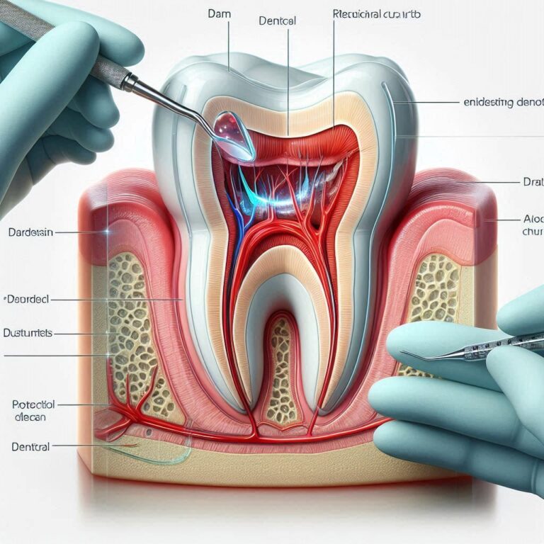
Imagine a detective arriving at a complex crime scene but being forced to work blindfolded. They could touch surfaces, listen for sounds, and ask questions, but they would be utterly missing the critical evidence hidden from plain sight—the fingerprint on a glass, the coded message in a drawer, the hair strand on a carpet. In many ways, a dentist performing a routine check-up without dental radiographs is that blindfolded detective. The human eye is an incredible tool, but it is limited to the surfaces it can see: the outer enamel of a tooth, the visible portion of the gums, the cheek mucosa. The true story of your oral health, however, is often written in the hidden chapters—the bone beneath the gums, the tight contacts between teeth, the roots anchored in the jaw, and the developing teeth in a child’s jaw.
This is where the science and art of dental radiology come into play, serving as the dentist’s indispensable “x-ray vision.” And among the most crucial, comprehensive, and commonly prescribed sets of diagnostic images is the one defined by the American Dental Association (ADA) Code D0102. This code is far more than a line item on a bill or a checkbox on a form; it is a foundational pillar of modern preventive and restorative dentistry. It represents a detailed map of your oral landscape, revealing the hidden contours, potential pitfalls, and silent problems that, if left undetected, could lead to significant pain, complex procedures, and compromised health down the line.
This article will serve as your definitive guide to the D0102 dental code. We will journey deep into its definition, explore the specific images it comprises, demystify the procedure, address paramount safety concerns, analyze its cost and value, and illuminate its irreplaceable role in crafting a healthy, functional, and beautiful smile. Our goal is to transform this alphanumeric code from an abstract concept into a clear understanding of a vital tool in safeguarding your well-being.

2. Decoding the Code: What Exactly is D0102?
Within the intricate system of the ADA’s Current Dental Terminology (CDT), every procedure, from a simple cleaning to a complex implant surgery, is assigned a unique code. This system ensures uniformity, simplifies insurance processing, and provides clarity for both providers and patients. The code D0102 falls under the category of “Diagnostic” services. Its official descriptor, as per the CDT, is: “periodic oral evaluation” – but this is a common point of confusion.
It is vital to understand that D0102 is not the clinical evaluation itself. The periodic oral evaluation, which is the physical examination performed by the dentist, has its own code (D0120). Instead, D0102 is specifically the code for the series of radiographic images taken to support that evaluation. Think of it this way:
-
D0120 (Periodic Oral Evaluation): The dentist’s visual examination, using tools like mirrors and probes to check your teeth and gums.
-
D0102: The set of X-ray images that provides the sub-surface data the dentist uses to inform their diagnosis during the D0120 exam.
The D0102 code is typically used for a series of intraoral radiographs taken during a recall or periodic examination. The most common configuration for a D0102 series is a set of bitewing radiographs, often supplemented with selected periapical images to form a complete overview. The exact number and type of films are determined by the dentist based on the individual patient’s needs and clinical situation. Its “periodic” nature means it is not the full-mouth series (D0210) typically taken at an initial visit, but a more focused set used to monitor changes over time since the last comprehensive set of images.
3. A Symphony of Images: The Specific Radiographs Included in D0102
A D0102 is not a single, monolithic image. It is a curated collection, each type of radiograph playing a specific role in the diagnostic orchestra. The two primary players are bitewings and periapicals.
Bitewing Radiographs (BWX)
These are the workhorses of the periodic exam. The name “bitewing” comes from the small paper or plastic tab (the “wing”) that the patient bites down on to hold the film or sensor in place. A single bitewing image captures the crowns of both the upper and lower teeth in a specific area of the mouth.
-
What They Show: Bitewings are unparalleled in their ability to reveal decay (caries) between the teeth (interproximal caries), which is completely invisible to the naked eye. They also provide an excellent view of the height of the alveolar bone (the bone that supports the teeth), allowing the dentist to monitor for bone loss associated with periodontal (gum) disease. Additionally, they can show the fit of existing fillings, crowns, and other restorations, checking for open margins or recurrent decay underneath.
-
Typical Series: A full series for an adult with all their teeth often consists of four bitewing films, capturing the left and right sides of both the front and back teeth.
Periapical Radiographs (PA)
“Periapical” literally means “around the apex (tip) of the root.” These images are designed to capture the entire tooth, from its absolute crown to the very end of its root(s) and the surrounding bone structure.
-
What They Show: PAs are essential for diagnosing problems inside the tooth (endodontic issues) or around the root tip. They are used to diagnose abscesses, infections, cysts, or other abnormalities in the periapical region. They are crucial for root canal therapy, as they show the number, shape, and length of the roots. They also are used to assess impacted teeth, fractures within the root, and the bone levels around a specific tooth in greater detail than a bitewing can provide.
-
In a D0102 Context: While a D0210 full-mouth series includes PAs of every tooth, a D0102 might include selected periapicals. For example, if a patient has a history of root canal treatment on one tooth, the dentist might take a PA of that specific tooth during a periodic exam to ensure it remains healthy, even if the rest of the exam is supported by bitewings.
Key Differences Between Bitewing and Periapical Radiographs
| Feature | Bitewing (BWX) | Periapical (PA) |
|---|---|---|
| Primary Purpose | Detect decay between teeth, monitor bone levels | View entire tooth, root tip, and surrounding bone |
| Structures Shown | Crowns of upper & lower teeth, crestal bone | Entire tooth (crown to root tip), periapical bone |
| Common Use | Periodic exams, checking for new cavities | Diagnosing abscesses, root fractures, root canal work |
| Typical D0102 Role | Core component of the series | Selected images based on specific patient needs |
The Panoramic Radiograph (PAN)
It is important to note that a panoramic X-ray (D0330) is not typically part of a standard D0102 code. A PAN is an extraoral film, meaning the film is outside the mouth, and it captures a single image of the entire jaws, teeth, sinuses, and nasal area. It is a fantastic screening tool for impacted wisdom teeth, jaw fractures, tumors, and development in children, but it lacks the fine detail of intraoral films like bitewings for detecting small cavities. A dentist may recommend both a PAN and a D0102 series for a truly comprehensive baseline evaluation.
4. The Diagnostic Powerhouse: Why Your Dentist Recommends a D0102
The recommendation for a D0102 series is never made arbitrarily. It is a clinical decision rooted in the principle of preventive care. Finding and treating a problem when it is small is almost always simpler, less expensive, and less invasive than addressing it once it has become symptomatic and advanced. The D0102 is the primary tool for achieving this. Here’s what it can reveal:
-
Interproximal Dental Caries: This is its superpower. A cavity between two molars can burrow deep into the dentin, dangerously close to the nerve, without causing any pain or being visible from the outside. The D0102 finds it when it’s still a small lesion that can be treated with a simple filling.
-
Recurrent Decay: Decay can sneak in at the margins of existing fillings, crowns, and bridges. The D0102 monitors these areas to ensure the integrity of the restoration is maintained.
-
Periodontal Bone Loss: Gum disease is a silent destroyer. It doesn’t usually cause pain until it is very advanced. Bitewings provide a measuring stick to compare bone levels from one visit to the next. Early detection of bone loss allows for interventions like deep cleanings (scaling and root planing) to halt its progression.
-
Pulp Pathologies and Periapical Infections: Selected periapical images can reveal dark shadows at the root tips, indicating an infection or abscess that requires root canal treatment or extraction.
-
Calculus (Tartar) Deposits: Heavy calculus deposits, especially below the gumline, can be visible on X-rays, aiding in diagnosis and treatment planning for cleanings.
-
Developmental Monitoring: In children and adolescents, these X-rays monitor the development and eruption of permanent teeth, ensuring there is enough space and identifying any potential impactions early.
-
Trauma: Hidden fractures in teeth or roots resulting from trauma, even years prior, can be identified.
Without the D0102, a dentist is making educated guesses about the health of over 50% of your tooth surfaces. It is the difference between proactive care and reactive care.
5. The D0102 Procedure: What to Expect During Your Appointment
If you are scheduled for a D0102 series, knowing what to expect can ease any anxiety. The process is straightforward and performed by a trained dental assistant or hygienist.
-
Preparation: You will be seated in the dental chair and a heavy lead apron with a thyroid collar will be placed over your chest and lap. This is a standard safety precaution that effectively shields your body from stray radiation.
-
Digital Sensor Placement: Modern dentistry has largely transitioned to digital radiography. Instead of plastic film packets, a small, smooth, and often slightly bulky electronic sensor is used. The assistant will gently place this sensor inside your mouth, positioned against the teeth to be imaged.
-
Positioning the Aiming Device: The X-ray machine has a long arm with a tube at the end. The assistant will position this tube just outside your cheek, lining it up precisely with the sensor inside. They use a device called a cone-beam collimator to ensure the beam is focused only on the target area.
-
The Exposure: You will be asked to remain very still. The assistant will step behind a protective wall or leave the room and activate the machine. The exposure itself is incredibly quick—often less than a second. You will hear a faint beep or buzz. There is no pain associated with the X-ray beam.
-
Image Review: With digital systems, the image appears on a computer screen within seconds. The dentist will review these images in high resolution, zooming in and enhancing contrast as needed, to make an accurate diagnosis.
The entire process for a four-bitewing series is typically completed in under five minutes.
6. Safety First: Understanding Radiation and Protective Measures
The word “radiation” can understandably cause concern. However, it is crucial to contextualize the exposure from dental X-rays.
The Dose is Extremely Low. We are all exposed to background radiation daily from the sun, soil, rocks, and air (cosmic and terrestrial radiation). This is known as background radiation exposure.
-
The effective dose from a full set of four digital bitewing images is approximately 0.005 mSv (millisieverts).
-
To put this in perspective, you receive a dose of about 0.008 mSv on a short 1-2 hour airplane flight due to increased cosmic radiation at altitude.
-
The average person in the U.S. receives about 3.0 mSv of background radiation exposure per year.
This means the radiation from a D0102 series is less than the radiation you are exposed to naturally in a single day. The diagnostic benefit of identifying a serious oral health problem early far outweighs the negligible risk associated with this minimal level of radiation.
Modern Safety Measures:
-
Lead Aprons: Standard practice for all intraoral X-rays.
-
Digital Technology: Digital X-ray sensors are far more sensitive than old-fashioned film, requiring up to 90% less radiation to produce a high-quality image.
-
Collimation: The beam is tightly focused only on the area of interest.
-
ALARA Principle: Dentists follow the “As Low As Reasonably Achievable” principle, meaning they only prescribe X-rays when there is a clear clinical need and they use the lowest possible exposure settings to get the necessary diagnostic information.
7. The Financials: Cost, Insurance, and Value of D0102
The cost of a D0102 series can vary based on geographic location, the dental practice’s overhead, and whether film or digital systems are used. On average, patients can expect a cost ranging from $75 to $250 for a set of four bitewing X-rays.
Insurance Coverage: Most dental insurance plans cover 100% of the cost of preventive services, which include periodic exams (D0120), cleanings (D1110), and periodic X-rays (D0102), when deemed medically necessary. They typically follow guidelines suggesting bitewing X-rays at 6-12 month intervals for high-risk patients and 12-24 month intervals for low-risk patients. It is always advisable to check with your insurance provider about your specific coverage.
The Value Proposition: While the out-of-pocket cost might seem like an expense, it is better viewed as a critical investment in your long-term health and financial well-being. The cost of a D0102 ($100-$200) is minuscule compared to the cost of treating problems it can uncover early:
-
Treating a small surface cavity: $150 – $300 (filling)
-
Treating a large cavity that has reached the nerve: $1,000 – $2,000 (root canal + crown)
-
Treating advanced gum disease: $1,500 – $3,000+ (per quadrant for deep cleaning)
-
Replacing a tooth lost to decay or gum disease: $3,000 – $5,000+ (implant)
The D0102 is the most cost-effective diagnostic tool in dentistry, capable of saving patients thousands of dollars and significant discomfort.
8. Case Studies: D0102 in Action – Real-World Diagnostic Scenarios
Case Study 1: The Silent Interproximal Cavity
Sarah, a 32-year-old with no dental pain, attends her 6-month recall. Her clinical exam is perfect—no visible decay, healthy gums. Her dentist takes a standard D0102 series as it has been 18 months since her last X-rays. The bitewing reveals a moderate-sized cavity between her upper first and second molar, halfway through the dentin layer. Sarah receives a simple composite filling at the same appointment. Without the D0102, this cavity would have continued to grow, likely resulting in a toothache and the need for a root canal and crown within a year.
Case Study 2: The Hidden Bone Loss
Mark, a 55-year-old, is a former smoker. He brushes diligently but has noticed some bleeding when he flosses. His clinical exam shows 4mm periodontal pockets in a few areas, but the full extent is unclear. His D0102 bitewing series, when compared to his X-rays from two years prior, shows clear, progressive horizontal bone loss across his entire posterior dentition. This objective evidence allows the dentist to diagnose moderate chronic periodontitis and recommend a series of scaling and root planing appointments to arrest the disease and prevent tooth loss.
Case Study 3: The Failed Old Restoration
Maria has a large silver amalgam filling from 15 years ago. The tooth feels fine. Her D0102 series shows a distinct dark line between the filling and the tooth structure on the X-ray, indicating a broken seal and recurrent decay undermining the filling. The dentist replaces the old amalgam with a new crown before the decay can infect the pulp, saving Maria from a future dental emergency.
9. Beyond D0102: How It Fits into the Complete Diagnostic Puzzle
The D0102 is a vital component, but it is part of a larger diagnostic ecosystem. Dentists use a “mix-and-match” approach based on a patient’s individual needs:
-
D0120 (Periodic Exam): The clinical counterpart to the D0102.
-
D0210 (Full Mouth Series – FMX): Typically 14-20 images including PAs of every tooth and bitewings. Taken at initial exams or when comprehensive baseline data is needed.
-
D0330 (Panoramic Film – PAN): Excellent for overall jaw structure, wisdom teeth, and screening.
-
D0380 (Cone Beam CT – CBCT): Provides 3D imaging for complex implant planning, orthodontic assessment, and surgical procedures. It delivers a higher dose than 2D X-rays and is only used when its specific diagnostic benefits are required.
The D0102 is the go-to code for the efficient and effective monitoring that occurs between these more comprehensive exams.
10. The Future of Dental Radiography: Digital Advancements and 3D Imaging
The field is constantly evolving. Digital sensors continue to improve, requiring even less radiation. Software now incorporates Artificial Intelligence (AI) that can algorithmically analyze X-rays to flag potential areas of decay, bone loss, or even signs of osteoporosis, acting as a powerful second opinion for the dentist. While 3D CBCT technology is becoming more common, the 2D D0102 series will remain the gold standard for routine interproximal and crestal bone assessment due to its unparalleled detail for these specific tasks, low cost, and minimal radiation dose.
11. Conclusion: Your Health is Worth the Picture
The D0102 dental code is far more than a billing number; it is a commitment to proactive, evidence-based oral healthcare. It provides an invaluable window into the hidden aspects of your dental health, enabling early detection of decay, disease, and other problems long before they become painful or expensive to treat. The procedure is quick, safe, and comfortable, with radiation exposure levels that are truly negligible, especially when weighed against the profound benefits. By understanding and consenting to this periodic diagnostic tool, you are actively partnering with your dentist to preserve your natural smile for a lifetime. Your health is truly worth the picture.
12. Frequently Asked Questions (FAQs)
Q1: My teeth feel fine and my dentist doesn’t see any problems. Why do I need X-rays?
A: Many serious dental issues, like decay between teeth or bone loss from gum disease, are completely painless and invisible to the naked eye in their early stages. X-rays are a preventive tool designed to find these problems before you feel them, allowing for simple, conservative treatment.
Q2: How often should I get a D0102 series?
A: There is no one-size-fits-all answer. The frequency depends on your individual risk factors: history of decay, gum health, age, diet, and dry mouth conditions. High-risk patients may need them every 6-12 months, while low-risk patients may only need them every 18-36 months. Your dentist will recommend an interval tailored specifically to you.
Q3: I’m pregnant. Can I still get dental X-rays?
A: Yes, but only if absolutely necessary. You should always inform your dentist if you are or might be pregnant. While the radiation dose is extremely low and focused on your mouth (far from the abdomen), dentists will typically postpone routine X-rays as a precaution. However, if you have a dental emergency requiring X-rays for diagnosis, they can be performed with double-lead shielding. The American Dental Association and American Congress of Obstetricians and Gynecologists affirm that with proper shielding, dental X-rays are safe during pregnancy.
Q4: What’s the difference between the small films you put in my mouth and the big machine that goes around my head?
A: The small films are for intraoral X-rays (like D0102 bitewings and periapicals), which provide extremely high detail of individual teeth and the bone immediately surrounding them. The big machine that rotates is for a panoramic X-ray (D0330), which provides a single broad-view image of the entire jaw, teeth, and surrounding structures but with less fine detail.
Q5: Are digital X-rays really better?
A: Yes. Digital systems offer significant advantages: they use up to 90% less radiation than traditional film, the images are available instantly, and the dentist can enhance them (zoom, contrast adjustment) on a computer screen to aid in diagnosis. They are also more environmentally friendly as they eliminate the need for chemical processing.
13. Additional Resources
-
American Dental Association (ADA): The definitive source on dental codes and guidelines. Their page on “Dental Radiographs” is an excellent patient resource.
-
ADA.org – Dental Radiographs (Note: This is a representative link format; please verify the exact URL on the ADA website).
-
-
U.S. Food and Drug Administration (FDA): Provides information on radiation safety in medical and dental imaging.
-
American Dental Hygienists’ Association (ADHA): Offers patient education materials on the importance of oral health maintenance, including the role of X-rays.
-
Your Dentist: Always your best resource. Do not hesitate to ask your dentist or hygienist to show you your X-rays on the screen and explain what they are looking for. A transparent dentist will be happy to educate you.
Date: September 2, 2025
Author: The Dental Codex Team
Disclaimer: This article is intended for informational purposes only and does not constitute professional medical or dental advice, diagnosis, or treatment plan. Always seek the advice of your qualified dentist or other certified health provider with any questions you may have regarding a dental condition or procedure. The information contained herein is based on current American Dental Association (ADA) guidelines as of the publication date; codes and descriptors are subject to change.