D0251 Dental Code: The Definitive Guide to the Extraoral Posterior Bitewing
Imagine a world where a routine dental check-up is not just about identifying cavities but about comprehensively mapping the intricate architecture of your oral health with unprecedented clarity and comfort. For decades, the dental industry has relied on a standard set of tools, with the intraoral bitewing radiograph standing as the undisputed gold standard for detecting decay between teeth. Patients worldwide are familiar with the slightly awkward process: the careful placement of a small, often uncomfortable, plastic-tabbed sensor inside the mouth, the patient biting down gingerly, and the hope that the image is captured correctly on the first try to avoid a repeat of the process.
But what if this paradigm could be shifted? What if we could obtain a diagnostically superior image without introducing anything into the patient’s mouth at all? This is not a glimpse into a distant future; it is the reality offered by the American Dental Association’s (ADA) Current Dental Terminology (CDT) code D0251 – the extraoral posterior bitewing. This code represents more than just a new billing number; it signifies a fundamental evolution in diagnostic imaging, blending cutting-edge technology with a deeply patient-centric approach. This article delves into the depths of cpt code D0251, exploring its technical specifications, clinical advantages, practical applications, and its potential to redefine preventive dentistry.

2. Decoding the Code: What Exactly is D0251?
To understand D0251, one must first be familiar with the CDT code set. Maintained by the ADA, the CDT provides a uniform language for reporting dental procedures and services to third-party payers (insurance companies). This standardization is crucial for accurate billing, claims processing, and data collection across the dental industry.
The code D0251 is formally defined in the CDT manual under the category of Diagnostic Imaging as:
“extraoral posterior bitewing(s)”
Its official descriptor is:
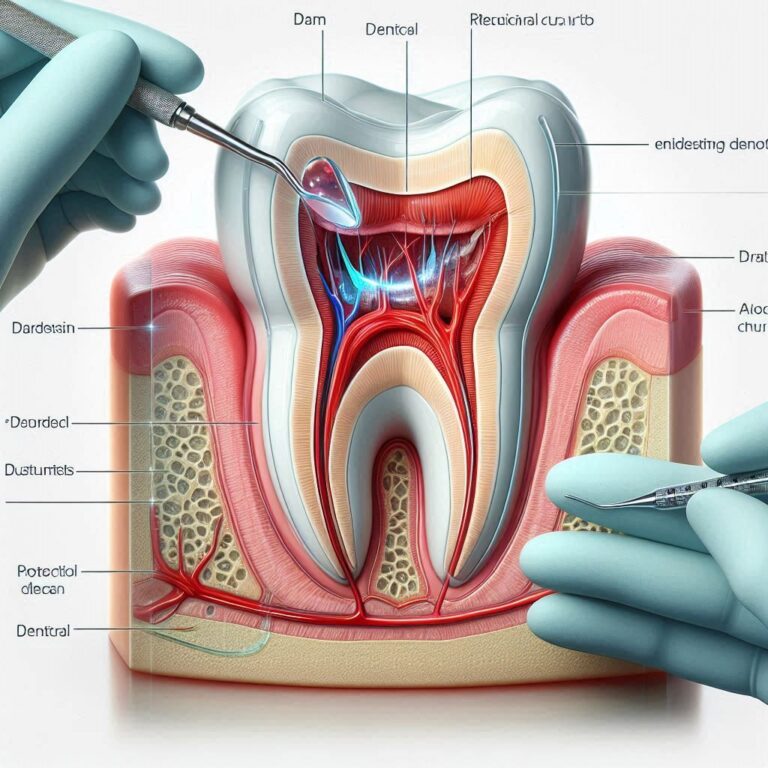
“An image(s) of the coronal portions of the teeth and alveolar bone taken using an extraoral imaging system, which depicts the posterior teeth in a bitewing orientation for the purpose of detecting interproximal caries and evaluating the alveolar crest.”
Let’s break down this definition into its core components:
-
Extraoral: This is the pivotal differentiator. The image is captured with the image receptor (the sensor or film) placed outside the oral cavity. This contrasts with intraoral radiography, where the receptor is placed inside the mouth.
-
Posterior: The image focuses on the premolar and molar regions—the back teeth where the vast majority of interproximal (between-teeth) decay occurs.
-
Bitewing Orientation: This refers to the specific angulation of the X-ray beam. The goal is to project the crowns of both the upper and lower posterior teeth onto the same image without significant overlapping, creating a “wing” where the patient bites. This view is specifically designed to open up the contacts between teeth, making decay visible.
-
Coronal Portions and Alveolar Bone: The image captures the crowns of the teeth (the parts visible above the gumline) and the supporting alveolar bone crest. This allows the dentist to assess not only for cavities but also for early signs of periodontal (gum) disease, such as bone loss.
-
Interproximal Caries: The primary diagnostic goal is the detection of dental caries (cavities) located on the proximal (side) surfaces of the teeth, areas that are completely hidden from visual examination during a clinical check-up.
In essence, D0251 is designed to provide the same diagnostic information as a traditional intraoral bitewing series but through an external imaging system, most commonly a panoramic X-ray unit or a Cone Beam Computed Tomography (CBCT) system equipped with specialized software.
3. The Rationale: Why Move Beyond the Intraoral Standard?
The intraoral bitewing (coded as D0270-D0274 depending on the number of films) has served dentistry exceptionally well. Its resolution is extremely high, and it is a cost-effective method. However, it has inherent limitations that the extraoral approach seeks to overcome:
-
Patient Discomfort and Gag Reflex: This is the most significant driver for alternative methods. For many patients, placing an intraoral sensor, particularly for posterior teeth, triggers a powerful gag reflex. This can turn a simple, minutes-long procedure into a stressful and traumatic experience, sometimes making it impossible to obtain a diagnostic image. D0251 eliminates this challenge entirely.
-
Anatomical Challenges: Patients with small oral cavities, large tongues, or prominent tori (bony growths on the palate or mandible) often present significant challenges for proper sensor placement. An extraoral technique bypasses these anatomical restrictions.
-
** Infection Control and Cross-Contamination:** While strict protocols are followed, intraoral sensors require barrier protection and meticulous cleaning and disinfection between each patient. An extraoral system, where the receptor never enters the patient’s mouth, inherently reduces the risk of cross-contamination.
-
Consistency and Technician Error: Positioning an intraoral sensor perfectly requires skill. Errors in horizontal or vertical angulation can lead to overlapping contacts or “cone cuts” (missing parts of the image), necessitating a retake and additional radiation exposure for the patient. Extraoral systems often use laser-guided positioning and head stabilizers, leading to more consistent, reproducible images with fewer retakes.
-
A Broader Diagnostic View: While a series of intraoral bitewings provides a detailed look at specific quadrants, a single extraoral bitewing projection can often capture an entire arch’s worth of posterior teeth on one image. This provides a more holistic view of the dentition, which can be beneficial for overall assessment and patient education.
4. A Comparative Look: D0251 vs. D0274 (The Traditional Bitewing)
The following table provides a direct, side-by-side comparison of the two procedures, highlighting their key differences.
D0251 vs. D0274 – A Comparative Analysis
| Feature | D0251 – Extraoral Posterior Bitewing | D0274 – Intraoral Bitewing (4 films) |
|---|---|---|
| Image Receptor Placement | Outside the oral cavity | Inside the oral cavity |
| Patient Comfort | High. Eliminates gag reflex and intraoral discomfort. | Variable. Can cause significant gagging and discomfort. |
| Ideal For | Patients with strong gag reflexes, small mouths, tori, special needs, or anxiety. | Patients with standard anatomy and no significant gag reflex. |
| Primary Technology | Panoramic X-ray unit or CBCT machine with bitewing software. | Digital sensor or phosphor plate with intraoral X-ray head. |
| Spatial Resolution | Lower (typically 5-10 line pairs/mm). Adequate for caries detection. | Higher (typically 12-20+ line pairs/mm). Gold standard for fine detail. |
| Field of View | Wider. Can image all posterior teeth on one side per image. | Narrower. Images 2-4 teeth per film/sensor. |
| Radiation Dose | Slightly higher than a full intraoral series (BW+PAs), but lower than a CBCT scan. | Very low. The standard for low-dose radiography. |
| Positioning | Laser-guided, head stabilized. Highly reproducible. | Manual sensor placement by technician. Prone to user error. |
| Infection Control | Simpler. Receptor never enters the mouth. | Complex. Requires single-use barriers and disinfection. |
| Primary Diagnostic Use | Detection of interproximal caries, assessment of alveolar crest height. | Detection of interproximal caries, assessment of alveolar crest height, detailed restoration margins. |
This table illustrates that the choice between D0251 and D0274 is not about one being universally “better” than the other, but about selecting the right tool for the specific patient and clinical situation.
5. The Technology Behind the Image: How Extraoral Bitewings Are Captured
The ability to create a “bitewing” view from outside the mouth is a feat of modern engineering and software. It is primarily achieved through two technologies:
1. Panoramic X-Ray Systems with Bitewing Software:
Most modern panoramic machines now offer a “bitewing mode” as a standard or optional software package. The process involves:
-
The patient is positioned in the machine using the standard chin rest and forehead stabilizer.
-
The operator selects the “Bitewing” program on the software interface.
-
The machine’s rotating arm, which contains the X-ray source and the image receptor, is programmed to move to a specific, stationary position focused on the posterior teeth.
-
Special bite blocks are often attached to the machine to help the patient maintain the correct jaw separation and orientation.
-
The X-ray exposure is made, capturing a focused image of the premolars and molars. Typically, two exposures are needed—one for the left side and one for the right.
2. Cone Beam Computed Tomography (CBCT) Systems:
CBCT units, known for their 3D imaging capabilities, can also be used to generate 2D reconstructions that mimic traditional radiographic views, including bitewings.
-
A small FOV (Field of View) CBCT scan is taken of the jaws.
-
Sophisticated software algorithms then reconstruct the 3D volumetric data.
-
The dentist or technician can then use the software to generate “synthetic” 2D images in any plane, including the perfect bitewing view. This is sometimes called “CBCT-derived bitewings” or “reformatted bitewings.”
The image quality, particularly the spatial resolution, of an extraoral bitewing is generally lower than that of a direct digital intraoral sensor. However, advancements in detector technology and software algorithms have significantly closed this gap. Studies have shown that the diagnostic accuracy of modern extraoral bitewings for detecting moderate to large interproximal caries is excellent and clinically acceptable, making them a reliable alternative.
6. Clinical Applications: Who is the Ideal Candidate for D0251?
The decision to use D0251 is a clinical one, based on the individual needs of the patient. Its applications are diverse and patient-focused.
-
The Gagging Patient: This is the prime candidate. For these individuals, D0251 can be a game-changer, allowing for necessary diagnostic imaging without the associated stress and trauma.
-
Patients with Special Needs: Children, elderly patients, or individuals with physical or cognitive disabilities who cannot tolerate or cooperate with intraoral sensor placement benefit immensely from the extraoral approach.
-
Patients with Anatomical Restrictions: Those with small mouths, prominent palatal or mandibular tori, or trismus (limited jaw opening) are excellent candidates.
-
Anxious and Phobic Patients: Dental anxiety is often tied to specific stimuli. Removing the uncomfortable sensor can reduce a significant trigger, improving the overall patient experience and fostering a more positive relationship with dental care.
-
Baseline Imaging and Monitoring: For patients with a low-to-moderate caries risk, extraoral bitewings can be a fantastic tool for routine monitoring. They provide a sufficient overview to track changes over time between more detailed examinations.
-
Periodontal Screening: While not a replacement for a full periodontal series of radiographs, the extraoral bitewing does include the alveolar bone crest. It can be useful for a preliminary screening of bone levels, particularly in the posterior regions.
It is crucial to note that D0251 may not be suitable in all cases. If a patient requires evaluation of the endodontic (root canal) status, periapical pathology, or the precise marginal fit of a crown, higher-resolution intraoral periapical radiographs (D0220-D0230) are still necessary. The dentist must always adhere to the ALARA principle (As Low As Reasonably Achievable) regarding radiation exposure, only prescribing radiographs when there is a clear clinical need.
7. The Patient Experience: From Preparation to Procedure
For a patient accustomed to traditional X-rays, the D0251 procedure will feel familiar yet distinctively easier.
-
Preparation: As with any X-ray, the patient will remove any metallic objects that could interfere with the image, such as glasses, jewelry, and removable dental appliances. A lead apron with a thyroid collar will be placed for protection.
-
Positioning: The patient will be guided to stand or sit within the panoramic/CBCT machine. They will place their chin on the chin rest and their forehead against the stabilizer. This ensures the head is perfectly aligned.
-
Bite Registration: The operator may attach a disposable bite block to the machine. The patient will be instructed to bite gently on this block, which holds the teeth in a slightly separated position, crucial for opening the interproximal contacts.
-
Exposure: The operator will leave the room and initiate the exposure from a control panel. The machine will emit a soft humming or buzzing sound as it positions itself and takes the image, which lasts only a few seconds. The patient must remain perfectly still.
-
Review: The image appears almost instantly on a computer screen within the operatory. The entire process for both sides is typically completed in under two minutes.
The feedback from patients who have struggled with traditional bitewings is overwhelmingly positive, often described as “so much easier” and “nothing like the old way.”
8. Interpretation and Diagnosis: Reading the Extraoral Bitewing
Interpreting an extraoral bitewing requires the same fundamental skills as reading an intraoral one, but with an awareness of its unique characteristics. The dentist will systematically examine:
-
Interproximal Surfaces: Scrutinizing each tooth contact point for radiolucencies (dark areas) that indicate demineralization and caries. The contrast and resolution are sufficient to detect lesions once they have progressed beyond the very earliest enamel stage.
-
Alveolar Bone Crest: Evaluating the height and density of the bone between the teeth. Healthy bone crests are typically located 1-2 mm apical to the cementoenamel junction (CEJ) of the teeth. Signs of horizontal or vertical bone loss are noted.
-
Existing Restorations: Checking the margins of fillings, crowns, and bridges for any signs of recurrent decay or open margins.
-
Anatomical Noise: The dentist must be aware that extraoral images can sometimes include superimposition of other structures, like the opposite side of the jaw or the cervical spine, depending on the projection. This requires experience to distinguish from pathology.
While the resolution is lower, the diagnostic yield for its intended purposes—caries and crestal bone assessment—is high and clinically valid.
9. The Financial Landscape: Coding, Billing, and Insurance Considerations
As a relatively new code (first introduced in the CDT 2020), D0251 is still gaining widespread recognition among insurance carriers. This can lead to confusion in billing.
-
Coding Accuracy: It is imperative that dental offices use the specific code D0251 for this specific procedure. Submitting a claim for an extraoral bitewing under an intraoral code (D0274) is incorrect and can be considered fraudulent.
-
Insurance Reimbursement: Coverage for D0251 varies significantly by insurance plan. Some plans may cover it at the same frequency and fee as intraoral bitewings. Others may not recognize it yet and may deny the claim. Some may consider it a “non-covered service.”
-
Patient Communication: This makes pre-treatment communication vital. If a dentist believes D0251 is the best option for a patient, the financial coordinator should pre-authorize the procedure with the insurer or, at a minimum, inform the patient that while it is the recommended service, it may not be covered, and they would be responsible for the fee. The office fee for D0251 is often slightly higher than for an intraoral series due to the use of more advanced equipment.
-
Documentation: The clinical notes must clearly justify the medical necessity of using the extraoral technique, e.g., “Due to patient’s extreme gag reflex preventing successful intraoral sensor placement, extraoral posterior bitewings (D0251) were taken for caries detection and monitoring.”
10. The Future is Extraoral: Emerging Trends in Dental Radiography
D0251 is not an endpoint but a signpost pointing toward the future of dental diagnostics. We are moving towards an ecosystem where advanced imaging is faster, more comfortable, and more informative.
-
AI-Powered Analysis: Artificial intelligence software is already being integrated into radiographic systems to automatically flag potential areas of caries, bone loss, and other pathologies. This will be equally, if not more, applicable to extraoral images, serving as a powerful second opinion for the dentist.
-
Dose Reduction: Technology will continue to drive radiation doses down, making extraoral imaging even safer for routine use.
-
Multi-Modal Imaging: The future may lie in a single, quick extraoral scan that can be reconstructed by software into a panoramic image, a set of bitewings, a cephalometric image, and even a limited 3D volume, all from one low-dose exposure. D0251 is a key step in this integrative direction.
11. Conclusion
The D0251 extraoral posterior bitewing code represents a significant and patient-centered advancement in dental diagnostics. It successfully addresses longstanding challenges like the gag reflex and anatomical limitations by leveraging modern imaging technology. While it does not replace the need for high-resolution intraoral radiographs in all scenarios, it establishes itself as a vital, comfortable, and diagnostically reliable tool for a wide range of patients. As technology evolves and insurance recognition grows, D0251 is poised to become a mainstream component of preventive dental care, ensuring that crucial diagnostic information is accessible to everyone, regardless of their physiological constraints.
12. Frequently Asked Questions (FAQs)
Q1: Is the radiation dose for an extraoral bitewing higher than for traditional bitewings?
A: Yes, the effective dose from a set of extraoral bitewings taken on a panoramic unit is generally higher than that of a set of digital intraoral bitewings but remains very low and well within safe limits. It is significantly lower than a CBCT scan. Your dentist follows the ALARA principle to ensure any radiation exposure is justified and minimized.
Q2: If I can tolerate regular X-rays, should I still get the extraoral kind?
A: For patients with no gag reflex or anatomical issues, high-resolution intraoral bitewings (D0274) remain the gold standard for detailing very early decay and fine margins. There is no clinical need to switch to extraoral if intraoral is well-tolerated and effective.
Q3: My insurance denied the claim for D0251. Why?
A: Some insurance plans have not yet updated their policies to include this newer code. They may see it as “not medically necessary” if their policy only lists intraoral codes. Always have your dental office check your benefits beforehand and be prepared for the possibility of out-of-pocket payment if the service is not covered.
Q4: Can extraoral bitewings detect everything that regular bitewings can?
A: They are excellent for detecting interproximal caries and evaluating bone levels. However, for diagnosing very early, incipient decay (which may just be watched) or for evaluating fine details around a crown margin, the superior resolution of an intraoral sensor may still be required. Your dentist will choose the appropriate tool for the diagnostic task.
Q5: Are extraoral bitewings used for children?
A: Absolutely. They are an excellent option for children who have difficulty holding still or tolerating intraoral sensors. Making the radiographic experience positive and non-threatening is crucial in pediatric dentistry.
13. Additional Resources
-
American Dental Association (ADA): The official source for the Current Dental Terminology (CDT) code set, including the complete descriptor and rules for use of D0251.
-
American Academy of Oral and Maxillofacial Radiology (AAOMR): Provides position statements and clinical recommendations on the use of all forms of dental radiography, including extraoral techniques.
-
Journal of the American Dental Association (JADA): Publishes peer-reviewed research studies on the efficacy and diagnostic accuracy of new dental technologies and procedures, including comparative studies of intraoral vs. extraoral bitewings.
Date: September 11, 2025
Author: Dr. Evelyn Reed, DDS
Disclaimer: This article is for informational purposes only and does not constitute professional medical or dental advice, diagnosis, or treatment plan. Always seek the advice of your qualified healthcare provider with any questions you may have regarding a dental or medical condition. The CDT codes are updated annually by the American Dental Association (ADA). Always verify the most current code set and descriptors for accurate billing and documentation.