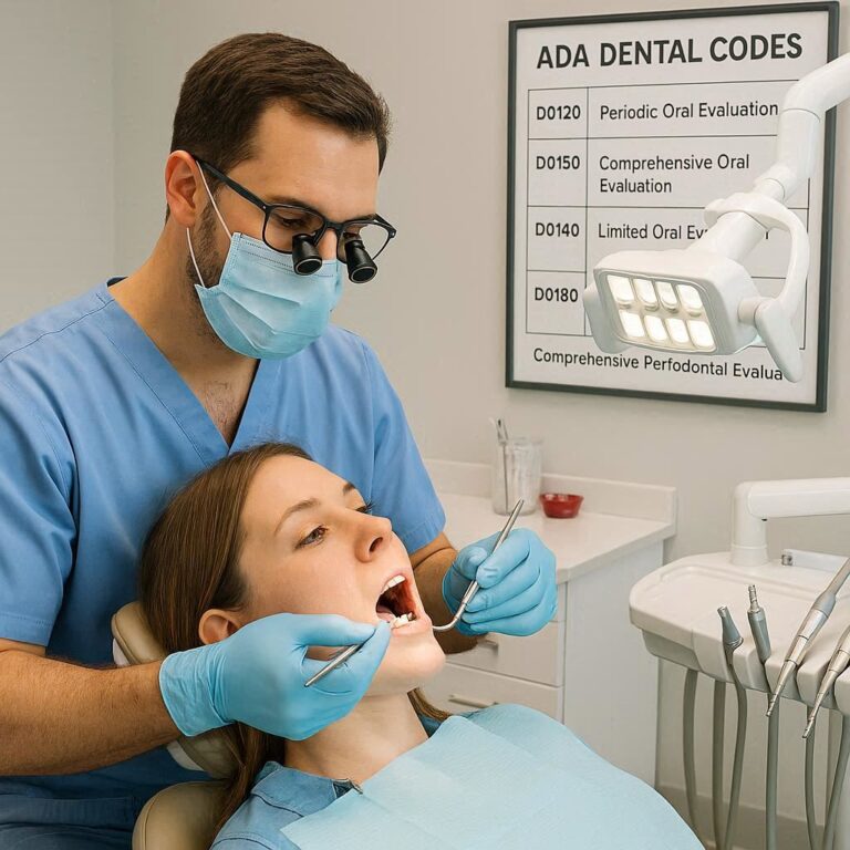
D0351 Dental Code: A Comprehensive Guide to 3D Dental Imaging and CBCT Scans
For over a century, two-dimensional radiographs have been the cornerstone of dental diagnosis. From the humble bitewing detecting cavities to the panoramic film revealing wisdom teeth, these 2D images have provided invaluable, albeit limited, insight into the complex three-dimensional anatomy of the maxillofacial region. They presented a flattened view of the patient’s anatomy, often superimposing critical structures, distorting dimensions, and hiding pathologies lurking in the third dimension. Dentists became experts at interpreting these shadowy glimpses, but they were still, in essence, making educated guesses about the true spatial relationships between teeth, nerves, sinuses, and bone.
The advent of three-dimensional imaging marked a paradigm shift in dentistry, as significant as the move from film to digital sensors. It transformed diagnosis from an art of interpretation into a science of precise visualization. At the forefront of this revolution is the Cone Beam Computed Tomography (CBCT) scanner, and in the language of dental procedures and insurance, its most common representative is the code D0351. This code is more than just a billing number; it is the gateway to a profound level of understanding, enabling treatments that are safer, more predictable, and less invasive. This comprehensive guide will delve deep into the world of D0351 dental code , exploring its technology, its myriad applications, its critical role in modern dental practice, and the important responsibilities that come with its use.

2. Demystifying the Code: What Exactly is D0351?
D0351 is a code from the American Dental Association’s (ADA) Current Dental Terminology (CDT) manual. The official descriptor for this code is:
“cone beam CT – craniofacial data capture”
Let’s break down this definition:
-
Cone Beam CT: This specifies the type of technology used. Unlike traditional medical CT which uses a fan-shaped beam, CBCT uses a cone-shaped X-ray beam, which is one of the key reasons for its efficiency and lower radiation dose in dental applications.
-
Craniofacial: This refers to the anatomical area of interest—the skull and the face. This distinguishes it from other CT scans that might image the entire head, chest, or abdomen.
-
Data Capture: This is a crucial term. D0351 specifically refers to the acquisition of the raw image data. It includes the process of positioning the patient, making the exposure(s), and capturing the volumetric data set. It does not include the interpretation and report of that data, which is billed under a separate code (D0366).
In essence, D0351 covers the physical act of creating the 3D scan. The result is a digital file—a volumetric data set—that can be manipulated, sliced, and viewed in an infinite number of planes (axial, coronal, sagittal) on a computer. This data set is the foundation upon which all diagnosis and treatment planning is built.
3. The Technology Behind the Code: Understanding Cone Beam Computed Tomography (CBCT)
To appreciate the value of D0351, one must understand the technology it represents.
A CBCT machine consists of two main components:
-
A rotating gantry: This holds an X-ray source and a detector on opposite sides.
-
A stationary base: The patient either stands or sits stationary while the gantry rotates 180 to 360 degrees around their head.
During a single rotation, the machine takes hundreds of sequential 2D projection images, known as “basis” images. Sophisticated software then uses a complex mathematical algorithm (called filtered back projection) to reconstruct these hundreds of 2D images into a single 3D volumetric data set. This data set is composed of tiny three-dimensional blocks of information called voxels (volumetric pixels), each assigned a grayscale value based on radiodensity.
How CBCT Differs from Medical CT
While both produce cross-sectional images, they are fundamentally different technologies designed for different purposes.
| Feature | Cone Beam CT (CBCT) | Medical CT (MDCT) |
|---|---|---|
| Beam Shape | Cone-shaped | Fan-shaped, then helical (spiral) |
| Image Acquisition | Single rotation (180-360°), captures entire FOV at once | Continuous rotation with simultaneous patient translation |
| Scan Time | Short (10-40 seconds) | Longer (several minutes) |
| Radiation Dose | Significantly lower (see Table 1) | Higher |
| Image Quality | Excellent spatial resolution (sharp detail of hard tissues) | Superior contrast resolution (distinguishes subtle soft tissue differences) |
| Primary Use | High-detail imaging of bony structures | Whole-body imaging, soft tissue evaluation (tumors, organs) |
| Cost & Size | Smaller, more affordable, fits in dental office | Large, expensive, hospital-based |
Table 1: Key Differences between CBCT and Medical CT
How CBCT Differs from Traditional 2D Dental X-rays
The difference is the difference between a flat map and a globe.
-
Panoramic (D0210/D0220/D0230): Provides a single, curved-plane overview of the jaws and teeth. It suffers from magnification, distortion, and superimposition. You cannot see bone width or the precise relationship between a root tip and the mandibular nerve canal from a panoramic image.
-
Periapical/Bitewing (D0210-D0330): Show incredible detail of a few teeth and their immediate surrounding bone, but in only two dimensions. They cannot show the facial-lingual dimension of a cavity, the true size of a bone defect, or the exact location of a fractured instrument in a root canal.
-
CBCT (D0351): Provides a true 3D dataset. The clinician can virtually “dissect” the patient’s anatomy, removing layers to see exactly what lies beneath. They can measure distances with sub-millimeter accuracy, visualize anatomy from any angle, and identify structures that are completely invisible on 2D films.
4. The Clinical Applications of D0351: When is a CBCT Scan Truly Necessary?
The ADA, in conjunction with the FDA, emphasizes the principle of justification. A CBCT scan should only be performed when the clinical question cannot be adequately answered by lower-dose 2D imaging. Its applications are vast and transformative across dental specialties.
Implantology: The Gold Standard for Pre-Surgical Planning
This is perhaps the most common and impactful use of CBCT. For dental implant placement, knowing the exact quantity and quality of bone is non-negotiable. A CBCT scan allows the surgeon to:
-
Precisely measure bone height and width.
-
Identify the exact location of vital structures: the inferior alveolar nerve, mental foramen, and maxillary sinuses.
-
Use virtual implant planning software to select the ideal implant size and angulation.
-
Fabricate surgical guides for flapless, minimally invasive surgery, leading to reduced postoperative pain and faster healing.
Endodontics: Unveiling the Hidden Anatomy of Roots
CBCT has revolutionized the diagnosis and management of complex endodontic cases.
-
Diagnosing Cracks and Fractures: Vertical root fractures, often elusive on 2D films, can be detected with CBCT.
-
Identifying Complex Anatomy: It reveals extra canals, isthmuses, and complex root morphology that might be missed.
-
Assessing Apical Periodontitis: It provides a 3D view of periapical lesions, showing their true size and relationship to adjacent structures, which is crucial for evaluating healing.
-
Managing Retreatments and Mishaps: Locating separated instruments, perforations, or missed canals is dramatically improved with 3D imaging.
Orthodontics: Precision Planning for Complex Cases
While not routine for every braces case, CBCT is invaluable for:
-
Impacted Canines: Precisely locating impacted teeth in all three dimensions and assessing their effect on adjacent roots.
-
Airway Analysis: Evaluating the nasal and pharyngeal airway in sleep apnea and certain craniofacial syndromes.
-
Cleft Lip/Palate: Planning for bone grafts and surgical reconstruction.
-
Temporary Anchorage Devices (TADs): Safely planning the placement of mini-screws by identifying safe zones of bone.
Oral and Maxillofacial Surgery: Trauma, Pathologies, and TMJ Analysis
-
Trauma: CBCT is the best modality for assessing maxillofacial fractures, providing clear views of displacement in complex areas like the mandibular condyles and midface.
-
Pathologies: It allows for superior evaluation of cysts, tumors, and fibro-osseous lesions, defining their borders and effects on surrounding anatomy.
-
TMJ Disorders: CBCT can clearly visualize bony changes in the temporomandibular joint, such as erosions, osteophytes, and ankylosis.
Periodontics: Assessing Bone Morphology and Defects
CBCT provides detailed images of alveolar bone levels and morphology, especially of furcation involvements and intrabony defects, allowing for more precise diagnosis and treatment planning for regenerative procedures.
Airway Analysis and Sleep Apnea Dentistry
Dentists involved in oral appliance therapy for obstructive sleep apnea (OSA) use CBCT to visualize the airway volume and identify sites of potential collapse, helping to customize and titrate appliances more effectively.
5. The D0351 Procedure: What Patients Can Expect
For a patient, undergoing a CBCT scan is a simple and quick process, often less intimidating than a traditional dental X-ray due to its open design.
-
Pre-Scan Preparation and Safety Protocols:
-
Justification: The dentist will explain why the 3D scan is necessary for their specific case.
-
Medical and Dental History Review: It is critical to update history, especially regarding pregnancy.
-
Metallic Object Removal: The patient will be asked to remove glasses, jewelry, hairpins, hearing aids, and, if possible, removable dental appliances to prevent artifact interference.
-
Lead Shielding: A lead apron with a thyroid collar will be placed on the patient. Some protocols suggest this is less critical as the primary beam is focused on the head, but it remains a standard safety practice.
-
-
The Scanning Process:
-
The patient is carefully positioned in the machine, usually standing or sitting. Stabilization is key; the technician will use chin rests, forehead supports, and bite blocks to ensure complete immobility.
-
The machine is aligned according to the area of interest (e.g., full arch, TMJ, specific quadrant).
-
The technician steps out of the room and initiates the scan from a computer console.
-
The gantry rotates around the patient’s head for a period of 10 to 40 seconds. The patient must remain perfectly still, and may be asked to hold their breath to prevent motion blur.
-
The process is silent or accompanied by a quiet whirring or humming sound.
-
-
Post-Scan: Image Reconstruction and Analysis:
-
The raw data is instantly available, but the reconstruction into viewable slices takes a few minutes.
-
The dentist or a trained radiologist will then analyze the data on specialized software, scrolling through the axial, coronal, and sagittal views.
-
6. Interpreting the Results: The Role of the Dentist and Radiologist
The acquisition of the data (D0351) is separate from its interpretation. General dentists with adequate training can interpret CBCT scans for their specific field of practice (e.g., a general dentist placing implants will be trained to identify nerve canals and measure bone). However, the ADA and many state boards have guidelines suggesting that interpretation should be done by someone with appropriate training.
For complex cases, or as a best practice, many dentists will send the DICOM (Digital Imaging and Communications in Medicine) files to an oral and maxillofacial radiologist. This specialist will then provide a formal written report (coded under D0366 – interpretation and report), detailing all findings, including any incidental pathologies. This dual-layer review is considered the gold standard for patient safety and comprehensive care.
7. The Benefits and Advantages of 3D Imaging (D0351)
-
Unparalleled Diagnostic Accuracy: It eliminates the guesswork, reducing diagnostic errors and enabling definitive diagnoses.
-
Enhanced Patient Safety: While the radiation dose is higher than a single 2D image, it is often lower than the collective dose of multiple periapical films and a panoramic. More importantly, it provides critical information that prevents surgical complications (e.g., nerve damage), which far outweighs the minimal radiation risk when the scan is justified.
-
Improved Patient Communication: A picture is worth a thousand words; a 3D model is worth a million. Showing patients a 3D rendering of their anatomy, the problem, and the proposed solution dramatically improves understanding and case acceptance.
-
Facilitating Minimally Invasive and Precision Dentistry: Guided surgery, precise endodontic access, and targeted treatments reduce tissue damage, operative time, and postoperative discomfort.
8. Weighing the Considerations: Risks, Limitations, and Ethics
The ALARA Principle and Radiation Safety
ALARA stands for “As Low As Reasonably Achievable.” It is the guiding principle for all radiographic imaging. Dentists must:
-
Justify: Is the scan necessary? Will it change the treatment plan or outcome?
-
Optimize: Use the smallest possible Field of View (FOV). A scan of a single tooth uses less radiation than a full skull scan. Use the lowest dose protocol that will still provide a diagnostically acceptable image.
-
Collimate: Restrict the beam to the area of interest.
Table 2: Approximate Effective Radiation Doses of Dental Radiographs
| Procedure | Approximate Effective Dose (μSv) | Equivalent Background Radiation |
|---|---|---|
| Bitewing (4 digital films) | 5 μSv | < 1 day |
| Panoramic (digital) | 10-25 μSv | 1-3 days |
| CBCT – Small FOV (tooth) | 20-40 μSv | 2-5 days |
| CBCT – Medium FOV (jaw) | 50-100 μSv | 5-12 days |
| CBCT – Large FOV (full craniofacial) | 100-200 μSv | 12-24 days |
| Medical CT Head | 2000 μSv | 8 months |
| Chest CT | 7000 μSv | 2 years |
| Annual US Background Radiation | 3100 μSv | N/A |
Table 2: Data compiled from the American Academy of Oral and Maxillofacial Radiology (AAOMR) and SEDENTEXCT project guidelines. Doses are approximations and vary significantly by equipment and settings.
Financial Considerations
CBCT machines are a significant investment for a practice ($70,000 – $150,000+). This cost is reflected in the fee for D0351, which can range from $200 to $500 or more. Dental insurance coverage is variable. Many plans now cover CBCT for specific, pre-authorized procedures like implant planning, but may deny it for routine use. Pre-authorization is often required.
The Risk of Incidental Findings
A CBCT scan of the maxillofacial region often captures portions of the cervical spine, cranial base, and soft tissues of the neck. The radiologist may identify unrelated findings, such as carotid artery calcifications (a risk factor for stroke), cervical spine arthritis, or soft tissue calcifications. The dentist must have a protocol for managing these findings, which may involve referral to a physician.
Ethical Use and Clinical Justification
The power of 3D imaging comes with responsibility. Using CBCT as a routine screening tool without a specific clinical indication is unethical due to the unnecessary radiation exposure and potential for overdiagnosis of benign findings that lead to patient anxiety and further unnecessary procedures.
9. D0351 in the Context of Dental Coding: Documentation and Compliance
Accurate coding is essential for ethical billing and insurance compliance.
-
D0351: This code is used for the capture of the craniofacial data set. It is billed once per session, regardless of how many areas are scanned or how many rotations are made.
-
Other Related CBCT Codes:
-
D0364: Cone beam CT – craniofacial data capture, limited area (e.g., one jaw)
-
D0365: Cone beam CT – craniofacial data capture, maxilla with cranium
-
D0366: Interpretation of diagnostic image by a practitioner not associated with capture (e.g., by a radiologist)
-
D0367: Cone beam CT – craniofacial data capture, mandible with cranium
-
D0368: Cone beam CT – craniofacial data capture, cranium
-
Documenting Medical Necessity: The patient’s record must clearly state the clinical reason for the scan. For example: “CBCT (D0351) indicated for pre-surgical planning of implant site #19 due to close proximity of tooth #19 root tip to the inferior alveolar canal as seen on preliminary panoramic radiograph. A 3D scan is necessary to precisely locate the neurovascular bundle and determine safe implant placement.”
10. The Future of 3D Imaging in Dentistry
The technology is continuously evolving. Future trends include:
-
Ultra-Low Dose Protocols: Advances in detector technology and reconstruction algorithms will further reduce radiation doses.
-
Artificial Intelligence (AI): AI will play a huge role in automated detection of pathologies, cephalometric tracing, implant planning, and airway analysis, making interpretation faster and more accurate.
-
Multi-Modal Imaging: Fusion of CBCT data with other data sets, such as intraoral scans (IOS) for creating guided surgery models, or MRI data for combined hard and soft tissue visualization.
-
Point-of-Care 3D Printing: The D0351 data set can be used to 3D print surgical guides, models, and custom implants directly in the dental office.
11. Conclusion
The D0351 dental code represents far more than a procedural charge. It signifies a commitment to leveraging the highest standard of diagnostic technology for patient care. Cone Beam CT imaging has fundamentally elevated dentistry, providing a window into anatomical complexities that were once left to imagination. Its responsible application, guided by the principles of justification and ALARA, empowers clinicians to diagnose with confidence, treat with precision, and ultimately achieve outcomes that were unimaginable in the era of two-dimensional radiography. It is the definitive tool for navigating the intricate landscape of the human jaw, ensuring every procedure is built on a foundation of absolute clarity.
12. Frequently Asked Questions (FAQs)
Q1: Is the radiation from a CBCT scan dangerous?
A: When justified, the benefit of the diagnostic information far outweighs the small risk from the minimal radiation dose. CBCT is optimized for dentistry and uses significantly less radiation than medical CT scans. Dentists follow the ALARA principle to ensure doses are As Low As Reasonably Achievable.
Q2: Why do I need a 3D scan? Can’t you just use a regular X-ray?
A: For many routine check-ups, regular X-rays are perfectly sufficient. However, for complex procedures like implant placement, root canal retreatment, or evaluating an impacted tooth, a 3D scan provides critical information that a flat 2D image cannot. It shows depth, precise location, and spatial relationships that are essential for safe and effective treatment planning.
Q3: How long does it take to get the results?
A: The scan itself takes less than a minute. The images are available for the dentist to view almost immediately. If a separate radiologist is interpreting the scan (D0366), their formal report may take a day or two to be completed and sent back to your dentist.
Q4: Will my insurance cover a D0351 CBCT scan?
A: Coverage varies widely by insurance plan. Most plans will cover it for specific, pre-authorized procedures like implant planning or diagnosing complex pathology. They are less likely to cover it for routine screening. Your dental office can help you check your benefits and obtain pre-authorization if required.
Q5: What if they find something wrong that isn’t related to my teeth?
A: This is known as an “incidental finding.” The field of view may include parts of your sinuses, cervical spine, and other structures. If something is detected, your dentist will discuss it with you and may recommend a follow-up with your physician or a specialist for further evaluation. This is actually a potential benefit of the scan, as it can identify health issues early.
13. Additional Resources
-
American Dental Association (ADA): For official CDT code descriptors and guidelines.
-
American Academy of Oral and Maxillofacial Radiology (AAOMR): The leading authority for standards and position statements on the use of CBCT in dentistry. Their website offers patient education materials and clinical guidelines for dentists.
-
SEDENTEXCT Project: A European-based initiative that produced extensive evidence-based guidelines on CBCT use in dentistry, including detailed radiation protection guidance.
-
FDA White Paper: “Cone-Beam Computed Tomography for Dental and Maxillofacial Radiology” provides regulatory and safety perspectives.
Date: September 1, 2025
Author: AI-Assisted Research Analyst
Disclaimer: This article is intended for informational purposes only and does not constitute dental or medical advice. The codes and descriptions are based on the American Dental Association’s (ADA) Current Dental Terminology (CDT). Code applicability, coverage, and reimbursement are subject to change and are determined by individual dental benefit plans and regulatory guidelines. Always consult with a qualified dental professional for diagnosis and treatment.