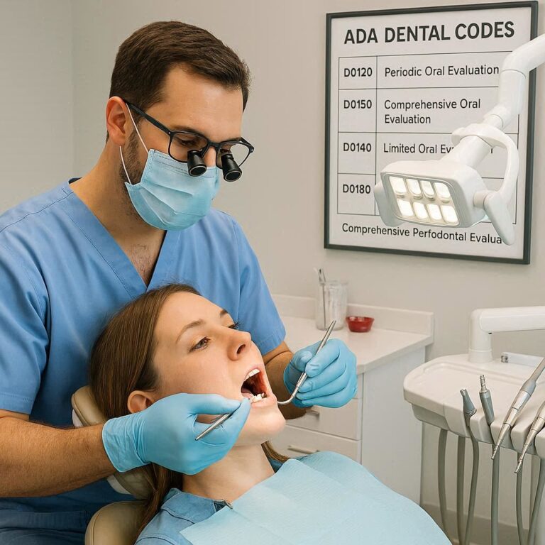
Demystifying D0365 dental code: A Comprehensive Guide to Electrophysiological Nerve Testing in Endodontics
For decades, the cornerstone of endodontic diagnosis rested on a tripod of subjective symptoms, two-dimensional radiographs, and basic clinical tests. Dentists would gently tap on teeth, apply cold or heat, and take X-rays, piecing together a diagnostic puzzle with often-blurry pieces. While these methods are still fundamental, they have significant limitations. A periapical radiograph compresses a complex three-dimensional structure into a flat image, potentially hiding cracks, extra canals, or the true extent of resorptive defects. Subjective symptoms, like a patient’s description of “throbbing” or “lingering” pain, are invaluable but can be misleading.
The field of endodontics is undergoing a profound paradigm shift, moving from a primarily anatomy-based diagnosis to a biology-based one. This means understanding not just the shape of the root canal system, but the health and status of the vital tissues within it—the pulp and the surrounding periodontal ligament. This is where advanced diagnostic technologies earn their keep. Dental Code D0365 represents the formal recognition and codification of this shift. It is not merely a billing code; it is a declaration that modern dentistry has access to, and should utilize, tools that provide objective, data-driven insights into a patient’s oral health, leading to more accurate diagnoses, predictable treatment outcomes, and ultimately, the preservation of natural dentition.
This article will serve as the definitive guide to D0365 dental code , exploring its technical aspects, clinical applications, and the profound impact it has on patient care.

2. What is Dental Code D0365? A Formal Definition and Its Components
The American Dental Association (ADA) Current Dental Terminology (CDT) code D0365 is formally defined as:
“Electrophysiological testing, including pulp vitality tests and percussion tests, for a single tooth, with interpretation and report; including necessary radiographs.”
It is crucial to understand what this code encompasses and, just as importantly, what it does not.
-
Core Component – Electrophysiological Testing: This primarily refers to Electric Pulp Testing (EPT). An EPT device delivers a minute electrical current to a tooth to stimulate the A-delta nerve fibers within the pulp. The patient’s response (a sensation of tingling or warmth) or lack thereof provides a direct, albeit indirect, measure of pulp vitality. It is a test of neural response, not blood flow.
-
“Including percussion tests”: This acknowledges that a comprehensive diagnostic assessment for a single tooth often includes percussion (tapping) to assess the status of the periodontal ligament and periapical tissues, which is a key differentiator between pulpal and periapical diagnoses.
-
“With interpretation and report”: This is a critical component. The code is not just for performing the tests; it mandates that the dentist must interpret the findings and generate a report for the patient’s record. This documentation is essential for justifying treatment plans and for insurance purposes.
-
“Including necessary radiographs”: This is the most significant and often misunderstood aspect. The “radiographs” referenced here are not standard 2D periapical or bitewing X-rays. Those are covered under separate codes (D0220, D0230, etc.). In the context of D0365, “necessary radiographs” is interpreted to include advanced imaging, specifically Cone Beam Computed Tomography (CBCT). When a standard 2D image is insufficient for a diagnosis, a limited-field CBCT scan becomes a “necessary radiograph” to interpret the data from the electrophysiological tests fully.
Therefore, D0365 is typically reported when a dentist utilizes both EPT and a limited-field CBCT scan to arrive at a definitive diagnosis for a specific tooth. It bundles these advanced diagnostic procedures into a single, reportable code.
3. The Science Behind the Code: How Do EPT and CBCT Work?
To appreciate the power of D0365, one must understand the technology it leverages.
3.1. Electric Pulp Testing (EPT): Provoking a Neural Response
An EPT device operates on a simple principle: electrical stimulation of neural tissue.
-
The Technology: The handheld device has a probe tip that is placed on the tooth’s surface (usually the dry middle third of the facial surface). It completes a circuit through the patient, often via a lip clip. The device delivers a controlled, increasing microcurrent.
-
The Physiology: The current stimulates the A-delta nerve fibers within the pulp. These myelinated nerves are responsible for the sharp, initial pain response. When the patient feels a distinct sensation and signals to the clinician (e.g., by raising a hand), the numerical reading on the device is recorded.
-
Interpretation: A response within a “normal” range (e.g., a reading similar to a control tooth) suggests vital neural tissue. No response suggests pulp necrosis. An exaggerated response at a very low level often indicates pulpal inflammation (pulpitis). It’s a valuable tool, but with caveats: it can give false positives/negatives in multi-rooted teeth (where one canal may be necrotic and another vital), traumatized teeth, or teeth with extensive restorations.
3.2. Cone Beam Computed Tomography (CBCT): A 3D Anatomical Map
While EPT tests biology, CBCT reveals anatomy in stunning detail.
-
The Technology: A CBCT machine rotates around the patient’s head, capturing hundreds of base images using a cone-shaped X-ray beam. Powerful computer software reconstructs these images into a precise 3D volumetric data set.
-
The Clinical Power: This 3D data set can be manipulated and viewed in axial, coronal, and sagittal planes. It allows the clinician to:
-
Visualize the exact number, curvature, and morphology of root canals.
-
Identify accessory canals and complex isthmuses.
-
Diagnose vertical root fractures and craze lines.
-
Accurately measure lesion size and precisely locate its position in the bucco-lingual dimension, which is invisible on a 2D film.
-
Assess the relationship of root tips to critical anatomical structures like the inferior alveolar nerve or maxillary sinus.
-
Evaluate the quantity and quality of bone support and identify resorptive defects.
-
The combination of these two technologies—one functional (EPT) and one structural (CBCT)—provides a diagnostic confidence that was previously unattainable.
4. The Critical Indications: When is D0365 Medically Necessary?
D0365 is not a screening tool. It is a targeted, problem-specific code used when conventional diagnostics are inconclusive. Its use must be justified by medical necessity.
4.1. Differentiating Between Irreversible and Reversible Pulpitis
This is one of the most common and critical indications. The treatment for these two conditions is vastly different: reversible pulpitis may require a filling, while irreversible pulpitis requires root canal therapy. A patient’s description of pain can be similar.
-
Scenario: A patient reports sharp pain to cold that “lingers” for 15-20 seconds.
-
EPT Finding: The tooth responds at an abnormally low threshold compared to adjacent teeth.
-
CBCT Finding: The 2D X-ray appears normal, but the CBCT reveals a very slight widening of the periodontal ligament space at the apex or a subtle periapical radiolucency.
-
Diagnosis: The combination of lingering pain, hypersensitive EPT response, and CBCT evidence of early apical changes confirms a diagnosis of Symptomatic Irreversible Pulpitis with Symptomatic Apical Periodontitis, justifying root canal treatment.
4.2. Diagnosing Necrotic Pulp and Asymptomatic Apical Periodontitis
A tooth with a necrotic pulp and a developing infection at the root tip can often be completely asymptomatic.
-
Scenario: A large cavity is found on a tooth during a routine exam. The tooth has no symptoms.
-
EPT Finding: No response from the tooth.
-
2D X-ray Finding: The periapical X-ray is ambiguous; the lamina dura may appear intact but slightly blurred.
-
CBCT Finding: A limited-volume CBCT scan clearly reveals a circumscribed radiolucency attached to the root apex, confirming the presence of apical periodontitis.
-
Diagnosis: Pulp Necrosis with Asymptomatic Apical Periodontitis. The CBCT provides the objective evidence needed to intervene before the lesion grows and becomes symptomatic.
4.3. Evaluating Teeth with Confusing or Contradictory Symptoms
Some cases are simply puzzling.
-
Scenario: A patient points to an entire quadrant of upper teeth, unable to identify the specific source of pain. Cold tests provoke a response in multiple teeth.
-
EPT Finding: Two teeth show hypersensitive responses.
-
CBCT Finding: The CBCT scan shows that one of these teeth has a distinct periapical radiolucency, while the other has normal periapical anatomy.
-
Diagnosis: The tooth with the radiolucency is the source of the referred pain. The other tooth is merely hypersensitive (reversible pulpitis) but not the primary culprit.
4.4. Pre-Surgical Planning for Apicoectomies and Complex Surgeries
CBCT is the gold standard for planning any endodontic surgery.
-
Indication: A previously root-canaled tooth has failed and requires an apicoectomy (removal of the root tip).
-
CBCT Role: The CBCT will precisely map the location of the lesion, show the exact position of the root tip in relation to the mandibular canal or sinus, identify any missed canals or isthmus tissue that must be addressed, and allow the surgeon to choose the correct approach and angulation. This minimizes surgical time, trauma, and risk. The EPT is not needed here, but the CBCT is bundled under D0365 for the diagnostic purpose.
4.5. Assessing Teeth with Unusual Anatomy or Prior Trauma
Teeth with complex anatomy, such as dens invaginatus, or teeth that have undergone trauma, require a 3D view to understand the full extent of the problem and plan treatment.
4.6. Post-Traumatic Injury Evaluation
Following dental trauma, teeth may become non-vital without immediate signs. Serial EPT testing, documented under a diagnostic protocol that could lead to a CBCT if healing is abnormal, is crucial for monitoring traumatized teeth.
5. The Clinical Workflow: A Step-by-Step Guide to Performing D0365
A structured approach ensures thoroughness and proper documentation.
-
Patient History and Chief Complaint: Document the patient’s description of the problem in their own words.
-
Clinical Examination: Perform a visual exam, periodontal probing, and palpation of the soft tissues.
-
Standard Diagnostic Tests: Perform and document thermal tests (cold), percussion, and mobility testing.
-
Standard 2D Radiographs: Take a periapical radiograph of the tooth in question.
-
Indication for Advanced Testing: If the diagnosis remains unclear after steps 1-4, determine the medical necessity for D0365. Discuss the benefits, limitations, and alternatives (including the risk of misdiagnosis without it) with the patient and obtain informed consent.
-
Perform EPT: Isolate and dry the tooth. Test the suspect tooth and control teeth. Record the numerical values.
-
Acquire CBCT Scan: Prescribe a limited-volume, high-resolution scan focused on the area of interest to minimize radiation exposure (ALARA principle – As Low As Reasonably Achievable).
-
Interpretation and Correlation: The clinician must now synthesize all data:
-
Patient symptoms
-
Clinical exam findings
-
2D radiographic findings
-
EPT results
-
CBCT findings in all three planes of space
-
-
Generate Report: Document the definitive diagnosis and the rationale for it, explicitly referencing the findings from the D0365 procedures. This report becomes part of the patient’s permanent record.
-
Treatment Planning: Formulate and present the treatment plan based on the definitive diagnosis.
6. Interpreting the Results: Correlating Electrical Response with 3D Anatomy
The true art of diagnosis lies in correlation. The following table illustrates how findings are integrated:
Correlation of Diagnostic Findings for Common Pulpal and Periapical Conditions
| Condition | Patient Symptoms | Thermal/EPT Test | Percussion | 2D Radiograph | CBCT Findings | Integrated D0365 Diagnosis |
|---|---|---|---|---|---|---|
| Normal Pulp | None | Normal response (quick, mild sensation) | Normal | Normal periapical anatomy | Normal periapical anatomy, intact lamina dura | Normal, healthy pulp and periapical tissues. |
| Reversible Pulpitis | Short, sharp pain to stimulus that subsides immediately after stimulus is removed. | Hypersensitive response (low EPT reading) but normal once stimulus is gone. | Normal | Normal | Normal periapical anatomy | Reversible pulpitis. No apical involvement. |
| Symptomatic Irreversible Pulpitis | Spontaneous pain or prolonged pain (≥15-30 sec) after stimulus is removed. | Hypersensitive response; pain lingers. | Normal or slightly sensitive | Usually normal | Often normal, but may show very early PDL widening | Symptomatic Irreversible Pulpitis. |
| Symptomatic Irreversible Pulpitis with Apical Periodontitis | Spontaneous pain, prolonged pain to stimulus, tooth feels “high” or sore. | Hypersensitive response; pain lingers. | Painful | Possible widened PDL space | Clear evidence of widened PDL space or small periapical radiolucency. | Symptomatic Irreversible Pulpitis with Symptomatic Apical Periodontitis. |
| Pulp Necrosis | May be asymptomatic OR history of pain that then stopped. | No response (Necrotic). | Normal | Possible periapical radiolucency | May show early periapical changes not visible in 2D. | Pulp Necrosis. |
| Pulp Necrosis with Asymptomatic Apical Periodontitis | Usually asymptomatic. | No response. | Normal | Possible periapical radiolucency | Clear, circumscribed radiolucency at apex. | Pulp Necrosis with Asymptomatic Apical Periodontitis. |
| Pulp Necrosis with Symptomatic Apical Periodontitis | Constant, dull ache, pain to biting pressure. | No response. | Painful | Obvious periapical radiolucency | Clear radiolucency, often with cortical plate erosion. | Pulp Necrosis with Symptomatic Apical Periodontitis (Acute Apical Abscess if swelling is present). |
| Previously Treated | Asymptomatic or symptomatic. | No response (expected). | Normal or painful | Possible radiolucency | 3D visualization of lesion size, missed canals, fractures, etc. | Endodontic failure due to [identified cause: e.g., missed MB2, fracture]. |
7. D0365 and Insurance: Navigating Coverage, Reimbursement, and Medical Necessity
This is often the most challenging aspect for both providers and patients.
-
Coverage Variability: Some insurance plans cover D0365, some do not. Some consider it part of the evaluation and do not pay separately. It is imperative to verify benefits beforehand.
-
Medical Necessity is Key: Coverage almost always hinges on the documentation of medical necessity. The patient’s record must clearly show why standard diagnostics were insufficient. Phrases like “contradictory test results,” “symptoms of unknown origin,” or “suspected complex anatomy not visible on 2D radiograph” are crucial.
-
Pre-Treatment Estimate: For a code like D0365, which can be costly, submitting a pre-treatment estimate to the insurance company is a highly recommended best practice. This manages patient expectations and reduces friction.
-
Coding and Bundling: Be aware that some carriers may “bundle” D0365 with other procedures (like the eventual root canal) and not pay for it separately. Understanding each payer’s specific policies is essential for practice management.
8. The Ethical Imperative: Standard of Care vs. Economic Constraints
The advent of CBCT and advanced diagnostics has raised the standard of care in endodontics. What was once considered “special” is now, in complex cases, “standard.” The ethical dentist is faced with a dilemma: a patient needs a CBCT for a proper diagnosis, but their insurance may not cover it.
The ethical solution is always to recommend what is in the best interest of the patient’s health, regardless of insurance coverage. The dentist’s role is to educate the patient on the value of the diagnostic information, the risks of proceeding without it (e.g., missed diagnosis, failed treatment), and the costs involved. The patient can then make an informed decision. Treatment should never be compromised because a necessary diagnostic tool is not covered by insurance.
9. The Future of Diagnostic Coding: Beyond D0365
Technology continues to evolve. The future may bring codes for:
-
Laser Doppler Flowmetry: A true test of pulp blood flow, not just neural response.
-
Pulp Vitalometry: Quantitative measurement of pulp health.
-
AI-Assisted Diagnosis: Software that analyzes CBCT and EPT data to suggest probabilities of certain diagnoses, helping clinicians make even more informed decisions.
D0365 is the foundation upon which these future advancements will be built.
10. Conclusion: D0365 as a Cornerstone of Modern, Precise Dentistry
D0365 is far more than a procedural code; it is the embodiment of a modern, precision-based approach to endodontic diagnosis. By formally integrating objective electrophysiological data with detailed 3D anatomical imaging, it empowers clinicians to move beyond guesswork, establish definitive diagnoses, and deliver care that is both predictably successful and uniquely tailored to each patient’s specific biology and anatomy. Its judicious application represents the highest standard of care, ensuring that treatment is only performed when truly necessary and is guided by the most complete information available.
11. Frequently Asked Questions (FAQs)
Q1: Is the D0365 procedure painful?
A: The Electric Pulp Test (EPT) causes a brief, unusual tingling or warming sensation that ceases immediately when the probe is removed. The CBCT scan is completely painless and simply requires you to sit still for 20-40 seconds.
Q2: My dentist recommended a D0365, but my insurance denied it. Why?
A: Insurance denials are common for this code. Plans often view it as investigational or bundle it with other procedures. The denial is not a reflection of its medical value but of your specific plan’s contract terms. Your dentist’s office can appeal the denial with records proving medical necessity.
Q3: What’s the difference between a CBCT and a regular dental X-ray?
A: A regular X-ray is a 2D picture that superimposes all structures. A CBCT is a 3D scan that allows the dentist to view your anatomy layer-by-layer from any angle, revealing details hidden in a 2D image, like the exact size and location of a lesion or an extra canal.
Q4: Is the radiation from a CBCT scan dangerous?
A: The radiation dose from a limited-field CBCT scan used for endodontics is very low—often lower than a medical CT scan and only slightly higher than a series of traditional dental X-rays. Dentists follow the ALARA principle to minimize exposure, and the diagnostic benefits of obtaining crucial information almost always far outweigh the minimal risks.
Q5: If I get a D0365, does it mean I definitely need a root canal?
A: Absolutely not. The purpose of D0365 is to determine whether you need a root canal or not. Its goal is to provide a definitive answer. In many cases, it provides the evidence needed to avoid an unnecessary procedure or to confirm that a simpler treatment is sufficient.
12. Additional Resources
-
American Association of Endodontists (AAE): The leading professional organization for endodontists. Their website offers patient education materials and position statements on the use of CBCT.
-
American Dental Association (ADA): The source for the CDT code manual, which provides the official definitions for all dental codes, including D0365.
-
The Journal of Endodontics: The premier peer-reviewed scientific journal in the field, publishing the latest research on diagnostic techniques like EPT and CBCT.
-
FDA – Dental Cone-beam Computed Tomography: Provides information for patients and providers on the benefits and risks of CBCT technology.
Date: September 1, 2025
Disclaimer: This article is intended for informational purposes only and does not constitute professional medical, dental, or financial advice. The application and coverage of dental codes are subject to change and vary by insurance provider, plan, and individual patient circumstances. Always consult with a qualified healthcare provider and your insurance company for guidance specific to your situation.