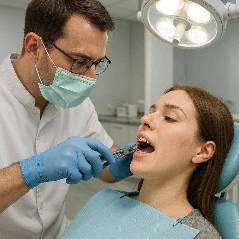
D0371 Dental Code: A Deep Dive into the 3D Conical Beam CT Scan in Modern Dentistry
Imagine an architect tasked with designing a skyscraper without knowing the exact composition of the ground beneath it, the location of buried utility lines, or the precise proximity of adjacent buildings. The project would be fraught with risk, guesswork, and potential for catastrophic failure. For decades, this was the reality for dentists performing complex procedures in the hidden landscape of the human jaw. They relied on two-dimensional X-rays—flat images that compressed intricate anatomy into a confusing superposition of structures, forcing clinicians to infer what lay beneath the surface.
The introduction of the D0371 dental code marked a paradigm shift, a revolution akin to the architect being handed a complete 3D geological survey, a utility map, and a laser-scanned model of the entire city block. D0371 is not just a billing code; it is the identifier for one of the most significant technological advancements in modern dental care: the Cone Beam Computed Tomography (CBCT) scan. This procedure has granted dentists a transformative power: the ability to see, measure, and navigate the oral and maxillofacial region with unprecedented clarity and precision, fundamentally improving diagnosis, treatment planning, and patient outcomes.
This article serves as the definitive guide to the D0371 dental code. We will embark on a detailed journey through its technology, its vast applications across every dental specialty, its benefits and limitations, and its profound impact on elevating the standard of care from an art of estimation to a science of exactitude.

2. Beyond the Code: Defining D0371 and Its Technological Marvel
The American Dental Association (ADA) Current Dental Terminology (CDT) code D0371 is formally defined as: “cone beam CT – maxillofacial image volume”. This succinct description belies the complexity and power of the procedure it represents.
At its core, a D0371 scan is a diagnostic imaging modality that provides three-dimensional (3D) information of the maxillofacial region, which includes the jaws, teeth, nasal cavity, sinuses, and often the temporomandibular joints. Unlike a traditional medical CT scanner that uses a fan-shaped X-ray beam and captures data in a series of axial “slices,” a CBCT unit utilizes a cone-shaped X-ray beam that rotates around the patient’s head in a single, 360-degree pass.
During this rotation, a detector on the opposite side captures hundreds of distinct 2D projection images, or “basis images.” Sophisticated computer algorithms then reconstruct these 2D images into a precise volumetric data set—a digital, 3D block of information. This volume can be virtually sliced in any plane (axial, coronal, sagittal) and manipulated on a computer screen to reveal anatomy without superposition, allowing for unparalleled analysis.
It is crucial to distinguish D0371 from other common dental codes:
-
D0210 (Intraoral – complete series of radiographic images): A series of 10-18 small, 2D films or sensors capturing the crowns and roots of all teeth.
-
D0220 (Intraoral – periapical first film) / D0230 (Intraoral – periapical each additional film): 2D images of individual teeth, focusing on the root tip and surrounding bone.
-
D0270 (Bitewing – single film) / D0274 (Bitewing – four films): 2D images showing the crowns of upper and lower teeth, primarily used to detect cavities between teeth.
-
D0330 (Panoramic film): A single, curved 2D image that shows all the teeth and the jaws in a single view, but with inherent magnification and distortion.
D0371 is in a league of its own, providing a true 3D dataset where the others provide only 2D representations.
3. The Evolution of Dental Imaging: From 2D Shadows to 3D Truth
The journey to D0371 is a story of relentless pursuit of diagnostic clarity.
-
The Dawn of Dental X-rays: In 1895, Wilhelm Röntgen discovered X-rays. Within weeks, dentist Otto Walkhoff took the first dental radiograph, using a 25-minute exposure. This began the era of seeing the invisible.
-
The Film Era: For most of the 20th century, film-based radiography was the standard. While revolutionary, it was limited by processing chemicals, density issues, and the static, 2D nature of the images.
-
Digital Radiography: The advent of digital sensors replaced film, offering instant images, lower radiation doses, and the ability to enhance images digitally. However, they still only provided 2D views.
-
The CT Bottleneck: Medical CT scanners provided 3D images but were impractical for dentistry. They were expensive, involved high radiation doses, and were located in hospitals, not dental offices.
-
The CBCT Revolution: The first CBCT unit designed specifically for dentomaxillofacial imaging was developed in the late 1990s in Europe. It addressed the shortcomings of medical CT by offering a compact size, significantly lower radiation dose, and dedicated dental software. The ADA formally acknowledged this technology with the creation of the D0371 code, cementing its role in clinical practice.
4. Deconstructing the Technology: How CBCT Works
Understanding the mechanics of a D0371 scan demystifies the process. The CBCT machine consists of three key components:
-
X-ray Source: Generates the cone-shaped beam of ionizing radiation.
-
Detector: A flat-panel detector that captures the X-rays after they pass through the patient’s tissues, collecting the hundreds of 2D basis images.
-
C-Arm or Rotating Gantry: The mechanical arm that connects the source and detector and rotates them in unison around the patient’s head.
The process is remarkably swift:
-
The patient is positioned seated or standing, with their head stabilized by a chin rest and forehead support. A lead apron is often placed for protection.
-
The machine is calibrated to the region of interest (e.g., full arch, mandible, maxillary sinuses).
-
The C-arm rotates around the patient in a complete or partial arc (often 360 degrees) in as little as 5 to 40 seconds. The patient must remain perfectly still.
-
During the rotation, the detector captures between 150 to 600+ individual low-dose basis images.
-
The raw data is sent to a computer workstation, where specialized software uses algorithms like Feldkamp-Davis-Kress (FDK) to reconstruct the 2D projections into a 3D volumetric cube, or “voxel map” (a 3D pixel).
This voxel data is the heart of the D0371 scan. It can be explored using multiplanar reformation (MPR), which allows the clinician to scroll through axial, coronal, and sagittal slices simultaneously. The software also enables 3D surface rendering, creating a photorealistic model of the skull and teeth.
5. A Universe of Applications: When is a D0371 Scan Necessary?
The power of D0371 lies in its versatility. It is not a routine screening tool but a targeted diagnostic instrument used when its 3D capabilities provide critical information that 2D images cannot.
Implantology: The Gold Standard for Precision
This is perhaps the most common application. For dental implants, knowing the exact bone dimensions is non-negotiable. A D0371 scan allows the clinician to:
-
Precisely measure the height, width, and density of the alveolar bone at the proposed implant site.
-
Identify the exact location of vital anatomical structures like the inferior alveolar nerve canal (in the lower jaw) and the maxillary sinuses (in the upper jaw), avoiding them during surgery.
-
Perform virtual implant placement using guided surgery software, designing a surgical stent that guides the drill to the exact planned position, angle, and depth. This minimizes surgical risk, improves success rates, and often allows for flapless surgery, reducing patient discomfort.
(Image: A side-by-side comparison of a panoramic X-ray showing a vague, ghostly outline of the mandibular nerve canal next to a CBCT cross-sectional slice where the nerve canal is clearly and distinctly visible, with precise measurements displayed.)
Endodontics: Illuminating the Root Canal System
The complex internal anatomy of teeth is a classic blind spot for 2D radiography. CBCT is invaluable for:
-
Diagnosing cracks and fractured roots.
-
Identifying extra canals (e.g., MB2 canals in upper molars) that, if missed, lead to treatment failure.
-
Assessing periapical pathologies and their true size and relationship to adjacent structures, like the sinus floor.
-
Managing endodontic retreatments and diagnosing persistent infections (apical periodontitis).
-
Evaluating root resorption (internal or external) and traumatic injuries.
Oral Surgery: Navigating Complex Extractions and Pathologies
-
Impacted Teeth: For impacted third molars (wisdom teeth), a D0371 scan reveals the tooth’s exact position in 3D space, its relationship to the nerve canal (allowing for risk assessment for paresthesia), and the morphology of its roots.
-
Pathologies: It provides definitive visualization of cysts, tumors, and other bony lesions, allowing for assessment of their extent, margins, and effect on surrounding structures.
-
Orthognathic Surgery: For corrective jaw surgery, CBCT data is used to create 3D virtual models of the skull for precise surgical planning and the fabrication of custom surgical guides.
Orthodontics: Beyond the Cephalometric Film
While cephalometric X-rays are 2D, CBCT offers a 3D ceph, eliminating superimposition errors and providing a more accurate analysis of skeletal relationships. It is particularly useful for:
-
Locating impacted canines and planning their surgical exposure and traction.
-
Assessing root angulation and parallelism throughout treatment.
-
Evaluating the buccal and lingual bone plates to avoid iatrogenic damage (e.g., dehiscence) during tooth movement.
-
Airway assessment in patients with sleep-disordered breathing.
Periodontics: Assessing Bone Loss in Three Dimensions
A D0371 scan can reveal the morphology of bony defects around teeth (e.g., furcation involvements, intrabony defects) with much greater accuracy than 2D films, which is critical for predicting prognosis and planning regenerative surgical procedures.
Temporomandibular Joint (TMJ) Analysis
CBCT provides exquisite detail of the bony components of the TMJ, allowing for evaluation of degenerative changes (osteoarthritis), condylar position, and morphology, which is crucial for diagnosing and treating TMJ disorders.
Airway Analysis and Sleep Apnea
A CBCT scan can be used to measure airway volume and identify anatomical obstructions that may contribute to obstructive sleep apnea (OSA).
6. The D0371 Procedure: What Patients Can Expect
For a patient, undergoing a D0371 scan is a simple and non-invasive experience.
-
Consultation: The dentist will explain why the scan is necessary, the benefits, and the alternatives.
-
Preparation: The patient will remove any metal objects (eyeglasses, jewelry, hairpins, hearing aids) that could cause artifacts. A lead apron with a thyroid collar will be placed.
-
Positioning: The patient will be carefully positioned in the machine. The technician will use laser lights or other guides to align the head so the region of interest is centered. Bite blocks, chin rests, and forehead supports are used to ensure complete immobility.
-
Scanning: The machine will rotate around the patient’s head. The patient will hear a faint whirring or humming noise but will feel nothing. The key is to remain perfectly still, and may be asked to hold their breath for a few seconds to prevent motion blur.
-
Completion: The entire process, from positioning to scan completion, typically takes less than five minutes. The data is then processed and available for the dentist to review.
7. Interpreting the Results: The Art and Science of Reading a CBCT Scan
A D0371 scan generates a vast amount of data. Interpreting it requires specialized training. Dentists, particularly oral and maxillofacial radiologists, undergo extensive education to read these scans. They navigate through the three anatomical planes, adjust contrast and brightness to differentiate between tissues, use tools to take precise measurements, and synthesize the 3D information to form a diagnosis. This interpretive skill is as critical as the technology itself.
8. Benefits and Advantages: Why D0371 is a Game-Changer
-
Accuracy: Eliminates distortion, magnification, and superimposition inherent in 2D radiography.
-
Comprehensive Diagnosis: Provides a complete view of the area of interest, revealing problems that are invisible on 2D films.
-
Improved Treatment Planning: Allows for virtual simulation of procedures, leading to more predictable and successful outcomes.
-
Minimally Invasive Surgery: Enables guided surgeries that are more precise, less traumatic, and have faster healing times.
-
Enhanced Patient Communication: The 3D models are a powerful visual aid for dentists to explain conditions and treatment plans to patients, improving understanding and informed consent.
9. Risks, Limitations, and Safety: A Balanced Perspective
Radiation Dose: Context is Key
The most significant concern with any radiographic procedure is ionizing radiation. It is vital to understand that the dose from a D0371 scan is not a single value. It varies dramatically based on:
-
Field of View (FOV): A small FOV scan of a few teeth involves a much lower dose than a large FOV scan of the entire skull.
-
Scanner Settings: Resolution (voxel size), mA, and kVp are adjusted by the clinician to use the lowest dose necessary to answer the diagnostic question (the ALARA principle – As Low As Reasonably Achievable).
Comparing Effective Doses of Dental Radiographs
| Procedure (ADA Code) | Approximate Effective Dose (µSv) | Equivalent Background Radiation |
|---|---|---|
| Bitewing X-rays (4 films) (D0274) | 5 µSv | < 1 day |
| Panoramic X-ray (D0330) | 10-25 µSv | 1-3 days |
| CBCT – Small FOV (D0371) | 20-50 µSv | 2-6 days |
| CBCT – Medium FOV (D0371) | 50-100 µSv | 6-12 days |
| CBCT – Large FOV (D0371) | 100-200 µSv | 12-24 days |
| Medical Chest CT | 7,000 µSv | 2 years |
| Annual US Background Radiation | 3,100 µSv | 1 year |
Table data synthesized from the American Dental Association and American Academy of Oral and Maxillofacial Radiology guidelines. Doses are approximations and can vary by equipment and settings.
As the table shows, while a D0371 scan involves a higher dose than a bitewing X-ray, it is still orders of magnitude lower than a medical CT scan. When used judiciously for a specific diagnostic purpose, the benefit of obtaining critical 3D information far outweighs the relatively small risk.
Artifacts and Diagnostic Challenges
CBCT images can be degraded by artifacts, which can mimic pathology or obscure anatomy. Common artifacts include:
-
Beam Hardening: Causes streaks and dark bands, often from metal restorations (amalgam, crowns, implants).
-
Motion Artifact: Blurring caused by patient movement during the scan.
-
Scatter: Reduces image contrast.
Furthermore, CBCT is excellent for visualizing bony structures but provides very poor soft tissue contrast compared to medical MRI or CT.
10. The Cost and Insurance Landscape of D0371
A D0371 scan is more expensive than traditional 2D imaging due to the high cost of the equipment, software, and specialized training required. Costs can range from $200 to $500+ depending on the size of the FOV and the geographic location.
Insurance coverage is variable. Most dental insurers will cover D0371 when it is deemed medically necessary. This typically requires a pre-authorization submission from the dentist, including a narrative and 2D X-rays justifying the need for 3D imaging (e.g., for implant planning, complex endodontic case, impacted tooth). Medical insurance may cover the scan if it is related to a medical diagnosis, such as TMJ disorder or sleep apnea.
11. Ethical Considerations and ALARA Principle in 3D Imaging
The power of D0371 comes with professional responsibility. The ethical dentist must always adhere to the ALARA principle and only prescribe a CBCT scan when the diagnostic yield justifies the radiation exposure. It should not be used as a routine screening tool. Guidelines from the American Academy of Oral and Maxillofacial Radiology (AAOMR) provide clear criteria for its use. The dentist must also be proficient in interpreting the images or must refer them to a specialist for interpretation to avoid misdiagnosis.
12. The Future of CBCT and D0371: AI, Integration, and Beyond
The technology behind D0371 is rapidly evolving:
-
Artificial Intelligence (AI): AI algorithms are being developed to automatically detect caries, periodontal bone loss, periapical lesions, and even screen for osteoporosis directly from CBCT scans, acting as a powerful second reader for the dentist.
-
Lower Doses and Higher Resolution: Advances in detector technology and reconstruction algorithms will continue to push the dose lower while improving image clarity.
-
Multi-Modality Integration: The fusion of CBCT data with other digital files—such as intraoral scans, facial photographs, and MRI data—creates a truly holistic “digital patient” for comprehensive treatment planning in digital smile design and complex rehabilitations.
-
Point-of-Care 3D Printing: The 3D models generated from D0371 scans can be instantly used to 3D print surgical guides, models, and custom implants at the chairside.
13. Conclusion: D0371 as a Pillar of Precision Dentistry
The D0371 dental code represents far more than a billing entry; it signifies a fundamental shift towards data-driven, precise, and minimally invasive dentistry. By providing a clear, three-dimensional window into the complex anatomy of the maxillofacial region, it empowers clinicians to diagnose with confidence, plan with accuracy, and treat with unparalleled predictability. While its use must be governed by the prudent principles of radiation safety and professional ethics, the CBCT scan has undeniably become an indispensable tool, elevating the standard of patient care and solidifying its role as a cornerstone of modern dental practice.
14. Frequently Asked Questions (FAQs)
Q1: Is a CBCT (D0371) scan the same as a medical CT or CAT scan?
A: No. While both produce 3D images, they use different technologies. CBCT is specifically designed for the maxillofacial area, uses a cone-shaped beam, and delivers a significantly lower radiation dose than a medical CT scan, which uses a fan-shaped beam and is designed for full-body imaging.
Q2: How often do I need a CBCT scan?
A: There is no routine schedule for D0371 scans. They are only performed when a specific diagnostic need arises that cannot be adequately addressed with 2D X-rays. Your dentist will recommend one based on your individual clinical situation.
Q3: I’m claustrophobic. Can I tolerate a CBCT scan?
A: Most patients find CBCT machines much easier to tolerate than medical CT or MRI scanners. You are not enclosed in a tunnel; the machine simply rotates around your head while you remain seated or standing. The procedure is also very quick, often lasting less than 30 seconds.
Q4: Who can read and interpret my D0371 scan?
A: The scan should be interpreted by a dentist who has received advanced training in CBCT interpretation. This could be your general dentist, a specialist (like an oral surgeon or endodontist), or an oral and maxillofacial radiologist. The level of training required is beyond standard dental education.
Q5: Can I get a copy of my CBCT scan?
A: Yes, you have a right to your medical and dental records. The CBCT data is stored in a standard digital format (DICOM), which can be provided to you on a CD or USB drive. You can then provide this to any other dental specialist you may see in the future, often eliminating the need for a repeat scan.
15. Additional Resources
-
American Dental Association (ADA): Guidelines for the use of CBCT in dentistry. https://www.ada.org
-
American Academy of Oral and Maxillofacial Radiology (AAOMR): The leading professional organization for experts in dental radiology. They publish official statements and clinical recommendations. https://www.aaomr.org
-
The Image Gently Alliance: Promotes radiation safety in imaging for children. Their “Image Gently in Dentistry” campaign provides resources for parents and clinicians. https://www.imagegently.org
-
National Council on Radiation Protection and Measurements (NCRP): Publishes Report No. 177, which provides detailed guidance on radiation protection in dentistry.
Date: September 2, 2025
Disclaimer: This article is intended for informational purposes only and does not constitute professional medical or dental advice, diagnosis, or treatment. Always seek the advice of your dentist or other qualified health provider with any questions you may have regarding a dental procedure or medical condition. The content is original and produced exclusively for this publication.