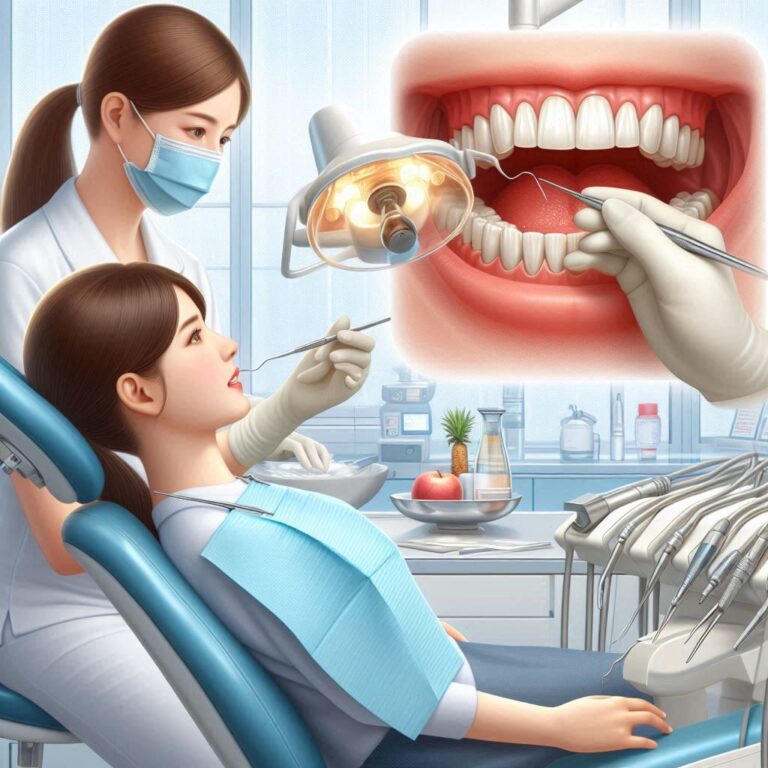
Understanding the D7640 Dental Code
The human jaw, particularly the mandible, is a marvel of biological engineering. It serves as the foundation for our lower teeth, facilitates crucial functions like chewing, speaking, and swallowing, and plays a significant role in defining facial aesthetics. However, the mandible, especially its anterior (front) portion, is vulnerable to injury, disease, and developmental abnormalities. When the integrity or function of the anterior mandible is compromised, surgical intervention becomes necessary. This is where the realm of oral and maxillofacial surgery comes to the forefront, encompassing procedures ranging from stabilizing simple fractures to complex reconstruction. Within this field exists a system of codes to classify procedures, one of which is the dental code D7640. While D7640 specifically pertains to a particular type of fracture treatment, understanding this code requires appreciating the broader spectrum of surgical procedures that can affect the anterior mandible, including osteoplasty and reconstruction.
This article aims to provide a comprehensive exploration of surgical interventions involving the anterior mandible. We will delve into the anatomy and function of this critical area, discuss the various conditions that necessitate surgery, meticulously define the dental code D7640 and its application, and then expand to cover the more intricate procedures of osteoplasty and reconstruction. We will walk through the surgical journey, from initial diagnosis and planning to the recovery process, touch upon potential challenges, and navigate the complexities of coding and insurance.
1. Anatomy and Functional Significance of the Anterior Mandible
The mandible, or lower jawbone, is a single, U-shaped bone that articulates with the skull at the temporomandibular joints (TMJ). Its structure is vital for both form and function. The anterior mandible specifically refers to the front portion, typically encompassing the area from one mental foramen to the other, housing the incisor and canine teeth. This region is a cornerstone of facial aesthetics, contributing significantly to the chin’s shape and projection.
Functionally, the anterior mandible is indispensable. It provides stable anchorage for the lower front teeth, which are crucial for incising (cutting) food. The bone’s density and shape in this area support the muscles of mastication and facial expression, enabling effective chewing, clear speech articulation, and a wide range of facial movements. The nerves and blood vessels supplying the lower lip and chin pass through the mental foramina located on the anterior mandible, making this region sensitive and vascularized. Any disruption to the structure or function of the anterior mandible can have profound impacts on a person’s ability to eat, speak, and on their overall facial appearance and self-esteem. Its prominence also makes it susceptible to direct impact and injury.
2. When the Anterior Mandible Requires Surgical Intervention: Indications and Causes
The need for surgical intervention on the anterior mandible arises from a variety of conditions, each presenting unique challenges and requiring tailored treatment approaches. These conditions can be broadly categorized as:
- Trauma and Fractures: This is perhaps the most common reason for acute surgical intervention. Direct blows to the chin or lower face can result in fractures of the anterior mandible. These fractures can range from simple, non-displaced cracks to complex, comminuted breaks involving multiple bone fragments. The presence of teeth in the anterior segment complicates fracture management, as proper alignment and stabilization are crucial for restoring the bite (occlusion).
- Congenital Deformities: Some individuals are born with abnormalities in the development of the mandible, which can affect the anterior region. Conditions like mandibular hypoplasia (underdevelopment) or prognathism (protrusion) can impact facial symmetry, bite alignment, and functional capabilities. Surgical correction often involves reshaping or repositioning the bone to achieve functional and aesthetic harmony.
- Acquired Defects from Disease or Tumor Removal: Pathologies such as cysts or tumors in the anterior mandible necessitate surgical removal of the affected bone. The resulting defect can range in size but often requires reconstruction to restore continuity, support for teeth, and facial contour. The extent of the defect dictates the complexity of the reconstructive procedure.
- Orthognathic Needs: In some cases of severe malocclusion (improper bite) related to skeletal discrepancies, the anterior mandible may need to be surgically repositioned to achieve proper alignment with the upper jaw. These procedures are part of orthognathic surgery and often involve precise cuts (osteotomies) to move segments of bone.
- Aesthetic Concerns Related to Function: While purely cosmetic procedures are distinct, some functional issues or deformities may have significant aesthetic components that are addressed during reconstructive or orthognathic surgery.
Understanding the underlying cause and the specific nature of the anterior mandibular issue is the first step in determining the appropriate surgical approach and the relevant dental or medical coding.
3. Decoding D7640: Closed Reduction of Mandibular Fractures
The dental code D7640 is specifically defined in the Current Dental Terminology (CDT) as “Mandible – closed reduction (teeth immobilized, if present)”. This code is used to report a particular method of treating fractures of the mandible.
What is Closed Reduction?
Closed reduction is a technique used to treat bone fractures where the fractured bone fragments are realigned without making a surgical incision to expose the fracture site. In the context of the mandible, this typically involves manipulating the jaw externally to bring the broken pieces back into their correct anatomical position.
How are Teeth Immobilized?
For anterior mandibular fractures, especially when teeth are present in the fractured segment, immobilizing the teeth is a critical part of achieving and maintaining the reduction. This immobilization, also known as maxillomandibular fixation (MMF) or intermaxillary fixation (IMF), involves stabilizing the lower jaw against the upper jaw. Common methods include:
- Arch Bars: Metal bars are wired or bonded to the teeth of both the upper and lower arches. Wires or elastic bands are then used to connect the upper and lower arch bars, holding the jaws together.
- Brackets and Wires: If the patient has orthodontic brackets, wires can be used to achieve MMF.
- IMF Screws: Small screws can be placed into the jawbone between the tooth roots, and wires or elastics are attached to these screws to connect the upper and lower jaws.
The goal of immobilizing the teeth is to use the existing dental occlusion as a guide for aligning the fractured bone segments and to provide stability during the healing process.
Indications for D7640
D7640 is typically indicated for specific types of mandibular fractures where:
- The fracture is relatively simple and non-displaced, or minimally displaced.
- The fracture line does not involve significant bone loss or comminution (shattering).
- The presence and position of teeth allow for effective immobilization and anatomical reduction using MMF techniques.
- The patient’s overall health and the nature of the fracture make a less invasive, closed approach feasible and appropriate.
This procedure is generally performed under local anesthesia, sometimes with intravenous sedation, depending on the complexity and patient factors. The period of immobilization can range from several weeks to a couple of months, during which the patient is typically restricted to a liquid or soft diet.
While D7640 is a vital code for reporting closed fracture treatment, it is important to recognize that not all anterior mandibular issues can be addressed with this method. More complex situations necessitate different surgical approaches and thus, different coding.
4. Beyond D7640: Anterior Mandibular Osteoplasty and Reconstruction
While D7640 deals with the closed reduction of fractures, many conditions affecting the anterior mandible require more involved surgical procedures classified under the broader categories of osteoplasty and reconstruction. These procedures involve actively reshaping, repositioning, or rebuilding the bone structure.
Defining Osteoplasty in the Anterior Mandible
Osteoplasty literally means “bone shaping.” In the anterior mandible, osteoplasty refers to surgical procedures aimed at modifying the contour, size, or shape of the bone without necessarily reconstructing a significant defect. This can involve:
- Bone Reduction: Carefully removing excess bone to correct irregularities, reduce prominence, or improve symmetry. This might be done after a healed fracture that resulted in a bump or unevenness, or as part of aesthetic contouring in specific orthognathic cases.
- Bone Addition/Grafting (Minor): Using small amounts of bone graft material to fill minor defects, smooth contours, or augment areas for improved shape.
Osteoplasty is often performed to address aesthetic concerns related to the bone’s shape or to improve the foundation for prosthetic devices like dentures or dental implants. It requires precise surgical technique to achieve the desired outcome while preserving vital structures.
(Consider placing a graphic illustrating bone reduction or augmentation in the anterior mandible.)
Defining Anterior Mandibular Reconstruction
Anterior mandibular reconstruction is a more complex set of procedures focused on rebuilding the bone structure when a significant portion has been lost or is severely deformed. This is commonly necessary after:
- Resection of tumors or cysts that require removal of a segment of the jawbone.
- Severe trauma resulting in comminuted fractures with bone loss that cannot be managed by simple reduction.
- Correction of significant congenital deformities where reshaping alone is insufficient.
The goal of reconstruction is to restore the continuity of the mandible, provide a stable base for teeth or dental implants, recover functional capabilities (chewing, speaking), and re-establish facial aesthetics.
Indications for Osteoplasty and Reconstruction
The indications for these more complex procedures are varied and often involve functional impairment or significant aesthetic disfigurement. Specific reasons include:
Surgical Techniques in Detail
Anterior mandibular osteoplasty and reconstruction utilize a range of sophisticated surgical techniques:
- Osteotomy: This involves making precise surgical cuts in the bone to separate segments that can then be repositioned. Different types of osteotomies are used depending on the specific goal, such as sagittal split osteotomy (though more common in the posterior mandible, modifications can be used anteriorly) or anterior mandibular osteotomy for genioplasty (chin reshaping).
- Bone Grafting: This is a cornerstone of reconstruction and often used in osteoplasty. Bone graft material is used to fill defects, augment bone volume, or bridge gaps. Graft sources include:
- Autogenous Grafts: Bone harvested from the patient’s own body (e.g., hip, tibia, or even other parts of the jaw). This is often the preferred method as it provides the patient’s own living bone cells, promoting better integration and healing.
- Allogeneic Grafts: Bone obtained from a human donor (typically processed and sterilized).
- Alloplastic Materials: Synthetic bone substitutes.
- Fixation: Once bone segments are repositioned or grafted bone is placed, rigid fixation is essential to stabilize the area during healing. This is commonly achieved using small titanium plates and screws, which hold the bone fragments securely in their new positions.
- Microvascular Free Flaps: For large reconstructive procedures, particularly after extensive tumor removal, a microvascular free flap may be used. This involves transplanting bone (often from the fibula in the lower leg or the scapula) along with its supplying blood vessels. These vessels are then surgically connected to blood vessels in the neck under a microscope, ensuring the transplanted bone remains viable and heals effectively.
Modern anterior mandibular surgery often incorporates advanced technologies like virtual surgical planning (VSP) and 3D printing. VSP allows surgeons to plan the procedure digitally on a 3D model of the patient’s jaw, improving precision and predictability. 3D printed guides and templates can then be created to assist the surgeon during the actual operation.
(Consider placing an image or graphic demonstrating bone grafting or plate and screw fixation.)
5. The Surgical Journey: From Consultation to Operating Room
The journey for a patient undergoing anterior mandibular surgery, whether it’s a closed reduction (D7640) or complex reconstruction, begins with a thorough evaluation and treatment planning phase.
- Initial Consultation and Diagnosis: The surgeon will take a detailed medical history, perform a clinical examination, and order imaging studies. For fractures, this typically involves X-rays (like panoramic and lateral oblique views) and potentially a CT scan for more complex breaks. For deformities or defects, CT scans are crucial for detailed 3D visualization. The diagnosis will determine the nature and extent of the problem.
- Treatment Planning: Based on the diagnosis, the surgeon develops a treatment plan. For simple fractures amenable to closed reduction (D7640), the plan focuses on the method of immobilization and the expected duration. For osteoplasty or reconstruction, the planning is far more intricate, involving decisions about the type of graft material (if needed), the specific osteotomies or reshaping techniques, and the type of fixation. Collaboration with other specialists, such as orthodontists (for bite alignment) or prosthodontists (for future tooth replacement), is common in complex cases. Virtual surgical planning may be utilized at this stage.
- Pre-operative Preparation: Patients undergo necessary medical evaluations to ensure they are fit for surgery. Specific instructions regarding fasting, medications, and oral hygiene will be provided. For procedures involving bone grafting from another site, there will also be preparation for the donor site surgery.
- The Surgical Procedure: The surgery is performed in a sterile environment, typically an operating room in a hospital or surgical center, depending on the complexity. Anesthesia will be administered (local with sedation for D7640, often general anesthesia for more complex procedures). The surgical steps vary significantly based on the planned intervention – from applying arch bars and wires for D7640 to making osteotomies, harvesting and shaping bone grafts, and fixing everything with plates and screws for reconstruction.
- Immediate Post-operative Care: Immediately after surgery, the patient is moved to a recovery area for monitoring. Pain management is initiated, and swelling and bruising are expected. For patients with MMF (common in D7640), communication will be challenging, and nutritional support (liquid diet) is provided. Patients undergoing more extensive reconstruction may have drains in place and require closer monitoring in a hospital setting for several days.
The surgical journey is a significant undertaking, requiring careful planning, skilled execution, and dedicated post-operative care for a successful outcome.
6. Recovery, Rehabilitation, and the Path to Healing
The recovery process after anterior mandibular surgery varies significantly depending on the type and complexity of the procedure performed.
- Initial Healing Phase: This phase typically lasts for several weeks. Pain and swelling are managed with medication. For patients with MMF (following D7640 or some other procedures), maintaining oral hygiene is challenging but crucial to prevent infection; special brushes and rinses are often used. A soft or liquid diet is necessary. Speech may be affected, especially with MMF.
- Diet Progression: As healing progresses and stability is achieved, the diet is gradually advanced from liquids to soft foods and eventually to a more normal consistency, as tolerated and advised by the surgeon.
- Removal of Fixation: For D7640 with MMF, the wires or elastics are typically removed after the anticipated healing period (usually 6-8 weeks), allowing the patient to gradually open their mouth and begin jaw movement. Plates and screws used in osteoplasty or reconstruction are usually left permanently unless they cause issues.
- Functional Rehabilitation: Physical therapy may be recommended, especially after complex reconstruction, to help restore jaw mobility, strength, and function. Speech therapy might also be necessary.
- Long-Term Healing and Osseointegration: If bone grafting was performed, the grafted bone needs time to integrate with the native bone (osseointegration). This process takes several months. For patients planning dental implants in the reconstructed area, there is typically a waiting period to ensure the bone is fully healed and stable.
- Potential for Further Procedures: In some reconstruction cases, additional procedures may be needed later, such as removal of excess bone, revision of contours, or placement of dental implants.
A typical, simplified recovery timeline might look like this:
(Consider placing a graphic illustrating jaw exercises during rehabilitation.)
The recovery period requires patience and adherence to the surgeon’s instructions. Regular follow-up appointments are essential to monitor healing and address any complications.
7. Potential Complications and Risks
Like any surgical procedure, anterior mandibular surgery carries potential risks and complications. While surgeons take every precaution to minimize these, patients should be aware of them.
- General Surgical Risks: These include infection at the surgical site, bleeding, adverse reactions to anesthesia, and blood clots.
- Risks Specific to Mandibular Surgery:
- Nerve Damage: The mental nerves, which provide sensation to the lower lip and chin, are located in the anterior mandible. Injury to these nerves during surgery can lead to temporary or permanent numbness or altered sensation.
- Non-union or Malunion: The bone fragments may fail to heal properly (non-union) or heal in an incorrect position (malunion), requiring further surgery.
- Hardware Complications: Plates and screws used for fixation can sometimes become loose, infected, or palpable, necessitating removal.
- Infection of Bone Graft or Reconstruction Site: This is a serious complication that can jeopardize the success of the procedure and may require removal of the graft or hardware.
- Relapse: In orthognathic cases, the repositioned bone segments can sometimes shift back towards their original position.
- Changes in Bite (Occlusion): Achieving perfect bite alignment can be challenging, and minor discrepancies may persist.
- Difficulty Opening Mouth (Trismus): Stiffness or limited ability to open the jaw can occur, often requiring physical therapy.
- Salivary Fistula: A rare complication where saliva leaks from a surgical incision.
- Aesthetic Dissatisfaction: Despite successful functional outcomes, the final aesthetic result may not fully meet the patient’s expectations.
Thorough discussion of these risks with the surgeon before the procedure is crucial for informed consent.
8. Navigating the Financial Landscape: Coding and Insurance (Including D7640)
Understanding the coding and insurance aspects of anterior mandibular surgery can be complex, as procedures may fall under either dental or medical insurance, depending on the nature of the condition and the surgical setting.
Dental Code D7640: As discussed, D7640 specifically refers to “Mandible – closed reduction (teeth immobilized, if present).” This is a dental code and is typically submitted to dental insurance. Coverage for fracture treatment varies among dental plans. It’s essential to verify benefits and understand plan limitations, deductibles, and co-pays. Pre-authorization from the dental insurance company is often required before the procedure is performed.
Beyond D7640: Osteoplasty and Reconstruction Coding: Procedures involving more complex osteoplasty and reconstruction of the anterior mandible may utilize different coding systems.
- Dental Codes: Some reconstructive or osteoplasty procedures might have corresponding dental codes within the D7000-D7999 series (Oral and Maxillofacial Surgery), although the specific codes will depend on the exact nature of the procedure (e.g., codes for bone grafting, excision of bone lesions, etc.).
- Medical Codes (CPT Codes): If the surgery is performed in a hospital or surgical center, or if the underlying cause is deemed medical (e.g., trauma requiring hospital admission, tumor removal), the procedures may be coded using CPT (Current Procedural Terminology) codes, which are used for medical billing. Procedures like complex mandibular reconstruction with bone grafts and microvascular techniques almost always fall under medical coding.
Key Considerations for Billing and Insurance:
- Medical vs. Dental Necessity: Insurance coverage often hinges on whether the procedure is deemed medically necessary (to treat a disease, injury, or congenital defect impacting function) or primarily cosmetic. While D7640 (fracture repair) is typically considered medically necessary, some osteoplasty procedures for aesthetic contouring might not be covered.
- Procedure Setting: Where the surgery is performed (dental office vs. hospital) can influence whether dental or medical codes are used and which insurance is primary.
- Documentation: Detailed and accurate clinical documentation, including operative reports, is critical for successful insurance claims processing for all types of anterior mandibular surgery.
- Pre-authorization: Obtaining pre-authorization from the relevant insurance company (dental or medical) before the surgery is highly recommended to understand coverage and potential out-of-pocket costs. This is particularly important for complex and expensive reconstructive procedures.
Navigating insurance for these procedures can be challenging. Patients should work closely with their oral surgeon’s office billing staff to understand the expected costs, coverage, and authorization process.
9. Advances in Anterior Mandibular Surgery
The field of anterior mandibular surgery is continuously evolving, driven by technological advancements and improved understanding of bone biology and healing.
- Virtual Surgical Planning (VSP): As mentioned earlier, VSP using 3D imaging allows for highly precise pre-operative planning, improving surgical accuracy and potentially reducing operating time and complications.
- 3D Printing: Custom 3D printed guides, templates, and even patient-specific implants are increasingly used to facilitate more accurate osteotomies, bone graft shaping, and placement.
- Biomaterials and Tissue Engineering: Research into novel bone graft substitutes and tissue engineering techniques holds promise for improving bone regeneration and reducing the need for harvesting the patient’s own bone in some cases.
- Minimally Invasive Techniques: While complex reconstruction often requires open surgery, there is a growing interest in minimally invasive approaches where feasible, which could potentially lead to smaller incisions and faster recovery times.
- Improved Fixation Devices: Advances in the design and materials of plates and screws continue to improve the stability and predictability of bone fixation.
These advancements contribute to improved outcomes, reduced patient morbidity, and enhanced functional and aesthetic results in anterior mandibular surgery.
10. Conclusion
Surgical intervention on the anterior mandible is a critical aspect of oral and maxillofacial surgery, addressing a range of conditions from fractures to complex defects. While dental code D7640 specifically describes the closed reduction of mandibular fractures with teeth immobilization, it represents just one piece of a broader surgical landscape. Procedures like osteoplasty and reconstruction, utilizing advanced techniques such as bone grafting and rigid fixation, are essential for restoring form and function to the anterior mandible after trauma, disease, or to correct developmental issues. Navigating the diagnostic, surgical, recovery, and financial aspects requires a thorough understanding of the procedures and involved processes.
11. FAQs
Q1: Is D7640 always performed under local anesthesia? A1: D7640 (closed reduction of a mandibular fracture) is often performed under local anesthesia, sometimes with intravenous sedation. However, the choice of anesthesia depends on the complexity of the fracture, the patient’s anxiety level, and their overall health status.
Q2: How long will my jaw be immobilized after a D7640 procedure? A2: The duration of maxillomandibular fixation (immobilization) after a closed reduction for a mandibular fracture typically ranges from 6 to 8 weeks. The exact time is determined by your surgeon based on the specific fracture and your healing progress.
Q3: Will I need a bone graft for an anterior mandibular surgery? A3: Whether you need a bone graft depends entirely on the nature of the surgical procedure. Bone grafting is commonly used in anterior mandibular reconstruction to fill defects where bone is missing. It may also be used in some osteoplasty procedures for minor augmentation. It is generally not required for a simple closed reduction of a fracture (D7640).
Q4: What is the difference between osteoplasty and reconstruction? A4: Osteoplasty involves reshaping or recontouring existing bone. Reconstruction involves rebuilding lost bone structure, often using bone grafts or other materials, when a significant defect is present. Reconstruction is typically a more complex procedure than osteoplasty.
Q5: Will insurance cover anterior mandibular surgery? A5: Coverage depends on your specific dental and/or medical insurance plan and whether the procedure is considered medically necessary to treat a functional impairment resulting from trauma, disease, or a congenital defect. Procedures for purely cosmetic reasons are generally not covered. It is crucial to obtain pre-authorization from your insurance provider before the surgery. D7640 for fracture repair is typically covered by dental insurance, but complex reconstruction often falls under medical insurance.