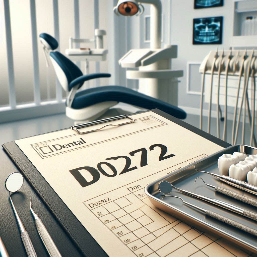
Dental Code D0272: Bitewing Radiographs in Modern Dentistry
Dentistry is a field that relies heavily on diagnostic tools to ensure accurate treatment planning and patient care. Among these tools, dental radiographs play a pivotal role in identifying issues that are not visible to the naked eye. One of the most commonly used radiographic techniques is the bitewing radiograph, coded as D0272 in the American Dental Association’s (ADA) Current Dental Terminology (CDT). This article delves into the intricacies of dental code D0272, exploring its significance, procedure, benefits, and advancements in modern dentistry. Whether you’re a dental professional, a student, or a curious patient, this guide will provide you with a comprehensive understanding of bitewing radiographs and their role in maintaining oral health.
2. Understanding Dental Code D0272
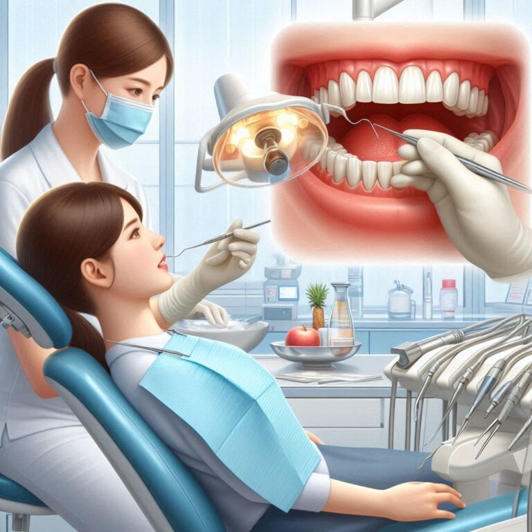
Dental code D0272 refers to the procedure of taking bitewing radiographs, which are a type of dental X-ray that focuses on the crowns of the upper and lower teeth. These radiographs are primarily used to detect interdental caries (cavities between teeth) and assess the health of the alveolar bone. The code is part of the ADA’s CDT, which standardizes dental procedures for billing and documentation purposes.
Bitewing radiographs are typically taken in sets of two to four images, depending on the patient’s dental arch size and the dentist’s requirements. The name “bitewing” comes from the technique used, where the patient bites down on a wing-shaped device to hold the X-ray film or sensor in place.
3. The Importance of Bitewing Radiographs in Dentistry
Bitewing radiographs are indispensable in modern dentistry for several reasons:
- Early Detection of Decay: They reveal cavities between teeth that are not visible during a clinical examination.
- Monitoring Bone Health: They help in assessing the level of alveolar bone, which is crucial for diagnosing periodontal disease.
- Evaluating Restorations: Dentists use them to check the integrity of fillings, crowns, and other restorations.
- Preventive Care: Regular bitewing radiographs are a cornerstone of preventive dentistry, enabling early intervention and reducing the need for extensive treatments.
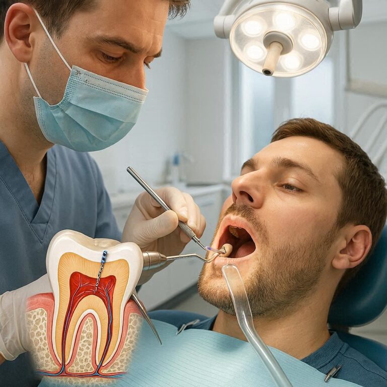
4. How Bitewing Radiographs Work
Bitewing radiographs use X-ray technology to capture images of the teeth and surrounding structures. The process involves the following steps:
- Positioning: The patient bites down on a bitewing tab or holder, which positions the X-ray film or digital sensor correctly.
- Exposure: The X-ray machine is activated, emitting a controlled burst of radiation that passes through the teeth and onto the film or sensor.
- Processing: The image is developed (in the case of traditional film) or digitally processed (in the case of digital radiography).
The resulting image shows the crowns of the teeth, the interdental spaces, and the alveolar bone in detail.
5. Indications for Bitewing Radiographs
Bitewing radiographs are recommended in the following scenarios:
- Routine dental check-ups for adults and children.
- Suspected interdental caries.
- Monitoring the progression of periodontal disease.
- Evaluating the fit of dental restorations.
- Assessing the impact of trauma on the teeth and supporting structures.
6. Benefits of Bitewing Radiographs
- High Diagnostic Accuracy: They provide clear images of interdental areas, enabling precise diagnosis.
- Minimal Radiation Exposure: Modern digital bitewing radiographs use significantly less radiation compared to traditional film X-rays.
- Non-Invasive: The procedure is quick, painless, and does not require any special preparation.
- Cost-Effective: They are relatively inexpensive compared to other imaging techniques like CT scans.
7. Limitations and Risks
While bitewing radiographs are highly beneficial, they do have some limitations:
- Limited Field of View: They only capture the crowns of the teeth and a portion of the alveolar bone.
- Radiation Exposure: Although minimal, there is still a small risk associated with radiation.
- Patient Cooperation: The procedure requires the patient to remain still and follow instructions carefully.
8. Technological Advancements in Bitewing Radiography
The field of dental radiography has seen significant advancements in recent years:
- Digital Radiography: Replaces traditional film with digital sensors, reducing radiation exposure and improving image quality.
- 3D Imaging: While not a replacement for bitewing radiographs, 3D imaging provides additional information for complex cases.
- AI Integration: Artificial intelligence is being used to enhance image analysis and improve diagnostic accuracy.
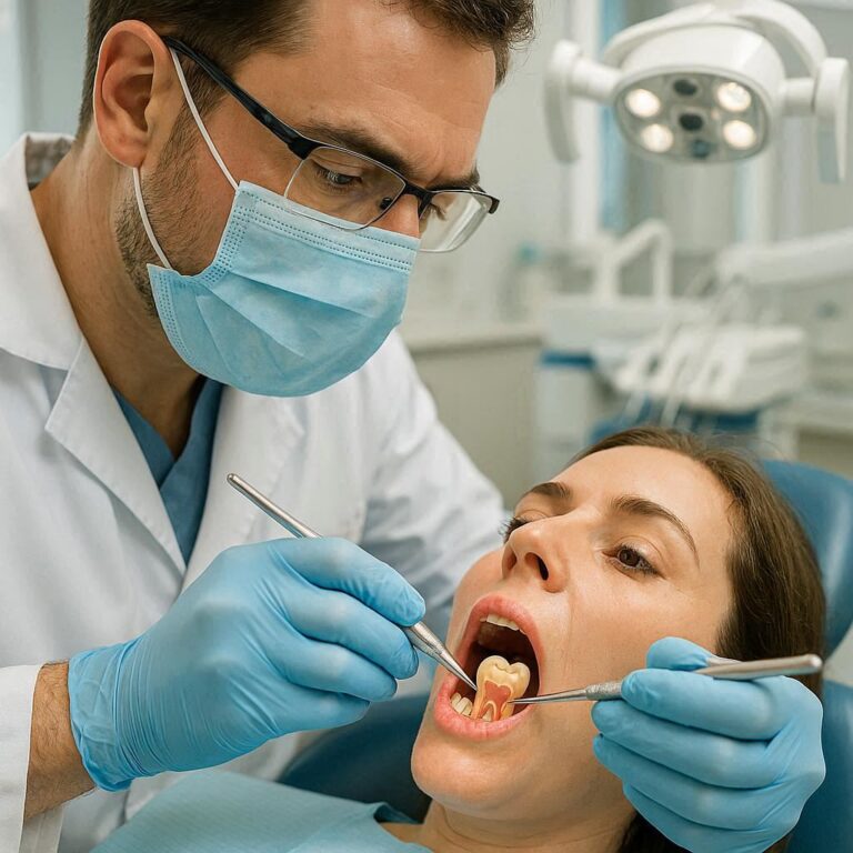
9. Step-by-Step Procedure for Taking Bitewing Radiographs
- Preparation: Explain the procedure to the patient and address any concerns.
- Positioning: Place the bitewing tab or holder in the patient’s mouth and ensure proper alignment.
- Exposure: Activate the X-ray machine and capture the image.
- Review: Check the image for clarity and retake if necessary.
10. Interpreting Bitewing Radiographs
Interpreting bitewing radiographs requires a trained eye. Dentists look for:
- Caries: Dark spots between the teeth indicate decay.
- Bone Loss: Reduced bone levels suggest periodontal disease.
- Restoration Integrity: Check for gaps or irregularities around fillings and crowns.
11. Common Challenges and Solutions
- Patient Discomfort: Use smaller sensors or pediatric-sized holders for children and sensitive patients.
- Poor Image Quality: Ensure proper positioning and exposure settings.
- Radiation Concerns: Educate patients about the minimal risks and benefits of the procedure.
12. Bitewing Radiographs vs. Other Dental Imaging Techniques
| Feature | Bitewing Radiographs | Panoramic X-rays | CT Scans |
|---|---|---|---|
| Field of View | Crowns and alveolar bone | Entire jaw and surrounding structures | 3D view of teeth and jaw |
| Radiation Exposure | Low | Moderate | High |
| Cost | Low | Moderate | High |
| Diagnostic Use | Caries and bone health | General overview | Complex cases |
13. The Role of Bitewing Radiographs in Preventive Dentistry
Bitewing radiographs are a cornerstone of preventive dentistry, enabling early detection and intervention. They help dentists:
- Identify issues before they become severe.
- Monitor changes over time.
- Educate patients about their oral health.
14. Legal and Ethical Considerations
- Informed Consent: Patients must be informed about the procedure and its risks.
- Radiation Safety: Follow ALARA (As Low As Reasonably Achievable) principles to minimize exposure.
- Record Keeping: Maintain accurate records for legal and diagnostic purposes.
15. FAQs
Q1: How often should I get bitewing radiographs?
A: Most dentists recommend bitewing radiographs once a year, but the frequency may vary based on your oral health.
Q2: Are bitewing radiographs safe for children?
A: Yes, they are safe for children, and pediatric-sized holders are available for comfort.
Q3: Can bitewing radiographs detect gum disease?
A: Yes, they can show bone loss associated with periodontal disease.
16. Conclusion
Bitewing radiographs, coded as D0272, are an essential tool in modern dentistry. They provide valuable insights into interdental caries, bone health, and restoration integrity, making them indispensable for preventive and diagnostic care. With advancements in technology, the procedure has become safer, faster, and more accurate, ensuring better outcomes for patients.