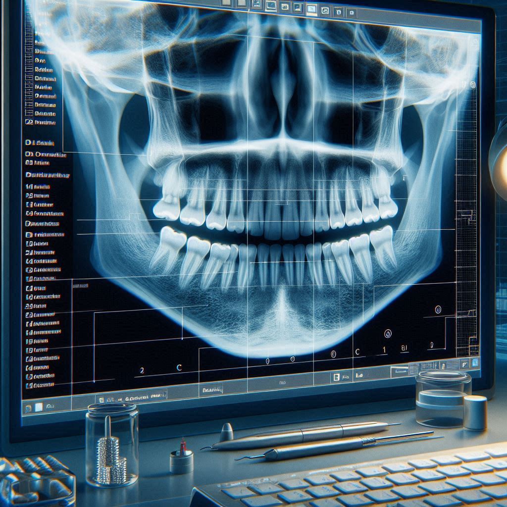
Dental Code Periapical X-Ray
Dental health is a critical component of overall well-being, and diagnostic tools like periapical X-rays play a pivotal role in maintaining it. Periapical X-rays are a type of intraoral radiograph that provides a detailed view of a specific tooth, including its root and surrounding bone structure. These images are indispensable for diagnosing issues such as abscesses, bone loss, and impacted teeth.
This article delves into the intricacies of dental codes associated with periapical X-rays, including CPT codes, scanning techniques, procedures, radiology, ICD-10 codes, and more. Whether you’re a dental professional, a student, or a patient seeking to understand the process, this guide offers a comprehensive and exclusive resource.
1. Dental Code Periapical X-Ray CPT Code
What is a CPT Code?
Current Procedural Terminology (CPT) codes are standardized codes used to describe medical, surgical, and diagnostic services. In dentistry, CPT codes help streamline billing and insurance claims.
CPT Code for Periapical X-Ray
The most commonly used CPT code for a periapical X-ray is D0220. This code specifically refers to the capture of an intraoral image of a single tooth, including its root and surrounding bone.
Common Dental CPT Codes
| CPT Code | Description |
|---|---|
| D0220 | Intraoral periapical first film |
| D0230 | Intraoral periapical each additional film |
| D0272 | Bitewing – single film |
| D0330 | Panoramic film |
Importance of Accurate Coding
Accurate use of CPT codes ensures proper reimbursement and minimizes claim denials. Dental professionals must stay updated with the latest coding guidelines to avoid errors.
2. Dental Code Periapical X-Ray Scan
What is a Periapical X-Ray Scan?
A periapical X-ray scan is a diagnostic imaging technique that focuses on capturing high-resolution images of a specific tooth and its surrounding structures. It is commonly used to detect:
- Dental abscesses
- Bone loss
- Root fractures
- Impacted teeth
Types of Periapical X-Ray Scans
- Traditional Film-Based X-Rays: Uses photographic film to capture images.
- Digital X-Rays: Utilizes electronic sensors and provides instant results with lower radiation exposure.
Advantages of Digital Scans
- Reduced radiation exposure
- Enhanced image quality
- Easy storage and sharing of images
3. Dental Code Periapical X-Ray Procedure
Step-by-Step Procedure
- Preparation: The patient is seated, and a lead apron is placed to protect against radiation.
- Positioning: The X-ray film or sensor is placed inside the mouth, near the tooth of interest.
- Alignment: The X-ray machine is positioned to focus on the targeted area.
- Capture: The X-ray is taken, and the image is processed.
- Review: The dentist examines the image for abnormalities.
Safety Measures
- Use of lead aprons and thyroid collars
- Limiting the number of X-rays to necessary cases
- Regular maintenance of X-ray equipment
4. Dental Code Periapical X-Ray Radiology
Role of Radiology in Dentistry
Radiology is the backbone of diagnostic dentistry. Periapical X-rays provide critical insights into conditions that are not visible during a routine examination.
Common Radiographic Findings
- Periapical Abscess: A pus-filled pocket at the root tip.
- Periodontal Disease: Bone loss around the tooth.
- Impacted Teeth: Teeth that fail to erupt properly.
Advancements in Dental Radiology
- Cone Beam Computed Tomography (CBCT): Provides 3D images for complex cases.
- Artificial Intelligence (AI): Enhances image analysis and diagnosis.
5. Dental Code Periapical X-Ray ICD-10
What is ICD-10?
The International Classification of Diseases, 10th Revision (ICD-10), is a coding system used to classify and code diseases, symptoms, and procedures.
ICD-10 Codes for Periapical X-Rays
- K04.7: Periapical abscess without sinus
- K04.6: Periapical abscess with sinus
- K04.5: Chronic apical periodontitis
Importance of ICD-10 Codes
ICD-10 codes are essential for accurate diagnosis, treatment planning, and insurance claims.
6. Dental Code Periapical X-Ray Contrast
Use of Contrast in Dental Imaging
Contrast agents are sometimes used in dental imaging to enhance the visibility of certain structures. However, their use in periapical X-rays is rare.
Indications for Contrast
- Suspected cysts or tumors
- Complex root canal anatomy
Risks and Precautions
- Allergic reactions to contrast agents
- Increased radiation exposure
7. Dental Code Periapical X-Ray Chest
Connection Between Dental and Chest X-Rays
While periapical X-rays focus on the teeth, chest X-rays are used to diagnose conditions affecting the lungs and heart. However, in rare cases, dental infections can spread to the chest, necessitating both types of imaging.
Case Study: Dental Infection Spreading to the Chest
A 45-year-old patient presented with a severe dental abscess. The infection spread to the chest, causing mediastinitis. Both periapical and chest X-rays were used to diagnose and manage the condition.
Conclusion
Periapical X-rays are an indispensable tool in modern dentistry, offering detailed insights into dental and periapical health. Understanding the associated codes, procedures, and radiology techniques is crucial for dental professionals and patients alike. By staying informed about CPT and ICD-10 codes, leveraging advanced imaging technologies, and adhering to safety protocols, dental practices can ensure accurate diagnoses and effective treatments.
FAQs
1. What is the difference between a periapical X-ray and a bitewing X-ray?
A periapical X-ray focuses on a single tooth and its root, while a bitewing X-ray captures the crowns of the upper and lower teeth in a single image.
2. How often should periapical X-rays be taken?
The frequency depends on the patient’s dental health. Generally, they are taken once every 1-2 years for routine check-ups or as needed for specific issues.
3. Are periapical X-rays safe?
Yes, periapical X-rays are safe, with minimal radiation exposure. Digital X-rays further reduce the risk.
4. Can periapical X-rays detect cavities?
Yes, they can detect cavities, especially those between teeth or below the gum line.
5. What should I do if my insurance denies a periapical X-ray claim?
Verify that the correct CPT and ICD-10 codes were used. If the codes are accurate, appeal the denial with supporting documentation.
Additional Resources
- American Dental Association (ADA): www.ada.org
- RadiologyInfo: www.radiologyinfo.org
- ICD-10 Code Lookup: www.icd10data.com