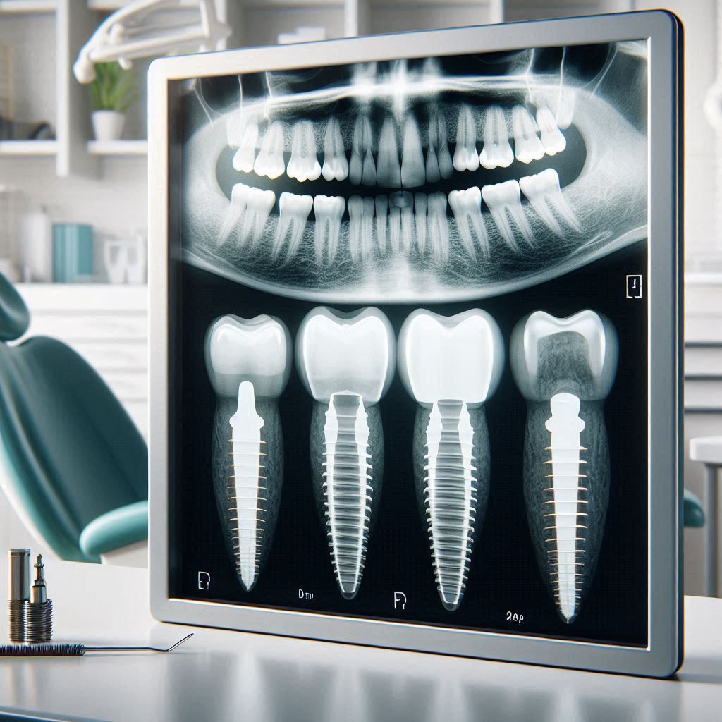
Dental Implant X-Rays
Dental implants have revolutionized modern dentistry, offering a permanent solution for missing teeth. However, their success heavily depends on precise planning and accurate radiographic assessment. Dental implant X-rays play a crucial role in evaluating bone structure, identifying vital anatomical structures, and ensuring optimal implant placement.
This comprehensive guide explores the various types of dental X-rays used in implantology, their clinical applications, and the latest advancements in imaging technology. Whether you’re a dentist, a dental student, or a patient considering implants, this article will provide valuable insights into the radiographic aspects of dental implantology.
2. The Importance of Dental Implant X-Rays
Before placing an implant, dentists must assess multiple factors:
- Bone quality and quantity – Is there sufficient bone to support the implant?
- Nerve proximity – How close is the inferior alveolar nerve?
- Sinus location – Is sinus lifting required for maxillary implants?
- Pathologies – Are there cysts, infections, or tumors that could affect healing?
Without proper radiographic evaluation, implant failure rates increase significantly. Studies show that CBCT scans reduce surgical complications by 30% compared to traditional 2D X-rays.
3. Types of Dental X-Rays Used in Implantology
Periapical X-Rays
- Focus on a single tooth and surrounding bone.
- Useful for assessing local bone density but limited in full-arch cases.
Panoramic X-Rays (OPG)
- Provides a broad view of the jaws, sinuses, and nerves.
- Best for initial screening but lacks 3D detail.
Cone Beam Computed Tomography (CBCT)
- Gold standard for implant planning.
- Delivers high-resolution 3D images with minimal radiation.
- Essential for guided surgery and virtual implant placement.
Comparison Table: Dental X-Ray Types for Implants
| Type of X-Ray | Best For | Limitations | Radiation Dose |
|---|---|---|---|
| Periapical | Single-tooth assessment | Limited field of view | Low |
| Panoramic (OPG) | Full-jaw overview | Distortions in magnification | Moderate |
| CBCT | 3D bone mapping | Higher cost | Low to Moderate |
4. Pre-Surgical Planning with Radiographic Imaging
Bone Density Assessment
- Type I (Dense bone) – Ideal for immediate loading.
- Type IV (Soft bone) – Requires longer healing periods.
Nerve and Sinus Mapping
- Mandibular implants must avoid the inferior alveolar nerve.
- Maxillary implants may need sinus augmentation if bone height is insufficient.
Virtual Implant Placement
- Software like SimPlant and BlueSkyBio allows precise digital planning.
- Enables fabrication of surgical guides for minimally invasive procedures.
(Insert high-quality image of a CBCT scan with implant planning overlay)
5. Step-by-Step Guide to Interpreting Dental Implant X-Rays
- Identify anatomical landmarks (mental foramen, maxillary sinus).
- Measure bone height and width (minimum 10mm height, 5mm width).
- Check for pathologies (cysts, infections, resorption).
6. Common Radiographic Challenges in Implant Dentistry
- Artifacts (metal streaks from existing restorations).
- Low bone volume (requiring grafting).
- Adjacent tooth root interference.
7. Advanced Imaging Technologies in Implantology
- AI-powered software predicts implant success rates.
- 3D-printed surgical guides improve accuracy.
8. Safety and Radiation Concerns
- ALARA Principle (As Low As Reasonably Achievable).
- Lead aprons and thyroid collars reduce exposure.
9. Case Studies
- Case 1: Full-arch restoration using CBCT-guided surgery.
- Case 2: Sinus lift with radiographic follow-up.
10. Future Trends
- Augmented Reality (AR) for real-time implant placement.
- Low-dose, ultra-high-resolution scanners.
11. Conclusion
Dental implant X-rays are indispensable for successful implantology. From 2D panoramic images to advanced CBCT scans, radiographic technology continues to evolve, improving precision and patient outcomes. By integrating AI and 3D planning, dentists can achieve predictable, long-lasting results.
12. FAQs
Q1: How often are X-rays needed for dental implants?
- Pre-op, during surgery (if guided), and post-op for follow-up.
Q2: Are dental X-rays safe?
- Yes, modern digital X-rays use minimal radiation.
Q3: Can implants be placed without X-rays?
- No, X-rays are essential for safe and accurate placement.