mini screw implants in orthodontics
- On
- InDENTAL
Imagine an architect designing a magnificent, complex structure but having no stable ground upon which to build its foundation. For decades, this was the fundamental challenge faced by orthodontists. The goal of orthodontic treatment is to move teeth through bone to achieve a functional bite and a beautiful smile. However, Newton’s third law of motion—for every action, there is an equal and opposite reaction—posed a constant and frustrating obstacle. To move a target tooth, an orthodontist had to push against another tooth (or group of teeth), often resulting in unwanted movement of these “anchor” teeth, a phenomenon known as anchorage loss.
This limitation forced compromises: extended treatment times, the use of cumbersome and patient-unfriendly appliances like headgear, and even the extraction of healthy premolars to camouflage the inability to control tooth movement perfectly. Then, a revolution occurred. The advent of Temporary Anchorage Devices (TADs), most notably Mini Screw Implants (MSIs), transformed the landscape of orthodontics. These unassuming titanium screws, typically no larger than a pencil eraser, provided what was once considered the holy grail of orthodontics: absolute anchorage.
This article delves deep into the world of mini screw implants, exploring their science, clinical application, and profound impact. We will journey from their fundamental design to the future of smart implants, providing a definitive guide for dental professionals, students, and curious patients alike. This is the story of how a tiny screw unlocked a new era of precision, efficiency, and possibilities in creating healthy, beautiful smiles.

Table of Contents
Toggle2. What Are Mini Screw Implants? Anatomy, Materials, and Design
2.1. The Anatomical Definition: More Than Just a “Mini-Screw”
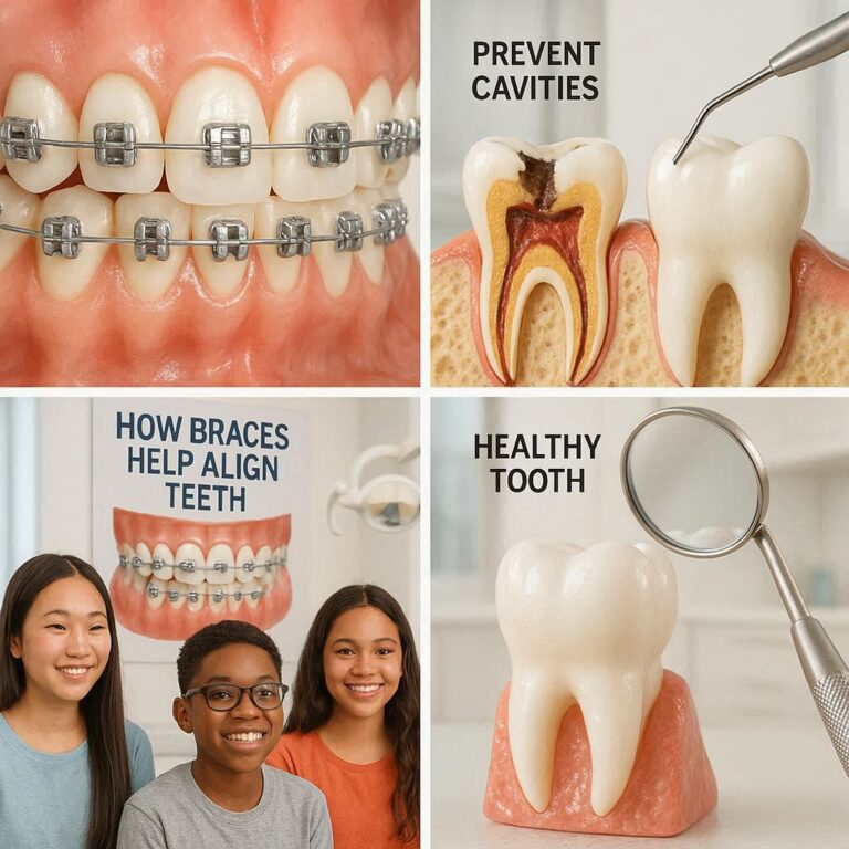
A Mini Screw Implant (MSI) is a small, biocompatible screw that is temporarily inserted into the alveolar bone (the bone that supports the teeth) or the palate to serve as a fixed, immovable point of anchorage for orthodontic tooth movement. Unlike dental implants used to replace missing teeth, MSIs are not designed to osseointegrate (fuse with the bone). Instead, they achieve stability through mechanical retention within the bone cortex, allowing for their easy removal once their anchorage purpose is fulfilled.
Key characteristics include:
-
Diameter: Typically ranges from 1.2 mm to 2.0 mm.
-
Length: Usually between 4 mm and 12 mm.
-
Temporary Nature: They are placed for the duration of specific tooth movements and are removed in a simple, straightforward procedure.
2.2. Biomaterials of Choice: The Titanium Advantage
The overwhelming material of choice for MSIs is medical-grade titanium alloy (Ti-6Al-4V). This preference is rooted in titanium’s exceptional properties:
-
Biocompatibility: Titanium is highly tolerated by the human body, eliciting minimal inflammatory or allergic responses.
-
High Strength-to-Weight Ratio: Despite their small size, titanium MSIs possess the necessary mechanical strength to withstand orthodontic forces without fracturing.
-
Corrosion Resistance: Titanium forms a passive oxide layer (TiO₂) on its surface, making it highly resistant to corrosion in the harsh oral environment.
Some manufacturers offer surface-treated (e.g., sandblasted, acid-etched) or stainless steel MSIs, but titanium remains the gold standard due to its proven track record and optimal balance of properties.
2.3. Engineering Excellence: A Deep Dive into MSI Design
The design of an MSI is a masterpiece of biomechanical engineering, with each component serving a specific purpose.
-
The Head: The topmost part that remains exposed in the oral cavity. It features a connection mechanism—often a slot, hole, or button—to which orthodontic forces (elastics, coils, wires) can be attached. Designs are streamlined to minimize soft tissue irritation.
-
The Shank/Neck: The transmucosal section that passes through the gum tissue. It is typically smooth to allow the gingiva to heal around it and prevent plaque accumulation.
-
The Body: The threaded portion that engages the bone. This is the primary source of stability. The thread design—including its pitch, depth, and shape (e.g., triangular, square, buttress)—is meticulously engineered to maximize insertion torque and distribute stress optimally within the bone.
-
The Tip/Apex: The leading end of the screw, designed to be self-tapping or self-drilling to facilitate easy insertion with minimal surgical trauma.
3. The Critical Need: Why Orthodontists Embraced Absolute Anchorage
3.1. The Historical Challenge: Anchorage Loss and Its Consequences
Traditional orthodontics relied on reactive forces. To pull canine teeth backwards into an extraction space, the orthodontist would push against the molars. However, this often resulted in the molars themselves moving forward, partially closing the space meant for the canines. This anchorage loss could lead to:
-
Incomplete correction of the bite.
-
Flaring of the front teeth.
-
Prolonged treatment time as the orthodontist struggled to “reclaim” lost space.
-
Less than ideal final results.
3.2. Conventional Anchorage Methods and Their Limitations
Before MSIs, orthodontists used various techniques to reinforce anchorage:
-
Extra-Oral Anchorage (Headgear): Effective but highly dependent on patient compliance. Many children and teenagers would not wear them for the required 12-14 hours per day, leading to treatment failure.
-
Intra-Oral Appliances: Devices like the Nance button or transpalatal arch. While compliance-independent, they are often bulky, can interfere with speech and hygiene, and still allow for some degree of anchorage loss as they are tooth-borne.
3.3. The Game Changer: Introducing Absolute Anchorage
MSIs provide absolute or stationary anchorage. Because they are anchored directly into the bone, they behave like a true immovable object. When a force is applied from an MSI to a tooth, the reaction force is dissipated into the skeletal structure without any movement of the anchor. This fundamental shift eliminates the problem of anchorage loss and allows the orthodontist to execute tooth movements that were previously extremely difficult or impossible.
4. The Orthodontic Armamentarium: Indications for Mini Screw Implants
The applications for MSIs are vast and have expanded the scope of what is treatable without surgery.
4.1. Intrusion: The “Impossible” Movement Made Reality
Intruding, or pushing teeth vertically upwards into the bone, requires significant force control. MSIs make this predictable.
-
Deep Bite Correction: Intruding the upper front teeth to level the bite without flaring them out.
-
Gummy Smile Correction: Intruding the upper molars to rotate the jaw closed, reducing excessive gum display.
-
Open Bite Treatment: Intruding over-erupted molars to allow the front teeth to close together.
[Image: A series of clinical photos showing the intrusion of upper anterior teeth using an MSI placed in the palate, connected via a power arm, to correct a deep bite.]
4.2. Molar Distalization: Creating Space Without Extractions
A common problem is dental crowding—not enough space for all the teeth. Instead of extracting premolars, an MSI can be placed to push the molars backwards, creating space for the alignment of the front teeth. This is a powerful non-extraction strategy.
4.3. Uprighting and Alignment: Correcting Tipped Teeth
Teeth that are tipped, often due to missing adjacent teeth, can be uprighted using an MSI as a pivot point, facilitating better restorative outcomes.
4.4. Closure of Extraction Spaces and Management of Midlines
MSIs ensure that when closing spaces left by extracted teeth, the anterior teeth retract fully into the space without the posterior teeth mesializing. They are also crucial for correcting dental midlines that are off-center.
4.5. Multidisciplinary Applications: Prosthodontics and Orthognathic Surgery
In cases where a tooth is missing, an MSI can be used to upright or move adjacent teeth to create an ideal space for a future implant or bridge. In surgical cases, they can be used for pre-surgical decompensation or to stabilize segments post-surgery.
Common Clinical Applications of Mini Screw Implants
| Clinical Problem | MSI Placement Site | Biomechanical Function | Key Advantage |
|---|---|---|---|
| Deep Bite | Palate / Between Upper Roots | Intrusion of anterior teeth | Avoids flaring of incisors; more predictable intrusion |
| Molar Distalization | Buccal shelf / Palate | Distal driving of molars to create space | Non-extraction treatment option; compliance-independent |
| Asymmetric Midline | Buccal interradicular | Unilateral retraction or protraction | Precise control to correct dental midline shifts |
| Open Bite | Buccal posterior region | Intrusion of over-erupted molars | Allows for autorotation of mandible and anterior bite closure |
| Uprighting Tipped Molars | Buccal or Palatal | Provides a vertical force to upright the tooth | Improves alveolar bone levels for future restoration |
5. The Clinical Protocol: From Treatment Planning to Successful Placement
5.1. Phase I: Meticulous Diagnosis and 3D Treatment Planning
Success begins long before the MSI is placed. A comprehensive evaluation includes:
-
Clinical Examination: Assessing bone quality, attached gingiva width, and oral hygiene.
-
Radiographic Analysis: Cone Beam Computed Tomography (CBCT) is the gold standard. It provides a 3D view of the proposed implant site, allowing the orthodontist to precisely measure bone width and height and identify the exact positions of tooth roots to avoid them during placement.
5.2. Phase II: Site Selection – The Art and Science of Anatomical Safety
Choosing the right location is paramount. Safe zones include:
-
Interradicular Space: The bone between tooth roots. CBCT is crucial to map safe pathways.
-
Buccal Shelf: The area distal to the lower second molar, which offers dense cortical bone.
-
Palate: The mid-palatal region offers excellent keratinized tissue and dense bone, ideal for intrusion mechanics.
-
Maxillary Tuberosity: The area behind the upper last molar.
5.3. Phase III: The Surgical Procedure – Step-by-Step
The placement is a minor surgical procedure performed under local anesthesia in the orthodontic office.
-
Anesthesia: Local anesthetic is administered. Often, only infiltration anesthesia is used without a nerve block, allowing the patient to provide feedback if the screw approaches a nerve.
-
Insertion: A self-drilling MSI is inserted directly through the mucosa using a slow-speed handpiece (15-30 rpm) with high torque control. A pilot hole may be drilled first in denser bone.
-
Placement Angle: The screw is typically placed at a 70-90 degree angle to the bone surface.
-
Verification: A periapical X-ray is taken immediately to verify the screw’s position relative to adjacent roots.
The entire process takes only a few minutes per screw.
5.4. Phase IV: Immediate Loading and Biomechanical Principles
A key advantage of MSIs is that they can be loaded with orthodontic force immediately. The orthodontist will attach a spring, elastic, or wire to the MSI’s head and to the tooth or archwire, applying a light, continuous force (typically 50-200 grams) to initiate the desired tooth movement.
6. Navigating Challenges: Risks, Limitations, and Failure Management
Despite high success rates (often >85%), failures and complications can occur.
6.1. Understanding Primary Stability: The Key to Success
The single most important factor for success is primary stability—the mechanical stability achieved at the moment of insertion. This is a function of bone density, screw design, and surgical technique. Low insertion torque can lead to mobility and failure.
6.2. Common Reasons for MSI Failure and How to Mitigate Them
-
Root Contact: The most common cause of failure. It can cause inflammation, pain, and mobility. Meticulous planning with CBCT is the best prevention.
-
Poor Oral Hygiene: Inflammation of the surrounding soft tissue (peri-implantitis) can lead to bone loss and screw loosening.
-
Overloading: Applying excessive force can exceed the bone’s tolerance, leading to resorption and failure.
-
Anatomical Factors: Low bone density or thin gingival tissue can compromise stability.
If an MSI fails, it is simply unscrewed and removed. The site heals quickly, and a new MSI can often be placed in an adjacent site after a short healing period.
6.3. Managing Soft Tissue Complications and Patient Discomfort
Mild discomfort and soft tissue inflammation are common for 24-48 hours post-placement, manageable with over-the-counter analgesics and chlorhexidine mouth rinses. The head of the screw can sometimes irritate the cheek; orthodontic wax is very effective at mitigating this.
7. The Patient Perspective: Experience, Care, and FAQs
7.1. What to Expect During and After the Procedure
Patients often report that the anticipation is worse than the procedure itself. The placement feels similar to a dental injection followed by a sensation of pressure. Post-operative pain is typically minimal and short-lived. The presence of the MSI becomes normal within a week.
7.2. Essential Oral Hygiene Practices for MSI Maintenance
Keeping the area clean is critical for success. Patients are instructed to:
-
Use a soft-bristle toothbrush.
-
Clean around the MSI head gently but thoroughly.
-
Use a chlorhexidine mouthwash or a water flosser to flush out debris.
-
Avoid playing with the screw with their fingers or tongue.
8. The Future is Now: Technological Advancements and Innovations
8.1. Guided Surgery: The Role of CBCT and 3D-Printed Stents
The future is in digital workflow. Using CBCT data and digital dental models, orthodontists can now design a virtual surgical guide that is 3D-printed. This guide fits over the teeth and has a precise metal sleeve that dictates the exact position, angle, and depth for MSI placement, eliminating guesswork and maximizing safety and efficiency.
8.2. Smart and Active Implants: The Next Frontier
Research is underway into “smart” MSIs that can deliver therapeutic agents to reduce inflammation or even implants with built-in sensors to monitor the orthodontic force being applied in real-time, ensuring it remains within optimal biological limits.
8.3. Biomaterial Innovations: Towards Bioresorbable Anchorage
The ultimate innovation would be a bioresorbable MSI that provides stable anchorage and then slowly dissolves over 12-18 months, eliminating the need for a removal procedure. Research into magnesium alloys and advanced polymers is actively exploring this possibility.
9. Conclusion: Summarizing the Impact of Mini Screw Implants
The introduction of mini screw implants represents the most significant advancement in orthodontic mechanics in the past half-century. By providing a source of absolute anchorage, they have empowered clinicians to achieve treatment goals with unprecedented precision, efficiency, and predictability. They have expanded the boundaries of non-surgical orthodontics, improved patient comfort by eliminating the need for compliance-dependent appliances, and consistently enabled superior clinical outcomes. As technology continues to evolve with guided surgery and new materials, the role of MSIs will only become more integral, solidifying their status as an indispensable tool in modern orthodontics.
10. Frequently Asked Questions (FAQs)
Q1: Does getting a mini screw implant hurt?
A: The procedure is performed under local anesthesia, so you should feel only pressure, not pain. After the anesthesia wears off, you may experience some mild soreness for a day or two, similar to a canker sore, which is easily managed with over-the-counter pain relievers.
Q2: How long do the screws stay in?
A: They are temporary devices. They remain in place only for as long as they are needed to achieve a specific tooth movement, which can range from a few months to over a year. They are removed in a simple procedure once their purpose is fulfilled.
Q3: What happens if the screw gets loose or fails?
A: If an MSI becomes loose, it is no longer effective and needs to be removed. This is a common and non-critical event. The site heals very quickly, and your orthodontist can often place a new screw in a slightly different location after a brief healing period.
Q4: Will the screw leave a hole in my gum or bone?
A: The hole in the gum closes and heals within a few days. The tiny hole in the bone fills in with new bone tissue over the following weeks, leaving no permanent damage.
Q5: Are mini screw implants safe?
A: When placed by a trained orthodontist using modern 3D imaging for planning, MSIs are extremely safe. The risk of serious complications is very low. The most common issue is mild, temporary inflammation around the screw, which is manageable with good oral hygiene.
11. Additional Resources
-
American Association of Orthodontists (AAO) – Patient Info on TADs: https://www.aaoinfo.org/ (Search for “TADs” or “temporary anchorage devices”)
-
Journal of Clinical Orthodontics: A leading publication featuring numerous case studies and technical articles on MSI applications. (Professional resource)
-
Seminars in Orthodontics – Volume on Temporary Anchorage Devices: A dedicated issue providing in-depth scholarly reviews on the science and use of MSIs. (Professional resource)
-
Cone Beam Computed Tomography in Orthodontics: 3D Diagnosis and Treatment Planning. (Academic Textbook)
Date: September 16, 2025
Author: Dr. Evelyn Reed, DDS, MS (Orthodontics)
Disclaimer: The information provided in this article is for educational and informational purposes only and does not constitute medical advice. Always consult with a qualified orthodontic specialist for diagnosis, treatment planning, and care tailored to your individual needs.

dentalecostsmile
Newsletter Updates
Enter your email address below and subscribe to our newsletter