The Complete Guide to peri implant infection treatment
- On
- InDENTAL IMPLANTS
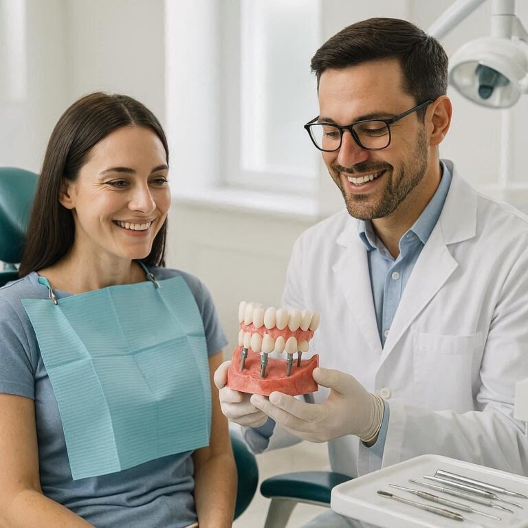
Dental implantology represents one of the most transformative advancements in modern dentistry, successfully restoring function, aesthetics, and quality of life for millions of patients worldwide. For decades, the focus was overwhelmingly on achieving osseointegration—the direct structural and functional connection between living bone and the surface of a load-bearing artificial implant. With success rates now routinely exceeding 95% in healthy patients, the field rightly celebrated this remarkable achievement. However, this very success has unveiled a new and formidable challenge: the long-term preservation of implant health. Just as a natural tooth is susceptible to periodontal disease, a dental implant is vulnerable to the inflammatory and destructive processes of the tissues that surround it. Peri-implant diseases, particularly peri-implantitis, have emerged as the primary threat to the long-term survival of dental implants, presenting a complex biological and clinical puzzle for practitioners and a significant source of morbidity for patients. This article delves deep into the world of peri-implant infections treatment, moving beyond superficial overviews to provide a exhaustive, evidence-based exploration of their etiology, diagnosis, and the sophisticated, multi-modal treatment protocols required to manage them effectively.

Table of Contents
Toggle2. Defining the Spectrum: Peri-Implant Health, Mucositis, and Peri-Implantitis
Understanding peri-implant disease begins with recognizing it as a spectrum, much like periodontal disease. The 2017 World Workshop on the Classification of Periodontal and Peri-Implant Diseases and Conditions established clear, consensus definitions that are critical for accurate diagnosis and communication.
-
Peri-Implant Health is characterized by an absence of clinical signs of inflammation (redness, swelling, bleeding on gentle probing BOP), and suppuration. Probing depths are stable and consistent with the health of the specific implant configuration. Health can exist around implants with reduced bone support, provided inflammation is controlled.
-
Peri-Implant Mucositis is a reversible, inflammatory lesion confined to the soft tissues surrounding an implant. The key clinical sign is BOP and/or suppuration on gentle probing with no evidence of additional bone loss beyond initial remodeling. It is a plaque-induced inflammation, and its resolution is achieved through the reinstruction of effective oral hygiene and professional debridement.
-
Peri-Implantitis is a pathological condition occurring in tissues around dental implants, characterized by inflammation in the peri-implant mucosa and progressive loss of supporting bone. The diagnosis requires:
-
Presence of bleeding and/or suppuration on gentle probing.
-
Increased probing depths compared to baseline measurements taken after the prosthetic phase.
-
Progressive bone loss visible on radiographs.
-
The distinction between initial bone remodeling post-loading and progressive bone loss is crucial. Bone loss that occurs after the first year of function is typically considered pathological.
3. The Microbial Culprits: Etiology and Biofilm Formation
The primary etiological agent for peri-implant diseases is microbial biofilm, a complex, organized community of bacteria that adheres to the implant and abutment surfaces. The composition of this biofilm shares similarities with that found in periodontal diseases but possesses unique characteristics that influence its pathogenicity and treatment.
The transition from health to disease is a shift in the microbial ecology from a predominantly commensal, Gram-positive aerobic flora to a pathogenic, Gram-negative anaerobic flora. Key pathogens implicated in peri-implantitis include:
-
Porphyromonas gingivalis
-
Tannerella forsythia
-
Treponema denticola (together known as the “red complex”)
-
Aggregatibacter actinomycetemcomitans
-
Fusobacterium nucleatum
The implant-soft tissue interface is fundamentally different from the tooth-soft tissue interface. The periodontal ligament, with its rich vascular supply and cellular activity, provides a robust defensive barrier. The implant, however, has a direct mucosal attachment via a hemidesmosomal seal, which is inherently weaker and more susceptible to breakdown. Once the biofilm matures and invades this seal, it triggers a host inflammatory response. The release of inflammatory mediators (e.g., IL-1β, TNF-α, PGE2) leads to tissue destruction and osteoclast activation, resulting in the apical migration of the biofilm and the characteristic bone loss of peri-implantitis.
4. Risk Factors and Contributors: Beyond Bacteria
While biofilm is the necessary cause, it is not sufficient alone. A multitude of risk factors and indicators influence a patient’s susceptibility to peri-implant infections.
Patient-Specific Risk Modifiers:
-
History of Periodontitis: This is the single strongest risk factor. Patients with a past history of chronic periodontitis have a significantly higher risk of developing peri-implantitis due to a susceptible oral microbiome and potentially a hyperinflammatory host response.
-
Poor Oral Hygiene: Inadequate plaque control is the primary modifiable risk factor.
-
Smoking: Nicotine causes vasoconstriction, impairing blood flow and the immune response in the peri-implant tissues, masking bleeding signs and hindering healing.
-
Uncontrolled Diabetes: Hyperglycemia impairs neutrophil function, collagen synthesis, and wound healing, increasing susceptibility to infection and progression of disease.
-
Genetic Factors: Certain genetic polymorphisms (e.g., in IL-1 gene cluster) are associated with a heightened inflammatory response to a bacterial challenge.
-
Medications: Drugs that cause xerostomia (dry mouth) or induce gingival hyperplasia can compromise oral hygiene.
Iatrogenic and Technical Risk Factors:
-
Cement Retention: Residual submucosal cement is a major contributor to peri-implant disease, acting as a potent plaque trap and irritant.
-
Poor Prosthetic Design: Overcontoured crowns, inadequate embrasure spaces, and poor implant positioning make effective hygiene impossible.
-
Lack of Keratinized Tissue: While debated, an adequate zone of keratinized mucosa is associated with better plaque control, less inflammation, and easier maintenance.
-
Occlusal Overload: Excessive biomechanical forces may contribute to bone loss, particularly in the presence of pre-existing inflammation.
5. The Diagnostic Imperative: A Multifaceted Approach
A thorough diagnosis is the foundation of effective treatment. It must be comprehensive and reproducible.
Clinical Assessment:
-
Probing: Must be performed with a light force (~0.25N) using a plastic or metal probe to avoid scratching the implant surface. Probing depths >5-6mm are a concern, but the change from baseline is more critical than an absolute number.
-
Bleeding on Probing (BOP): A primary indicator of inflammatory activity. Its consistent absence is a reliable predictor of stability.
-
Suppuration: Expressing pus is a clear sign of active infection.
-
Mobility: Implant mobility is a late sign signifying advanced bone loss and failure of osseointegration.
Radiographic Evaluation:
-
Periapical Radiographs: The gold standard for assessing bone levels. They must be standardized (using a paralleling technique with a film holder) to allow for accurate longitudinal comparison. Bone loss is measured from a stable reference point, often the implant-abutment interface.
-
Cone Beam Computed Tomography (CBCT): Not a routine diagnostic tool but invaluable in surgical planning. It provides 3D visualization of the defect morphology (e.g., supracrestal, intrabony, circumferential), the extent of bone loss, and proximity to vital structures.
Adjunctive Diagnostic Tools:
-
Bacterial DNA Tests: Can identify the presence and proportion of specific periodontal pathogens.
-
Inflammatory Marker Analysis: Testing crevicular fluid for levels of IL-1β, MMP-8, and other markers may offer insights into disease activity and host response, though this is primarily used in research.
6. The Non-Surgical Armamentarium: First-Line Defense
The treatment of peri-implant mucositis and the initial management of peri-implantitis always begin with non-surgical therapy. The goal is to disrupt and remove the biofilm and calculus without damaging the implant surface.
Mechanical Debridement:
-
Traditional Scalers: Titanium or carbon-fiber scalers are used to avoid scratching the implant surface, but access to implant threads and complex geometries is limited.
-
Ultrasonic/Sonic Scalers with Plastic or Titanium Tips: More effective at biofilm disruption. The energy and irrigation provide a valuable lavage effect. Care must be taken to avoid plastic tip wear, which can leave deposits on the surface.
-
Air Polishing Devices: A cornerstone of modern non-surgical care. These devices use a controlled stream of air, water, and low-abrasive powder (e.g., glycine, erythritol) to efficiently and gently remove biofilm. Erythritol powder has shown excellent efficacy in biofilm removal and has some inherent antibacterial properties.
Antimicrobial Adjuncts:
-
Local Delivery Antibiotics: For isolated, persistent pockets, locally administered antibiotics (e.g., minocycline microspheres, doxycycline gel) can provide high localized concentrations of antimicrobials directly at the site of infection, overcoming the access limitations of mechanical therapy.
-
Systemic Antibiotics: Reserved for cases with acute infection, suppuration, and systemic involvement (e.g., fever). They are never a standalone treatment and must always be combined with thorough mechanical debridement. Common regimens include amoxicillin with metronidazole.
-
Chlorhexidine Gluconate: The gold standard antiseptic for chemical plaque control. Used as a mouthwash or applied locally via irrigators or gels.
The Critical Role of Patient-Mediated Maintenance: Treatment success is entirely dependent on the patient’s ability to control biofilm daily. Reinstruction in oral hygiene techniques tailored to implants (soft brushes, interdental brushes, water flossers) is non-negotiable. Motivation and frequent recall visits are essential.
7. Surgical Intervention for Peri-Implantitis: Regaining Lost Terrain
When non-surgical therapy fails to resolve inflammation and arrest disease progression, surgical intervention is indicated. The surgical approach is chosen based on defect morphology, aesthetic location, and patient factors. The universal first step is flap elevation for direct visual access and surface decontamination.
Surface Decontamination:
This is the most critical and challenging step. The goal is to render the contaminated implant surface biocompatible again for tissue reattachment or regeneration. No single method has proven universally superior. Common techniques include:
-
Mechanical: Titanium brushes, plastic curettes, ultrasonic scaling.
-
Chemical: Irrigation with saline, chlorhexidine, or hydrogen peroxide.
-
Air-powder abrasive systems.
-
Laser systems (e.g., Er:YAG, Diode).
Resective Surgery (Apically Positioned Flap):
This is the most predictable surgical technique for eliminating pockets. It involves:
-
Osteoplasty: Recontouring the bone to eliminate the defect morphology.
-
Apical positioning of the flap to the newly recontoured bone crest.
The result is a shallower probing depth, facilitating patient hygiene and professional maintenance. The trade-off is tissue recession, which can be aesthetically compromising in the anterior zone.
Regenerative Surgery:
The goal is not just to arrest disease but to reconstruct the lost bone support. This involves:
-
Thorough debridement and decontamination.
-
Application of a bone graft (autograft, allograft, xenograft, or alloplast) to fill the defect.
-
Coverage with a barrier membrane (resorbable or non-resorbable) to guide regeneration.
While case reports show success, regenerative outcomes for peri-implantitis are less predictable than for periodontitis due to the challenge of achieving true re-osseointegration on a previously contaminated surface. The literature shows significant variability in bone fill results.
The Combination Approach:
A pragmatic approach often involves elements of both. A defect may be partially grafted to gain some bone, while the bony walls are recontoured to create a maintainable architecture.
8. Adjunctive and Emerging Therapies: The Future of Treatment
Research is continuously exploring new modalities to improve decontamination and healing.
-
Laser and Photodynamic Therapy (aPDT): Lasers (Er:YAG, Er,Cr:YSGG) are effective for debridement and have bactericidal effects. aPDT involves applying a photosensitizer to the pocket, which is then activated by a specific wavelength of light, producing cytotoxic oxygen species that destroy bacteria without damaging host tissue. They are powerful adjuncts but not magic bullets.
-
Systemic Modulation: The use of host modulators like sub-antimicrobial dose doxycycline (SDD) to inhibit matrix metalloproteinases (MMPs) that destroy connective tissue is being investigated as an adjunct to control the host’s destructive response.
-
Probiotics: The concept of orally administering beneficial bacteria to modulate the peri-implant microbiome is a promising area of research for both prevention and adjunctive treatment.
9. The Algorithm of Care: A Structured Treatment Protocol
A systematic, staged approach is essential for managing peri-implantitis, as outlined in the table below.
Staged Protocol for the Management of Peri-Implantitis
| Stage | Objective | Procedures | Re-Evaluation (6-8 weeks post-op) |
|---|---|---|---|
| 1. Cause-Related Therapy | Eliminate etiology, control inflammation | 1. Comprehensive diagnosis & risk factor analysis. 2. Supervised oral hygiene reinstruction. 3. Thorough non-surgical debridement (mechanical + air polishing). 4. Use of local antimicrobials if indicated. 5. Address prosthetic issues (e.g., remove excess cement, modify prosthesis). |
• Probing depths (PD) • Bleeding on Probing (BOP) • Suppuration • Radiograph for baseline |
| 2. Surgical Therapy | Access, decontaminate, and correct defects | If BOP/PD persist & bone loss progresses: • Regenerative Surgery: For intrabony defects in aesthetic areas. • Resective Surgery: For suprabony defects or non-aesthetic areas. • Combination Therapy: As required. |
• Clinical parameters (PD, BOP) • Radiographic bone level assessment (6-12 months) |
| 3. Supportive Maintenance Therapy (SPT) | Prevent disease recurrence | 1. Strict, individualized recall program (3-6 month intervals). 2. Reassessment of clinical parameters at each visit. 3. Reinforcement of oral hygiene. 4. Professional re-debridement. |
• Continuous monitoring for stability. |
10. The Unfortunate Reality: When to Retrieve an Implant
Despite best efforts, not all implants can be saved. Implant removal should be considered when:
-
There is advanced bone loss (>50-60% of the implant is surrounded by bone loss), leaving insufficient support.
-
The defect morphology is untreatable (e.g., a deep, circumferential defect).
-
The implant is mobile.
-
Repeated surgical interventions have failed to arrest the disease.
-
The patient is unable or unwilling to comply with the necessary intensive maintenance protocol.
Removal techniques include reverse torque explanation (if some osseointegration remains) or trephination to remove the implant and a core of surrounding bone.
11. The Cornerstone of Success: Long-Term Maintenance and Prevention
The most effective “treatment” for peri-implantitis is its prevention. A proactive, lifelong maintenance protocol is essential for every implant patient, especially those with risk factors. This involves:
-
Individualized Recall Intervals: Typically every 3-6 months.
-
Systematic Reassessment: At each visit, probing, BOP, and suppuration must be checked. Annual radiographs are recommended for comparison.
-
Professional Prophylaxis: Regular debridement with appropriate instruments.
-
Continuous Reinforcement: Oral hygiene must be constantly evaluated and reinforced.
12. Conclusion: A Synthesis of Science and Strategy
Peri-implantitis is a complex, biofilm-driven disease demanding a nuanced and multifaceted treatment approach. Success hinges on accurate diagnosis, strict control of modifiable risk factors, and a structured protocol beginning with non-surgical therapy. Surgical intervention, while challenging, can arrest progression and, in some cases, regenerate lost support, but long-term stability is irrevocably tied to a disciplined, lifelong maintenance regimen. The ultimate goal shifts from mere survival of the implant to the preservation of its health and function over decades.
13. Frequently Asked Questions (FAQs)
Q1: Is peri-implantitis painful?
A: In its early and chronic stages, peri-implantitis is often not painful. This “silent” nature is why regular check-ups are crucial. Pain can occur in acute phases or with the formation of an abscess, but its absence does not indicate health.
Q2: Can a dental implant get infected years after placement?
A: Absolutely. Peri-implantitis is typically a slowly progressing chronic disease. An implant that has been stable for many years can develop the condition if oral hygiene lapses, if the patient’s systemic health changes (e.g., new medication causing dry mouth), or if other risk factors emerge.
Q3: Is peri-implantitis treatable without surgery?
A: Peri-implant mucositis is reversible with non-surgical treatment. For peri-implantitis, non-surgical therapy is the essential first step and can successfully manage the disease in some cases, particularly if caught early. However, its primary goal is often to reduce inflammation before surgery and to manage the disease long-term; it may not eliminate the deep pockets caused by bone loss, which often require surgical intervention for definitive management.
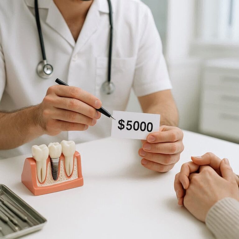
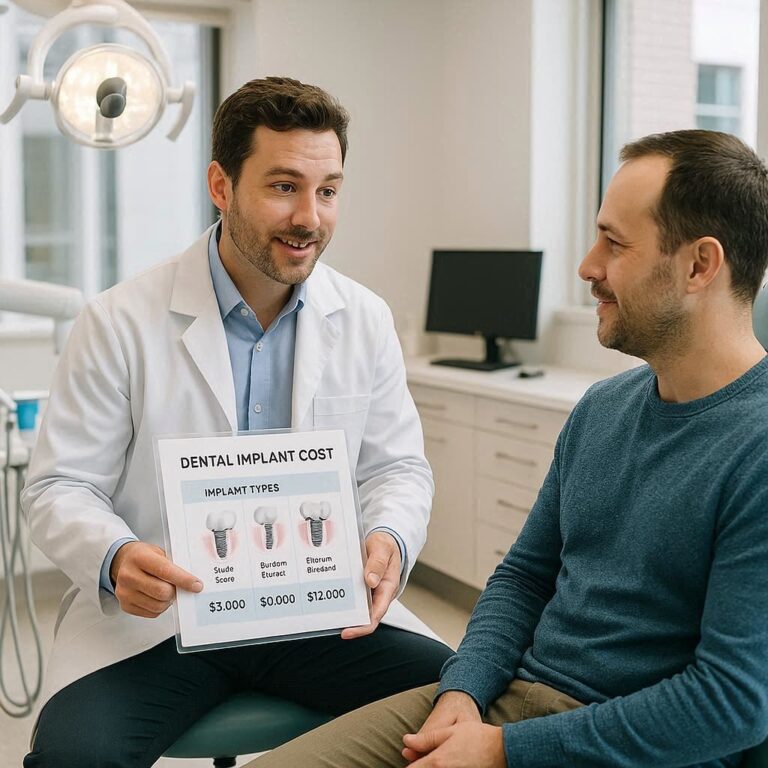
Q4: How much does treatment for peri-implantitis cost?
A: Costs vary widely based on geographic location, the specialist providing care, and the complexity of the treatment. Non-surgical therapy may cost a few hundred dollars, while regenerative surgery can cost several thousand dollars per implant. Dental insurance coverage for these procedures is often limited.
Q5: If my implant is removed due to peri-implantitis, can I get a new one?
A: Usually, yes. After the infected implant is removed, the site must heal. Often, bone grafting is required to rebuild the lost bone before a new implant can be placed. The underlying risk factors (like a history of periodontitis) must be thoroughly addressed before considering re-implantation.
14. Additional Resources
-
Academy of Osseointegration (AO): https://www.osseo.org/ (Provides consensus statements and educational resources)
-
American Academy of Periodontology (AAP): https://www.perio.org/ (See their patient resources on peri-implant diseases)
-
The 2017 World Workshop on the Classification of Periodontal and Peri-Implant Diseases and Conditions: The definitive source for current definitions and diagnostic criteria. (Full manuscripts published in the Journal of Periodontology and Journal of Clinical Periodontology).
-
Schwarz, F., Derks, J., Monje, A., & Wang, H. L. (2018). Peri-implantitis. Journal of Periodontology, 89 Suppl 1, S267-S290. A key review article summarizing the evidence.

dentalecostsmile
Newsletter Updates
Enter your email address below and subscribe to our newsletter