Prolia and Dental Implants: Navigating Safety, Risk, and Coordination of Care
Imagine sitting in a dental chair, excited about finally replacing a missing tooth with a permanent implant. Then, the dentist pauses while reviewing your medical history. “I see you’re taking Prolia,” they say. “We need to have a very important conversation before we proceed.”
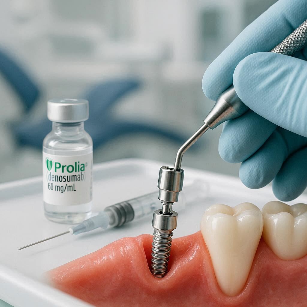
That moment of pause is not just bureaucracy. It is a potentially life-altering safety check. The intersection of Prolia (denosumab) and dental implant surgery represents one of the most nuanced and high-stakes crossovers between medicine and dentistry. The concern is not theoretical. It is anchored in the biological reality of how your jawbone heals, or more precisely, how it might fail to heal under the influence of a powerful drug.
This guide aims to unpack every layer of this complex topic. We will move far beyond the surface-level warnings. We will explore the science of bone turnover, dissect the statistics behind the rare but devastating condition known as Medication-Related Osteonecrosis of the Jaw (MRONJ), and provide a roadmap for navigating informed consent. Whether you are a patient seeking clarity or a clinician refining your protocol, this article serves as your comprehensive, human-centered reference point.
The High Stakes of Ignoring the Connection
You must understand why this topic warrants a 15,000-word deep dive. The jawbone is not a static chunk of calcium. It is a living, breathing, constantly remodeling organ. Every night when you sleep, cells called osteoclasts dissolve tiny micro-cracks in your skeleton, and cells called osteoblasts fill them in with new matrix. Prolia fundamentally paralyzes the “demolition crew” (osteoclasts). In the spine and hips, this protects against fracture. In the jaw, which turns over bone ten times faster than other skeletal sites, this creates a vulnerability.
When a dentist drills into a jaw that has lost its biological remodeling capacity, the bone can simply give up. It does not heal. It dies. This is MRONJ. And in the context of Prolia, the drug’s mechanism of action creates a unique timing issue not seen with other osteoporosis medications. The drug stays in the system for exactly six months, binding to receptors with an iron grip before abruptly releasing its hold. Miss this therapeutic window, and the risk profile shifts dramatically. The stakes are not just a failed implant. The stakes are exposed, necrotic bone in the mouth, chronic pain, and potential jaw fracture.
What is Prolia? Understanding the Mechanism of Action
To grasp the dental guidelines, you must first become intimately familiar with the drug at the center of the discussion. Prolia is not a traditional pill. It is not a once-a-week tablet that you take with breakfast. It is a biologic agent, a human monoclonal antibody delivered via a subcutaneous injection, usually in the arm, every six months.
The RANKL Connection
The active ingredient, denosumab, works like a precision-guided decoy. Your body uses a signaling pathway called RANK/RANKL to trigger the production and activation of osteoclasts. Imagine the osteoblast cells shouting “send in the demolition crew!” through a megaphone. That megaphone is RANKL (Receptor Activator of Nuclear factor Kappa-B Ligand).
Denosumab binds to and inhibits RANKL. It effectively smashes the megaphone. Without the signal, the stem cells never receive the command to mature into active osteoclasts. Within 24 hours of a Prolia injection, the markers of bone resorption in your blood plummet to nearly undetectable, postmenopausal levels.
This is a marvel of biotechnology for a patient with severe osteoporosis. A spine crumbling silently can be stabilized. A femur threatening to snap can be reinforced. However, this suppression is not “balanced.” There is no off-switch for the osteoblasts immediately. For a short period, there is an uncoupled bone remodeling process where formation outpaces resorption, increasing density. But long-term, a deeply suppressed state of low-turnover bone sets in.
“Denosumab is not metabolized by the liver or kidneys. It is cleared by the reticuloendothelial system. This is why the six-month duration of action is so precise and predictable, but also why the rebound effect upon discontinuation is so sharp.”
Indications Beyond Osteoporosis
While predominantly known for postmenopausal osteoporosis, Prolia is also prescribed for:
- Men with osteoporosis at high fracture risk.
- Patients with glucocorticoid-induced osteoporosis.
- Patients undergoing androgen deprivation therapy (prostate cancer) or aromatase inhibitor therapy (breast cancer) to combat treatment-induced bone loss.
- Note: The higher-dose formulation, XGEVA (120mg vs. 60mg Prolia), is used for bone metastases and giant cell tumor of bone. While the mechanism is identical, the risk profile for MRONJ is substantially higher with the oncologic dose. This guide will differentiate risk profiles where necessary, but the core principles of surgical caution apply to both.
Understanding this mechanism is the foundation of safety. You are not looking at a nutritional deficiency. You are looking at a targeted, systemic blockade of a fundamental skeletal maintenance process. The jaw, with its thin mucosal covering and constant bacterial exposure, is the canary in the coal mine for this skeletal suppression.
The Jawbone Under the Microscope: Why This Site is Uniquely Vulnerable
Why does MRONJ happen almost exclusively in the jaws? You might break a toe or fracture a wrist while on Prolia, but those sites heal normally (albeit slowly) with standard orthopedic care. The jaw is a unique evolutionary structure that operates under specific environmental stressors that the rest of the skeleton never faces.
High Bone Turnover Rate
The alveolar bone that holds your teeth has a blood supply and turnover rate that is vastly accelerated compared to the iliac crest or the femoral shaft. The constant pressure of mastication, the micro-movement of teeth, and the presence of the periodontal ligament create a high-traffic zone of cellular activity. This makes the site exquisitely sensitive to any agent that suppresses osteoclast function. The “remodeling suppression” is not uniform across the body; it is concentrated where remodeling is most active.
The Thin Biologic Barrier
Consider a hip replacement. The incision is deep, through layers of fat and muscle, and closed in a sterile, layered fashion. Now consider the oral cavity. A dental implant abutment, or even a healing extraction socket, is separated from the outside world by a millimeter or two of gingival tissue. The bacterial load is massive. The oral cavity is a warm, wet ecosystem of over 700 bacterial species. When the oral mucosa breaks down over dead bone—bone that cannot remodel to fight off the biofilm—infection takes hold. The bone does not die because of the infection; the bone dies because of the drug, and then the infection colonizes the dead scaffold.
Cumulative Micro-Damage
Every time you clench your teeth or chew a piece of steak, you create micro-cracks in the jawbone. In a healthy state, osteoclasts immediately target these cracks and resorb them, paving the way for new, strong bone. In a patient on Prolia, those micro-cracks sit there. They propagate. They coalesce. Eventually, the cumulative structural fatigue exceeds the material strength of the unresorbed bone. When a dentist places an implant, they create a controlled macro-damage event. If the mechanism to clear the micro-damage around the osteotomy is non-functional, the entire sequela of necrosis begins.
Defining the Danger: What is Medication-Related Osteonecrosis of the Jaw (MRONJ)?
Before 2003, this disease had no name. Dentists saw patients, particularly cancer patients on intravenous bisphosphonates, with bizarre non-healing sockets and exposed yellow-white bone that persisted for months. They called it “bis-phossy jaw,” a nod to the 19th-century “phossy jaw” of matchstick factory workers. Now, the nomenclature has standardized to MRONJ, recognizing that the pathway spans multiple drug classes, including denosumab, and angiogenesis inhibitors.
According to the American Association of Oral and Maxillofacial Surgeons (AAOMS) position paper, the case definition is strict. You cannot throw the term around loosely. The diagnosis requires three simultaneous truths:
The Three Pillars of Diagnosis
- Current or previous treatment with antiresorptive or antiangiogenic agents.
This is the exposure variable. Prolia (denosumab) squarely fits this category. The timeline of exposure relative to the dental event is the critical variable we will analyze later. - Exposed bone or bone that can be probed through an intraoral or extraoral fistula(e) in the maxillofacial region.
This is the clinical picture. It is not just pain. It is visible, palpable dead bone. Sometimes it is a sharp, irregular edge where the gum has receded. Sometimes it is a deep pocket where a probe hits gritty, smooth bone that does not bleed. - Persistence for more than eight weeks.
This is the temporal filter. A dry socket or a delayed healing site at four weeks is not MRONJ. It is a problem, but it is not this diagnosis. The eight-week cut-off distinguishes normal physiological crisis from complete cessation of healing.
Staging the Severity
Once diagnosed, MRONJ is not a binary condition. It exists on a spectrum, and the stage dictates the treatment approach. Understanding the staging helps a patient contextualize their situation.
| Stage | Clinical Description | Visible Signs | Symptom Profile |
|---|---|---|---|
| Stage 0 | No clinical evidence of necrotic bone, but non-specific symptoms exist. | No exposed bone. May have deep periodontal pockets, loosening of teeth that were previously stable, or altered sensation. | Dull, aching bone pain in the jaw that radiates. Thickening of the periodontal ligament space (visible on X-ray). |
| Stage 1 | Exposed and necrotic bone, or fistula probing to bone. Asymptomatic. | Visible “cheesy” or yellow-white dead bone in the mouth. No granulation tissue. | No signs of infection. The patient is comfortable despite the grim appearance. |
| Stage 2 | Exposed and necrotic bone, or fistula. Infected. | Redness, swelling, purulent drainage. The soft tissue around the exposed bone looks angry and inflamed. | Pain, tenderness, halitosis (bad breath). This is active infection complicating the necrosis. |
| Stage 3 | All features of Stage 2, plus extension beyond the alveolar bone. | Pathologic fracture, extra-oral fistula (draining to the cheek or chin), extension to the floor of the mouth or maxillary sinus. | Severe, narcotic-resistant pain. Significant swelling compromising airway in rare, aggressive cases. |
“The clinical reality of MRONJ is often a disconnect between what the patient feels and what the dentist sees. Stage 1 can look terrifying—a chunk of missing gum—but pain is often absent because the nerve endings are dead. Stage 2, however, brings the toxic combination of dead bone and live bacteria.”
Comparative Drug Risk: Bisphosphonates vs. Prolia
Patients and dentists often mentally lump all “bone drugs” into one basket. This is a serious clinical error. The pharmacokinetics, receptor binding, and reversibility of Prolia are fundamentally different from oral or IV bisphosphonates. To make an informed decision about a dental implant, you must understand the difference between the drug that binds to bone for ten years and the drug that clears the body in six months.
The Skeletal Half-Life Trap
Oral bisphosphonates like alendronate (Fosamax) attach to the hydroxyapatite crystals in bone. They sit there, dormant, for the life of the bone remodeling unit. When an osteoclast eats that bone, it ingests the bisphosphonate, which then causes cellular apoptosis (suicide). If you stop taking alendronate today, active drug remains in your skeleton, capable of damaging osteoclasts, for a decade.
Prolia is different. It circulates in the blood. It does not bind to bone mineral. It binds to the RANKL cytokine. The half-life of denosumab in the serum is approximately 25-30 days. After the six-month dose, the drug levels are at their peak for about two months, then gradually decline. By month six, serum levels are often near zero in many patients. This is the game-changer. The drug effect is reversible in a time frame that allows for elective surgical planning.
The Rebound Resorption Phenomenon
Here lies the crucial, often overlooked distinction. When you stop a bisphosphonate, the skeletal hold is gradually released over years. There is no sudden spike in bone breakdown. When the Prolia injection wears off, and the RANKL decoy disappears, the megaphone comes back online—angrily. The osteoclasts, which have been dormant for six months, surge back. A temporary period of rapid bone loss follows. Vertebral fracture risk can actually spike during this “holiday” if not managed with an alternative agent.
This has profound implications for implant timing. You do not want to place an implant when the bone is at its maximally suppressed, nearly “dead” state. But you also do not want to place it during the chaotic, catabolic rebound phase where the bone is dissolving rapidly. The sweet spot exists in a strategic window.
Prolia and Dental Implants: The Core Guidelines
We now arrive at the engine room of the discussion. Can you get a dental implant while on Prolia? The answer is almost never a simple “yes” or “no.” It is a risk-stratified, timeline-dependent, shared-decision-making calculation. The guidelines that follow synthesize recommendations from the AAOMS, the American Dental Association (ADA), and the International Task Force on Osteonecrosis of the Jaw.
The Primary Elective Rule: Avoid If Possible
The foundational guideline is stark: Elective oral surgery, including implant placement, should be avoided in patients receiving antiresorptive therapy unless the benefits outweigh the risk of necrosis. A dental implant is, by definition, an elective procedure. You do not need a dental implant to survive. You can choose a bridge or a removable partial denture, which, while less ideal for function, carry a zero percent risk of MRONJ. An ethical dentist will always present the non-surgical alternative as the baseline “safe harbor.”
The Critical Duration-Based Stratification
The risk of MRONJ is not static. It changes based on how long the patient has been exposed to Prolia. This temporal relationship is the most critical piece of data in your treatment planning checklist.
Less Than 3 Years of Therapy
The bone has been suppressed, but the cumulative biological debt of micro-damage is likely still manageable. The osteoclasts are dormant but not yet completely absent. The “over-suppressed bone disease” state has not fully set in.
- Guideline: Proceed with extreme caution. Comprehensive informed consent is non-negotiable. The patient must understand that the baseline risk of implant failure is higher, and the specific risk of MRONJ, while still low, is present.
- Surgical Protocol: Atraumatic technique is mandatory. Gentle drilling under copious saline irrigation to prevent thermal necrosis. Primary, tension-free closure of the soft tissue to shield the bone from saliva. A “flapless” approach, if clinically feasible and guided by 3D imaging, reduces vascular disruption.
- Post-Operative Care: Chlorhexidine gluconate 0.12% rinses twice daily. Weekly follow-ups for the first month to ensure soft tissue closure holds.
More Than 3 Years of Therapy
This is the “red zone.” After three to four years of continuous RANKL suppression, the alveolar bone is densely sclerotic. Histological samples often show empty lacunae—bone cells have died off within their little caves. The bone is technically alive (vascularized) but acellular and brittle. It has lost its capacity to repair micro-damage. The cumulative micro-crack burden has started to outweigh the structural integrity.
- Guideline: Placing an implant at this stage, without a drug holiday, is high-risk and broadly discouraged by expert consensus. The risk of MRONJ jumps significantly. The osteotomy site has a reduced capacity to integrate the titanium implant (osseointegration).
- The Reality Check: If a patient with 5 years of Prolia history demands an implant, most university-based oral surgeons will refuse or mandate a strict, lengthy drug holiday coordinated with the prescribing rheumatologist or endocrinologist. The risk calculus is simply too unfavorable.
A Note on Oncologic Dosing (XGEVA)
Patients receiving the 120mg monthly dose for metastatic cancer are in a different risk universe. The cumulative dose is 12 times higher annually than the osteoporosis dose. The incidence of MRONJ in this population can range from 1% to 15% or higher, particularly if they are also on anti-angiogenic drugs or steroids. In these patients, dental implants are generally considered contraindicated. The palliative care needs of the cancer usually take priority, and the surgical creation of a non-healing wound is a life-quality disaster.
The Drug Holiday: A Strategic Window of Opportunity
The concept of a “drug holiday” is unique to denosumab among antiresorptives in terms of its feasibility and biological rationale. Because Prolia does not store in the skeleton, stopping the drug actually clears the biologic effect. This creates a window where a dental implant can be placed into a bone that has some functional osteoclasts, mimicking normal physiology.
How Long Should the Holiday Last?
This is the million-dollar question, and the literature does not provide a single universal answer. It is a consensus-driven estimate based on pharmacodynamics.
- The Consensus Range: A minimum of 3 months of “washout” after the last dose was due.
- The Rationale: You received your Prolia shot on January 1st. The drug is active, with high RANKL binding, until roughly the end of June (month 6). At that point, you would normally receive your next injection. To clear the serum levels effectively and allow osteoclast genesis, most centers advocate waiting until at least month 9 (3 months after the missed dose). Some surgical centers prefer waiting until month 10 or 11 (4-5 months of holiday) for high-risk cases.
- The Surgical Window: The safest theoretical window is between 4 and 5 months after the last scheduled dose. At this point, drug levels are low, resorption has restarted (as evidenced by the rise in CTX levels), but the chaotic “rebound” spike has not yet peaked.
CTX Testing: A Biomarker, Not a Crystal Ball
You will read about the “CTX test” (C-terminal telopeptide) on many internet forums. It is a serum test measuring a fragment of Type I collagen released during bone resorption. The theory: If CTX is above a certain threshold, the bone is “remodeling enough” to heal.
“I often see patients clutching a CTX result asking, ‘Is this number good enough for an implant?’ My answer is always the same: The number reassures us, but it does not guarantee safety. The absence of osteoclasts in the local jaw microenvironment is not perfectly reflected in systemic blood work. A high CTX is good. A low CTX is a red flag. But a high CTX is not a license to drill carelessly.”
- Interpretation: Values below 100-150 pg/mL suggest significant suppression. Values above 300 pg/mL suggest a functional remodeling cycle. For patients on Prolia, CTX is suppressed near zero while the drug is active. The “bounce” into a safe range usually takes 6 to 9 months from the last injection.
- Practical Use: It is a trending tool. Don’t just test once. Test at month 6 (when you skip the shot), and again at month 8 and month 9. A rising trajectory is the green light you are looking for.
The Danger of Rebound Fractures
A drug holiday is not a “free vacation.” You must balance the dental implant desire against the catastrophic risk of a vertebral compression fracture. If your patient had four vertebral fractures prior to Prolia and now has a DEXA T-score of -4.0, a 9-month drug holiday could collapse their spine. The dental implant wish, in this scenario, must yield to the medical survival of the skeleton. This decision is not the dentist’s to make. It is the prescribing physician’s call, and the dentist must receive a written medical clearance that explicitly acknowledges the risks of delayed Prolia administration.
Practical Pre-Surgical Protocol: A Roadmap for Safety
If the decision is made to proceed—after medical clearance, risk acceptance, and surgical optimization—the “how” matters as much as the “when.” The surgical technique for a Prolia patient is not the standard technique. You are operating on a compromised organ system.
Pre-Operative Phase
- Medical Consult Letter: The dentist must send a structured letter to the prescribing physician. It should state: “I am evaluating patient X for dental implant placement. They have been on Prolia (60mg q6mo) for Y years. I request your guidance on the risk of an elective drug holiday of 4 months. If you deem the skeletal risk acceptable, please provide clearance to defer the next injection until [Date]. We will coordinate the surgery for [Surgical Date].”
- 3D Cone Beam CT (CBCT) Scan: Do not rely on a panoramic X-ray. You must assess the trabecular pattern. Sclerotic, dense, “marble-like” bone is a warning sign. Look for evidence of subclinical osteonecrosis (thickened lamina dura, poor cortication). This imaging also allows for virtual implant placement, ensuring the implant is encased in the best available bone without requiring excessive fenestration or fenestration repairs.
- Periodontal Stabilization: Eliminate active periodontal disease. Any pre-existing inflammatory burden doubles the risk of post-operative complications. A “deep cleaning” (scaling and root planing) should be completed weeks before the implant surgery to allow for soft tissue healing.
- Prophylactic Antibiotic Debate: Evidence is mixed, but many expert surgeons begin a broad-spectrum antibiotic (e.g., Amoxicillin/Clavulanate) 24 hours pre-operatively and continue for 7 days post-operatively for patients on >2 years of Prolia. Clindamycin is an alternative for allergic patients. The goal is to sterilize the field during the initial clot formation.
Intra-Operative Techniques
- Atraumatic Elevation: Full-thickness flaps must be elevated gently. Rough tearing of the periosteum destroys the minor blood supply coming from the soft tissue to the cortical plate. A clean, sharp dissection is non-negotiable.
- Cold Surgery: Use new, sharp drills under copious, chilled sterile saline. Thermal necrosis is the fastest way to kill bone that is already struggling to survive. Keep the drill speed within the manufacturer’s recommended range (usually low-speed, high-torque for osteotomies) and never “pump” the drill dry.
- Primary Closure: The implant must be submerged (two-stage approach). Do not use a healing abutment that pokes through the gum. You must bury the implant completely under the gum tissue and suture it closed watertight. Tension-free closure means you may need to advance a flap or score the periosteum to allow the gum to stretch over the buried implant without pulling open. The suture material should be non-resorbable (e.g., Gore-Tex or 4-0 Prolene) and left in place for 14-21 days—twice as long as normal—to ensure the connective tissue barrier is robust.
Post-Operative Monitoring
A normal implant patient returns in two weeks for a suture check and then three months later for the uncovering. A Prolia patient requires a much tighter schedule. Weekly checks for the first month are standard. Look for the “white bump” of bone spicules. Look for sloughing of the wound edges. If the wound breaks down and bone is exposed at week 3, immediate intervention (chlorhexidine irrigation, strict hygiene) is required to try to salvage the site before it becomes frank Stage 2 necrosis.
Comparative Table: Healing Capacity by Drug Status
To visualize the stark contrast in bone health and healing capacity, the table below compares a normal healthy patient, a patient on active Prolia, and a patient on a structured drug holiday. This comparison is essential for patient education, as the layperson often thinks bone is simply “hard white stuff” that heals like drywall.
| Biological Feature | Normal Healthy Bone | Active Prolia (>12 months therapy) | Prolia Drug Holiday (4 months off) |
|---|---|---|---|
| Osteoclast Count | Normal, responsive to micro-damage. | Severely depleted; functionally absent in alveolar sites. | Partially recovered; showing early resorption activity. |
| Serum CTX Level | 300-600 pg/mL (normal range). | <100 pg/mL (deeply suppressed). | 150-350 pg/mL (rising trajectory). |
| Osseointegration Capacity | Predictable; 95-98% success rate. | Unpredictable; risk of “encapsulation” by acellular bone instead of active integration. | Moderately predictable; success rates approach healthy patients in some studies. |
| Micro-Crack Repair | Immediate, targeted resorption of micro-damage. | Absent; micro-cracks propagate into macro-cracks. | Resumes slowly; some residual damage backlog may persist. |
| Healing after Osteotomy | Inflammatory granulation tissue forms within 48 hours. | Minimal inflammation; “dry” bone healing with fibrous tissue instead of bone bridge. | Delayed, but vascular granulation tissue present. |
| MRONJ Risk | Near zero. | Stage 1-2 MRONJ risk: 1-3% (osteoporosis dose); >5% (oncologic dose). | Significantly reduced, but not zero. Risk approximates <0.5%. |
This table is not academic. It translates directly to the clinical reality. When you look at the “Active Prolia” column, you see a bone environment that is architecturally intact but biologically inert. Placing an implant into that bone is essentially placing a screw into a petrified log. It may hold initially, but any stress or infection can crack it without repair.
The CTX Test: A Deep Dive into Biochemical Monitoring
The C-terminal telopeptide (CTX) test has become a buzzword, almost a talisman, in the world of antiresorptive dentistry. Patients demand it, and dentists order it, sometimes without fully understanding its specific utility and glaring limitations in the context of denosumab therapy. We must clear the fog around this blood marker.
What CTX Actually Measures
During bone resorption, collagen Type I, the main structural protein of bone, is chopped into fragments. The C-terminal end of one of those fragments is the CTX crosslink. It leaks into the blood and is excreted by the kidneys. It is a beautiful, elegant marker of how fast bone is being eaten away. Unlike a one-time biopsy, a blood test gives you a systemic snapshot of the resorption rate across the entire skeleton. In a normal postmenopausal woman not on meds, this number fluctuates with circadian rhythms (higher in the early morning) and food intake (suppressed after eating).
The CTX Paradox in Prolia
Here is where many clinicians stumble. With bisphosphonates, CTX drops, stays low for years, and only climbs very slowly after stopping. It’s a sluggish, long-term indicator. With Prolia, the CTX dynamic is a cliff drop, followed by a plateau, followed by a rocket launch.
- Days 1-150 post-injection: CTX is slammed flat, often below 50 pg/mL. The resorption machinery is asleep.
- Month 6 (when next dose is due): CTX remains suppressed. There is no “slow leak” of recovery yet.
- Month 7-8: The drug clears the plasma. RANKL is unblocked. The sleeping osteoclast precursors awaken. CTX suddenly spikes.
- Month 9-11: The rebound can overshoot normal baseline, entering a state of high resorption.
Therefore, a single random CTX test is almost useless for a Prolia patient. If taken the day the drug holiday starts (month 6), it will look terrifyingly low, but it does not mean the drug holiday is “failing.” It means the drug is working. You must trend the test.
The Lab Protocol for Prolia Dental Clearance
- Baseline: Obtain a CTX test right before the skipped dose is due (Month 6).
- Holiday Progress: Re-test at Month 8 (2 months off).
- Decision Point: Re-test at Month 9 or 10 (3-4 months off).
- The Criterion: The absolute number is less important than the doubling or tripling from the baseline suppressed value. If CTX goes from 80 pg/mL to 250 pg/mL, you have biological evidence of functional resorption. This is your green light.
Note for the informed patient: “We are not looking for ‘normal’ bone here. We are looking for ‘recovering’ bone. A CTX of 250 might still be below the population average, but it represents a massive activation of osteoclasts compared to the zero activity of six months ago. That active reversal is the environment needed for the implant threads to fuse.”
Visualizing the Drug Holiday Window: The Timeline Chart
The concept of the “window” is the crux of the entire Prolia implant protocol. A chronological visualization removes the abstract math. Imagine the Prolia injection as a heavy steel gate slamming shut on bone resorption. The drug holiday is the act of propping that gate open just enough to sneak the surgery through before the stampede of osteoclasts creates a rebound fracture risk environment.
Interpreting the Chart:
The graph makes explicit what words struggle to convey. The overlap of the “Safe Surgical Window” and the “Rising Recovery” CTX phase is minimal—roughly 45 to 60 days. Before this window, the gate is still locked. After this window, the gate is wide open, and the stampede is in full swing. This visual tool should be shared with the patient and the medical consultant. It frames the coordination problem accurately: The dentist must schedule the surgery like a spaceship docking with a moving, orbiting platform.
Managing Patients Currently on Prolia with Unplanned Dental Issues
So far, we have discussed the ideal elective scenario. But life is messy. What about the patient who shows up with a cracked molar and an abscess while actively on Prolia (month 2 of their injection cycle)? The infection is acute. The tooth must come out. This is not an elective implant; it is an urgent extraction with a high-risk profile.
The Emergency Extraction Protocol
The guidelines shift here. You cannot wait for a 4-month drug holiday. The sepsis will kill the bone just as surely as the drug. In fact, active infection is a co-factor for MRONJ. The infected socket acts as a superhighway for bacteria into the suppressed marrow.
- Antibiotic Load: Start antibiotics immediately, pre-operatively if possible. Penicillin V or Amoxicillin is standard; Metronidazole may be added for anaerobic coverage.
- Atraumatic Extraction: Section the tooth. Do not use excessive elevators that crush the buccal plate. Use a periotome and surgical burs to sever the ligament and gently lift the roots. Preserve every milligram of alveolar bone.
- Socket Graft Debate: Some experts place a resorbable collagen plug (no particulate graft, which can act as a nidus for bacteria) to stabilize the clot. Others rely solely on cross-mattress sutures to hold the clot.
- Mucosal Closure: Again, primary closure is vital. Mobilize a buccal fat pad flap if the defect is large.
The Bridge Decision: To Implant or Not?
After the extraction site heals (and you hold your breath for eight weeks to ensure MRONJ does not appear), the patient asks about the implant. This is your moment of reckoning. If the patient is willing, the responsible path is often to place a bridge or a removable appliance for now, let the patient coordinate a formal drug holiday for 6-9 months, and then place the implant. The “immediate implant placement” concept (placing the implant at the same time as the extraction) is absolutely contraindicated in a patient on active Prolia. The dual wounds (extraction socket + osteotomy) dramatically increase the surface area of exposed, vulnerable bone.
The Importance of Multidisciplinary Coordination: The Medical-Dental Handshake
One of the most recurring failures in these cases is the silo effect. The rheumatologist writes the Prolia script. The dentist plans the implant. Neither talks to the other. The patient, caught in the middle, often assumes that because the dentist is willing to do the procedure, it must be safe, or that because the doctor prescribed the drug, they must have considered the teeth. This is how tragedies happen.
The “Shared Risk” Letter
The dentist must refuse to proceed without a formal, signed, and dated letter from the prescriber. This is not a “clearance” in the sense of offloading legal liability (though that is a component). It is a medical necessity check. The conversation must address:
- Current Fracture Risk Profile: Is this patient’s DEXA score stable, or are they a “brittle” osteoporotic who will fracture if the drug is stopped?
- Duration of Holiday: The rheumatologist must explicitly approve the specific proposed length of the holiday (e.g., “I agree to a 5-month delay in the next Prolia injection”).
- Alternative Bridging Therapy: In high-risk patients, some physicians might prescribe a non-bisphosphonate “bridge” (like teriparatide, an anabolic agent) during the holiday to protect the spine while the jaw wakes up. This is advanced endocrinology and requires deep specialist collaboration.
The Patient as the Conduit
You, as the patient, are the CEO of your health in this scenario. You cannot delegate the communication. You must physically carry the letter from the dentist to the doctor. You must schedule the appointments so the sequencing aligns. When the rheumatologist says, “Just skip your next Prolia shot,” and the dentist says, “Let’s wait until October,” you must confirm those dates align with the 4-5 month window. If there is a misunderstanding, you end up with surgery in the middle of the high-risk rebound phase.
A Detailed Guide for Patients: Questions to Ask Your Doctors
If you are a patient reading this, feeling overwhelmed, you are not alone. The medical system often speaks in acronyms and hurried consultations. Arm yourself with the specific, probing questions below. Write them down. Bring the list to every appointment. A good doctor or dentist will welcome these questions; they show you are an engaged participant in the risk calculus.
Questions for Your Prescribing Physician (Rheumatologist/Endocrinologist/Oncologist)
- “Based on my fracture history and current DEXA score, what is the specific risk if I delay my Prolia injection by 4 to 6 months?”
- “Is there a bridging medication, like a low-dose anabolic agent, that could protect my spine during this dental surgery window?”
- “If you approve the drug holiday, what is the earliest date I can safely take my next dose if we need to abort the holiday due to back pain?”
- “Can you communicate directly with my oral surgeon regarding the acceptable length of the delay, and can you see me for a spinal check-up halfway through the holiday?”
Questions for Your Dental Surgeon (Implantologist/Oral Surgeon)
- “What is your personal clinical experience with placing implants in patients who have been on Prolia for a similar duration to mine? Have you managed a case of MRONJ personally?”
- “Will you use a flapless guided surgery technique to minimize bleeding and bone exposure?”
- “Can I see the 3D CBCT scan with you? Please show me the bone density and explain if there are any early signs of necrosis.”
- “What is your exact emergency protocol if I call you at 2 AM a week after surgery with a fever and severe jaw pain? Who will I speak to?”
- “If this implant fails due to MRONJ, what does the salvage therapy look like, and who pays for it?”
“There is no such thing as a foolish question when the potential outcome is a dead jawbone. The most dangerous patient is the silent one who assumes the experts have it all figured out without their input. The safest patient is the informed one who verifies the plan at every junction.”
Dental Implant Alternatives: Safety-First Options
Sometimes, the right answer is “no.” A dental implant is a luxury of normal physiology. It is an incredible technology that mimics nature, but it requires nature to do the heavy lifting of healing. If the risk of MRONJ is deemed unacceptable by the medical-dental team, the patient faces the psychological blow of not receiving their preferred restoration. However, safe and functional alternatives exist. This section frames those alternatives not as “poor substitutes,” but as prudent strategic solutions.
The Conventional Bridge (Fixed Partial Denture)
A bridge relies on the adjacent teeth for support. It does not require a hole in the jawbone.
- Advantages: Zero risk of MRONJ. The surgery is limited to tooth preparation (if crowns are needed on adjacent teeth) or a resin-bonded (Maryland) bridge, which involves no drilling into the dentin layer at all. A Maryland bridge uses metal or ceramic wings bonded to the back of the adjacent teeth.
- Disadvantages: It requires loading the adjacent teeth, which may be sound and healthy. The patient must floss under a bridge, and the long-term risk of decay on the anchor teeth is present.
- Prolia Patient Suitability: Excellent. This is often the first-line recommendation for a single missing tooth in a high-risk patient.
Removable Partial Denture (RPD)
An acrylic and metal “plate” with a fake tooth attached.
- Advantages: Non-surgical. Inexpensive. Repairs are easy. No drilling of any kind. It also replaces the gum volume lost to atrophy, which bridges sometimes cannot achieve aesthetically.
- Disadvantages: It moves. It clicks. It covers the palate (if upper) and can trigger a gag reflex. Many patients perceive a removable denture as a sign of aging or failure.
- The Reframing: A skilled clinician can frame the RPD as a “temporary, reversible stability appliance.” This allows the patient to function while waiting for a longer, safer drug holiday. You can always convert to an implant later if the medical risk profile improves.
The Resin-Bonded Bridge (Maryland Bridge)
This is a minimally invasive fixed option. The false tooth has small “winglets” that are bonded to the tongue-side of the neighboring teeth. Minimal tooth preparation is needed.
- Suitability: Highly specific to cases with healthy enamel on the adjacent teeth and a shallow bite.
- Prolia Patient Benefit: This is the “gold star” alternative. It achieves the fixed, non-removable feel of an implant without penetrating the gingiva or bone.
Treatment of MRONJ: What If the Prevention Fails?
Despite the best guidelines, MRONJ can still occur. A micro-movement of the implant thread, a denture rubbing the ridge, or a spontaneous sequestration can trigger the breakdown. If the patient presents with exposed bone at the 9-week mark, the protocol shifts from prevention to management. The goal is no longer “get the implant to integrate.” The goal is “stop the spread of the necrosis.”
Stage-Specific Management
The AAOMS stages drive the therapy. However, a crucial paradigm shift happened in the last decade: “less is more.” Aggressive surgical resection in the acute phase often destabilizes the entire quadrant. Conservative management is the backbone.
Stage 0 (Non-exposed, but pain)
- Action: Strict surveillance. Chlorhexidine mouthwash. Analgesics. Remove any obvious source of trauma (adjust the opposing tooth, smooth a sharp denture flange). A course of broad-spectrum antibiotics (Amoxicillin/Metronidazole) may be prescribed if deep periodontal pockets are present.
Stage 1 (Exposed, Non-infected)
- Action: Chlorhexidine 0.12% irrigations. No attempt to “peel off” the dead bone. The sequestrum (dead bone) is acting as a biological band-aid, protecting the deeper marrow. The body will slowly, over many months, attempt to separate it. A mobile, loose bone spicule can be gently removed without anesthesia once it’s “floating” freely.
Stage 2 (Exposed, Infected)
- Action: This is the primary management phase for most dental-related cases. Oral antibiotics. Penicillin VK or Doxycycline. Chlorhexidine rinses. Gentle debridement to remove sloughing, loose, “cheesy” bone. The goal is to convert Stage 2 back to Stage 1 (asymptomatic exposure). The implant, if it is loose, is acting as a foreign body nidus. It must be removed. If the implant is rigidly integrated but the bone around it is breaking down, salvage is almost impossible.
Stage 3 (Pathological Fracture)
- Action: Refer to a tertiary maxillofacial center. Resection with a margin into healthy, bleeding bone. Reconstruction with a vascularized free flap (e.g., fibula bone from the leg) is often required. This is a life-altering, disfiguring surgery that alters speech, swallowing, and aesthetics.
The Role of Pentoxifylline and Tocopherol (PENTO)
A notable adjunctive therapy gaining traction involves Pentoxifylline 400mg twice daily and Tocopherol (Vitamin E) 1000 IU daily. This combination aims to reduce inflammation and fibrosis. Studies (mainly retrospective) show some promise in healing mucosal coverage over exposed bone. It is not a cure, but a metabolic “softener” of the hostile environment.
Coordination Timeline: A Step-by-Step Workflow
This section translates the entire guideline into a bulletproof chronological workflow. Print this timeline. Stick it on the wall of the consulting room. It integrates the dentist, the patient, and the physician into a single track.
Phase 1: The Evaluation (Time: -12 months before surgery)
- Patient presents wanting an implant.
- Medical history reveals Prolia use. Date of last injection noted.
- Dentist: “Mrs. Jones, we need to pause here. We need a team meeting.”
- Dentist: Sends a consultation request to the prescribing physician. Ask for fracture risk assessment and holiday feasibility.
Phase 2: Medical Optimization (Time: -9 months before surgery)
- Physician: Approves a 4-6 month holiday. Schedules a spinal check-up in 4 months to ensure no pain.
- Patient: Receives their final scheduled injection. This is the “last dose before the window.”
Phase 3: The Biological Window Opens (Time: -6 months before surgery)
- Patient: The Prolia injection is now due, but it is intentionally skipped. The drug holiday starts.
- Lab Work 1: Baseline CTX test.
Phase 4: Washout and Surgical Slot (Time: -3 months before surgery)
- Patient: Still no Prolia.
- Lab Work 2: Trend CTX. Is it rising?
- Dentist: Takes a CBCT scan. Examines trabeculation. Begins virtual surgical planning.
Phase 5: The Surgery (Time: 0)
- Patient: Exactly 4.5 months after the missed dose. CTX has rebounded to >200 pg/mL.
- Dentist: Atraumatic surgery. Primary closure. No healing abutment. 2-week suture stay.
Phase 6: The Re-Integration (Time: +1 month post-op)
- Dentist: Checks soft tissue. Soft tissue is 100% closed. No bone exposure.
- Physician: The patient receives their Prolia re-injection. The drug holiday officially ends. The skeleton is re-protected.
Phase 7: Uncovery and Restoration (Time: +4 to 6 months post-op)
- Dentist: The implant has integrated under the protected gum. A second minor surgery (uncovery) places a healing abutment. This is much lower risk as the bone has already fused.
- Final crown delivered.
The Role of Anabolic Agents: A Bridge to the Future
The landscape is shifting. For years, the conversation was binary: suppress resorption (Prolia) or risk fracture. The emergence of anabolic agents—drugs that build bone up rather than stopping its breakdown—offers a tantalizing bridge strategy.
Teriparatide (Forteo) and Abaloparatide (Tymlos)
These are fragments of parathyroid hormone (PTH). While intermittent PTH is anabolic, continuous PTH is catabolic. A daily injection (for up to 2 years) stimulates osteoblasts robustly.
The Prolia Holiday Hack:
A growing body of expert opinion suggests the following sequence for a high-risk patient who absolutely needs an implant:
- Take Prolia as scheduled.
- Wait 6 months for the Prolia to wear off.
- Instead of leaving the skeleton unprotected during the dental holiday, start Teriparatide.
- Teriparatide stimulates bone formation. It does not just protect against the rebound loss; it actively builds a more resilient jawbone.
- Place the implant into this anabolic, actively forming bone environment.
- Complete implant integration.
- Stop Teriparatide. Resume Prolia.
This protocol is not yet standard of care. It is expensive and insurance-intensive. However, for the patient with a T-score of -4.0 who just lost a front tooth, this combo-therapy represents the pinnacle of personalized medicine. It is a conversation worth having with an endocrinologist who specializes in bone health.
Case Study: A Cautionary Tale of Uncoordinated Care
To ground these guidelines in reality, let’s look at a composite case, drawn from common clinical patterns. Names and minor details have been altered for privacy, but the clinical sequence is factual.
The Patient: Susan, a 68-year-old retired teacher. On Prolia for 4 years for severe osteoporosis. No previous fractures.
The Scenario: Susan’s lower right molar cracked. Her general dentist extracted it and, to “save her from a second surgery,” immediately placed a dental implant the same day. A bone graft was packed around the implant. The surgeon used a one-stage approach with a healing abutment sticking through the gum.
The Timeline:
- Week 2: Susan complained of a “sour taste” and throbbing pain. The gum around the healing abutment receded slightly, showing the grayish bone graft particles.
- Week 6: The pain intensified. The gum had receded completely, leaving the implant threads visible like a screw in a piece of driftwood. The surrounding bone was white and hard.
- Week 10: An oral surgeon diagnosed Stage 2 MRONJ. The implant was loose because it had never integrated. It was simply a foreign body in dead bone.
- Resolution: The implant and bone graft were removed. Susan required 6 weeks of antibiotics and chlorhexidine rinses. The defect healed with a soft tissue concavity, but the bone never regenerated. She now wears a removable partial denture over a permanently thinned jaw ridge.
The Autopsy of the Failure:
This disaster happened because three fundamental rules were broken simultaneously:
- No Drug Holiday: The procedure was done at peak Prolia suppression.
- Traumatic Surgery: Immediate extraction and implant placement created an enormous wound surface.
- Non-Submerged Technique: The open healing abutment created a direct bacterial superhighway to the suppressed bone.
Lesson Learned: An immediate implant is a convenience, not a medical necessity. For a Prolia patient, it is a ticking time bomb. Staging the procedures (extraction, wait, holiday, implant) is the only defensible path.
Informed Consent: Documenting the Risk Conversation
A signature on a generic form is insufficient. The consent process for an implant in a Prolia patient must be documented in granular detail. If the case goes wrong, the primary defense is not the fact that a risk was mentioned; the defense is the record that the patient understood and accepted this specific altered healing capacity.
Components of the Specialized Consent Addendum
The standard dental implant consent form discusses bleeding, infection, and nerve damage. The “Prolia Addendum” should address these points in plain language:
- Altered Bone Biology: “I understand that Prolia (denosumab) significantly reduces the jaw’s ability to heal and remodel itself. This is different from a normal, healthy bone healing process.”
- Risk of Non-Integration: “I understand that the implant has a higher chance of failing to fuse with my jawbone (osseointegration failure), which may require its removal.”
- Risk of MRONJ: “I understand the specific risk of Medication-Related Osteonecrosis of the Jaw (MRONJ), which involves death of the jawbone. I have seen pictures of this condition and understand that it can cause exposed bone, pain, infection, and possible jaw fracture.”
- Alternative Options Refused: “My dentist has explained non-surgical alternatives, including a bridge or a removable partial denture. I have chosen to proceed with the implant despite the increased risks.”
- Drug Holiday Protocol: “I accept the treatment plan involving a [X]-month delay in my Prolia injection, and I understand that skipping this dose may increase my risk of a spinal or hip fracture.”
This level of documentation is not about legal paranoia. It is about ensuring the patient truly grasps the gravity. If they are unwilling to sign a detailed list like this, they are not ready for the surgery.
The Genetic and Systemic Factors That Amplify Risk
Why does one patient on Prolia for a decade sail through an extraction, while another on the drug for two years develops a raging Stage 3 necrosis? The drug alone is not the full story. Individual susceptibility factors create a “perfect storm.”
The Inflammatory Milieu
Pre-existing diabetes mellitus and long-term corticosteroid use are massive risk multipliers. Both conditions independently suppress wound healing and angiogenesis. Steroids thin the oral mucosa, making it easier for bone to become exposed. A diabetic patient on Prolia has a compounded risk profile; their microvasculature in the jaw is already struggling.
Genetic Polymorphisms
Pharmacogenomics is entering the conversation. Single nucleotide polymorphisms (SNPs) in the RANK gene or the OPG gene may render certain individuals hypersensitive to the RANKL blockade. A patient with a specific genetic variant might experience a near-total wipeout of osteoclast activity on a standard 60mg Prolia dose, whereas another person maintains a tiny residual population. This is not clinically testable in a standard office today, but it explains the unpredictability that haunts the statistics.
Local Anatomy
The lingual surface of the mandible (the tongue side of the lower jaw) has a thin, delicate periosteum and sharp internal ridges (mylohyoid ridge). The cortical bone there is thick and poorly vascularized. An implant placed too far lingually in this area is a classic setup for MRONJ. The outer buccal side is often vascularized by the periosteum; the inner side is reliant on endosteal vessels, which are sparse. Surgical planning must respect these anatomical “dead zones.”
Navigating the Six-Month Cycle: The Calendar is Your Lifeline
We have established the 4-5 month window. But practical logistics matter. The Prolia patient lives on a strict 6-month heartbeat. A missed dose requires immediate re-coordination with the prescribing office, the pharmacy (specialty pharmacy shipments), and often insurance prior authorizations.
The Insurance Trap
A Prolia holiday often throws a wrench into Prior Authorization (PA) cycles. A PA usually lasts for 6 or 12 months. If the patient misses a dose, the old PA may expire. The new PA request must state the drug was “held for dental surgery,” which a medically-naive insurance algorithm might flag as a “gap in therapy,” leading to a denial. The patient and the rheumatology office must be ready to fight this denial with letters of medical necessity. The 9-month wait for the surgery might stretch to 12 months simply due to administrative red tape.
The “Missed Dose” Call
When a patient misses the dose and walks into the 7th month, they often receive automated calls: “You are past due for your Prolia injection.” This can induce panic. The patient must be pre-programmed to ignore this automated marketing and trust the medical plan. A note in the chart, a letter in the patient’s hand, and a clear line of communication back to the rheumatologist’s nurse prevent a panicked, off-schedule injection that destroys the washout window.
The Specialist’s Toolkit: Atraumatic Surgery Technologies
If a surgeon decides to proceed, the specific tools and technologies chosen can shift the risk profile from “high” to “moderate.” The days of free-hand drilling with a 2D X-ray are over for these cases.
Piezosurgery
A piezoelectric device uses ultrasonic micro-vibrations to cut mineralized tissue selectively. It will slice through bone but stop vibrating against soft tissue (blood vessels, nerves). This technology is revolutionary for Prolia patients. The cutting is cooler. The micro-vibration causes less maceration of the osteotomy margin. The bone cells at the edge of the cut, already stressed, are not subjected to the 200-rpm tearing force of a standard drill. The result is a cleaner, biologically “quieter” wound with less necrotic debris.
Low-Level Laser Therapy (LLLT)
Photobiomodulation, using a specific wavelength of red or near-infrared light, has shown promise in promoting wound healing and reducing inflammation. Applying LLLT to the surgical site immediately post-operatively and every 48 hours thereafter may increase blood flow, stimulate mitochondrial activity in the surviving osteoblasts, and reduce the volume of edematous fluid that can force the sutures apart. While not a cure for MRONJ, it is a low-cost, non-invasive support tool that adds a layer of biological encouragement.
Platelet-Rich Fibrin (PRF)
The patient’s own blood is drawn, spun in a centrifuge, and a fibrin clot rich in platelets and leukocytes is harvested. This “PRF plug” is packed into the osteotomy site or placed over the implant. It functions as a biological bandage, releasing growth factors (PDGF, TGF-beta) over several days. For a Prolia patient, this localized blast of healing signals is a direct antidote to the systemic silencing of the remodeling signal. It does not create osteoclasts, but it strongly supports the soft tissue closure and fibroblast invasion, which is the critical first line of defense against bone exposure.
Post-Restoration: Long-Term Implant Survival in the Prolia Patient
Success is not just about getting the crown on. It is about the 5-year, 10-year, and 20-year mark. The Prolia patient’s bone will undergo a different aging process around the implant threads compared to a normal patient.
Peri-Implantitis Vulnerability
Peri-implantitis is the infection and inflammation of the tissue around an implant, leading to progressive bone loss. In a healthy patient, osteoclasts mediate this bone loss in response to the bacterial biofilm. In a Prolia patient, the bacterial biofilm is still present, but the bone loss mechanism is blunted. This sounds like a benefit, but it is a masking effect. The infection simmers without radiolucent warning signs (the “saucerization” on X-ray). By the time bone loss is visible, the inflammatory infiltrate is massive, and the implant is often a lost cause due to soft tissue breakdown rather than bone loss. The patient must be on an aggressive 3-month periodontal maintenance recall. There is no “passive” monitoring here.
The Long-Term Holiday Dilemma
The patient receives their implant, it heals, and it is restored. Now, they resume Prolia. They are back to square one regarding future extractions or accidents. The implant, however, is integrated. Does the new drug resumption weaken the bone around the implant? The answer is nuanced. The bone directly touching the titanium surface (the interfacial bone) is likely stable. However, the trabecular bone deeper in the jaw is again being suppressed. The patient must carry a medical alert card explicitly stating: “DENTAL IMPLANT present. On Prolia. MRONJ risk. No elective bone-level surgery without specialist consult.” The card must travel in the wallet next to the driver’s license.
FAQ: Prolia and Dental Implants
1. Can I get a dental implant if I am currently taking Prolia?
Technically, yes, but standard guidelines strongly recommend against it without a strategic pause. You should coordinate a 4-5 month drug holiday with your prescribing physician to allow your jawbone to regain its healing capacity before surgery.
2. Why does Prolia affect my jawbone differently than the rest of my bones?
The jawbone has a very high turnover rate and a thin protective gum barrier against the bacteria-rich mouth. Prolia blocks the cells that repair micro-damage, and the jaw is where that damage accumulates fastest.
3. What is a “drug holiday” and why is it necessary?
A drug holiday is a deliberate, temporary pause in your Prolia injections to let the medication wash out of your system. It is necessary because Prolia remains active for about 6 months. Waiting a few months after that clears the drug and re-activates the bone cells required for the implant to fuse.
4. Does a CTX blood test guarantee my implant will be safe?
No, it is a useful indicator, not a guarantee. A rising CTX value confirms that bone turnover is restarting, but it cannot fully predict the local healing environment in your specific jaw site.
5. What happens if I choose not to take the drug holiday and proceed with the implant anyway?
You are accepting a significantly elevated risk of the implant failing to heal and, more critically, of developing a non-healing wound of exposed dead bone (MRONJ) that can be very difficult to treat.
Additional Resource: A Direct Link for Professionals
For clinicians seeking the latest consensus-driven position on this topic, the definitive document is the American Association of Oral and Maxillofacial Surgeons (AAOMS) Position Paper on Medication-Related Osteonecrosis of the Jaw.
This document is updated periodically and represents the gold standard for diagnostic criteria, staging, and management strategies referenced throughout this guide.
Access the AAOMS MRONJ Position Paper Here
(Note: The link above directs to the primary source document that governs clinical practice for this specific condition in the United States and is widely referenced internationally.)
Conclusion
The intersection of Prolia therapy and dental implant surgery is defined by a single biological truth: a jaw that cannot remodel cannot heal a surgical wound safely. The core guideline is coordination, insisting on a multidisciplinary plan that creates a 4-5 month drug holiday window to restore osteoclast function before any elective bone penetration. While non-surgical alternatives like bridges remain the safest default, a disciplined protocol involving CTX monitoring, atraumatic piezo-surgery, and strict primary closure can responsibly mitigate risk when an implant is pursued. Ultimately, the commitment to a deliberate timeline and shared decision-making between the patient, physician, and dentist is the only reliable defense against the devastating consequence of jaw necrosis.
Disclaimer: This article is for informational purposes only and does not constitute medical or dental advice. The content is intended to support, not replace, the relationship between a patient and their healthcare providers. Always consult a qualified physician and dentist regarding your specific risk profile, medication management, and surgical options.