What Are the Most Common Complications of Dental Implants?
You have invested time, money, and hope into your dental implant. The surgery went well. The healing progressed as expected. The crown looks and feels natural. For months or years, you enjoy the confidence and function that your implant provides. Then something changes. A subtle sensation, a slight movement, a twinge of discomfort. The possibility of complications enters your mind, unwelcome but persistent. Dental implant complications, while uncommon in well-executed cases, do occur. Understanding what can go wrong, how to recognize problems early, and what can be done about them transforms fear into preparedness. Most implant complications are manageable when identified and addressed promptly. Some are preventable entirely through proper planning and technique. This guide examines the most common complications of dental implant treatment, their causes, their management, and most importantly, how to minimize your risk of experiencing them.
Understanding Complication Rates and Context
Before examining specific complications, it is essential to understand the context in which they occur. Dental implants have remarkably high success rates, but no medical procedure is without potential problems.
The Difference Between Success and Survival
The dental literature distinguishes between implant survival and implant success. Survival simply means the implant remains in the mouth. Success requires meeting specific criteria including absence of mobility, absence of persistent pain or infection, less than 0.2 millimeters of annual bone loss after the first year, and absence of radiographic radiolucency around the implant.
An implant that survives but does not meet success criteria may have significant bone loss, chronic soft tissue inflammation, or esthetic compromise. The patient may be unaware of these issues because the implant still functions. Survival rates reported in marketing materials are typically higher than success rates as strictly defined in the scientific literature.
Understanding this distinction helps patients interpret the statistics they encounter. An implant with a 95 percent survival rate may have a lower success rate when more stringent criteria are applied. The goal should be success, not merely survival.
Early vs. Late Complications
Complications are categorized by when they occur relative to implant placement. Early complications occur during the surgical procedure, the initial healing period, or the early restorative phase. These include surgical complications such as bleeding, nerve injury, and infection, as well as failure of osseointegration.
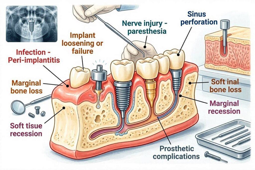
Late complications occur after the implant has been restored and in function, sometimes years after successful integration. These include peri-implantitis, mechanical complications such as screw loosening or fracture, and late implant failure. The causes and management of early and late complications differ significantly.
The risk profile changes over time. Early complications are primarily related to surgical factors and the patient’s initial healing response. Late complications are related to the patient’s long-term maintenance, occlusal forces, and the durability of the restorative components.
Risk Factors That Predispose to Problems
Several patient factors increase the risk of implant complications. Smoking is the most significant modifiable risk factor, increasing implant failure rates two to three times compared to non-smokers. The vasoconstrictive and cytotoxic effects of tobacco impair healing and ongoing tissue health.
A history of periodontitis increases the risk of peri-implantitis, as the same bacterial pathogens and host susceptibility that led to natural tooth loss can affect implants. Patients with periodontitis history require meticulous maintenance and more frequent professional recall.
Poorly controlled diabetes impairs healing and increases infection risk. Hemoglobin A1c levels above 7 percent are associated with higher complication rates. Well-controlled diabetes allows implant success rates approaching those of non-diabetic patients.
Bruxism and clenching generate excessive occlusal forces that can cause mechanical complications and potentially contribute to bone loss around implants. Patients with parafunctional habits require protective measures including night guards and carefully designed restorations.
Surgical Complications
Complications can occur during the implant placement procedure itself or in the immediate post-operative period. These complications are largely related to surgical technique, anatomical factors, and patient health.
Bleeding and Hematoma
Some bleeding is normal after implant surgery, typically presenting as pink-tinged saliva that resolves within 24 hours. Significant bleeding is uncommon but can occur if a blood vessel is damaged during the surgical procedure.
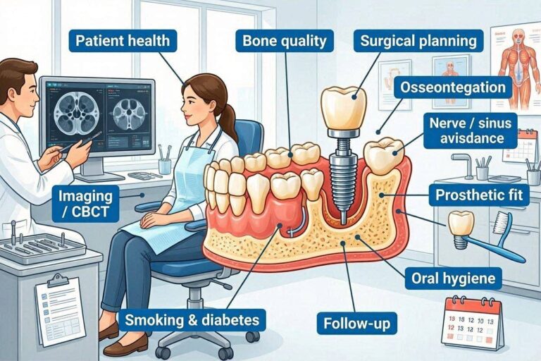
The floor of the mouth contains vessels that, if injured during mandibular implant placement, can cause bleeding into the sublingual space. This expanding hematoma can compromise the airway, creating a surgical emergency. Preoperative CBCT imaging identifies the position of these vessels, and careful surgical planning avoids them.
Patients on anticoagulant or antiplatelet medications require management protocols established in consultation with their physician. Some medications can be temporarily discontinued. Others require the surgeon to use local hemostatic measures. The risk of bleeding must be balanced against the risk of discontinuing medically necessary anticoagulation.
Post-operative bleeding that persists despite gauze pressure, or bleeding that is bright red and significant in volume, requires prompt professional attention. Most bleeding complications are managed with local measures including pressure, hemostatic agents, and suturing.
Nerve Injury
Nerve injury is one of the most serious implant complications because it can cause permanent sensory changes. The inferior alveolar nerve, which runs through the lower jaw and provides sensation to the lower lip and chin, is the nerve most commonly at risk.
The mental nerve, a branch of the inferior alveolar nerve that exits the lower jaw near the premolars, can be injured during implant placement in that region. The lingual nerve, which provides sensation to the tongue, is at risk during surgical procedures in the posterior mandible.
Nerve injury can occur through direct trauma from the surgical drill or implant, compression from a hematoma or edema, or thermal injury from inadequate irrigation during drilling. Direct nerve transection is the most severe form of injury and typically results in permanent sensory deficit.
Prevention through careful CBCT planning is the best management for nerve injury. The implant should maintain a safety margin of at least 2 millimeters from the nerve canal. If the available bone height is insufficient, shorter implants or nerve repositioning procedures may be considered.
When nerve injury occurs, prompt recognition and management are essential. Microsurgical nerve repair may be attempted for direct nerve injury. Sensory disturbances that result from compression or inflammation often resolve spontaneously over weeks to months. Patient counseling and support are important components of management.
Damage to Adjacent Teeth
Adjacent natural teeth can be damaged during implant placement if the implant is positioned too close to the tooth roots. The damage may involve the periodontal ligament, the root surface, or the pulp tissue.
The minimum safe distance between an implant and an adjacent tooth root is 1.5 to 2 millimeters. This distance preserves the blood supply that enters the tooth through the periodontal ligament and prevents direct trauma to the root surface.
Damage to an adjacent tooth may not be apparent immediately. The tooth may become symptomatic weeks or months after implant placement, presenting with pain, sensitivity, or signs of pulp necrosis. Root canal treatment or extraction may be necessary.
Prevention through careful treatment planning, including CBCT imaging and surgical guides, minimizes the risk of adjacent tooth injury. The implant should be positioned with adequate clearance from adjacent roots, and the surgical guide should be checked for accuracy before the implant site is prepared.
Intraoperative Implant Complications
Several complications can occur during the implant placement procedure itself. The implant may not achieve adequate primary stability, particularly in soft bone. The surgeon may choose a different implant size or alter the treatment plan if primary stability is insufficient.
The implant may be placed in a suboptimal position or angulation, compromising the esthetic and functional outcome. Immediate recognition and correction, possibly including implant removal and replacement, may be preferable to proceeding with a compromised position.
The implant site may be contaminated with saliva, bacteria, or debris during the procedure. Strict aseptic technique minimizes this risk. If contamination occurs, the site should be thoroughly irrigated, and the implant may be removed and replaced with a new sterile implant.
The implant may fracture during placement, though this is rare with modern implant designs. Excessive insertion torque, particularly in dense bone, can exceed the implant’s structural limits. The surgeon should follow the manufacturer’s torque recommendations and use appropriate technique for the bone quality encountered.
Healing Complications
The period between implant placement and restoration is biologically active but clinically quiet. Complications during this phase can compromise osseointegration and lead to early implant failure.
Failed Osseointegration
The most significant healing complication is failure of the implant to achieve osseointegration. Instead of bone forming directly on the implant surface, fibrous tissue encapsulates the implant. The implant lacks the rigid fixation required for function and must be removed.
Failed osseointegration may be discovered at the second-stage surgery when the implant is uncovered and found to be mobile. It may present during the healing period with persistent pain, swelling, or drainage. Radiographs show a radiolucent line around the implant where bone should contact the surface.
Causes of failed osseointegration include surgical trauma such as bone overheating during site preparation, bacterial contamination at the time of surgery or during healing, micromovement of the implant due to inadequate primary stability or premature loading, and compromised healing due to patient factors such as smoking or uncontrolled diabetes.
Treatment requires implant removal, site debridement, and a healing period of three to six months before a replacement implant can be placed. Bone grafting may be needed if the failed implant has left a significant bone defect. The success rate for replacement implants is favorable when the causes of the initial failure have been addressed.
Post-Operative Infection
Infection during the healing period can compromise osseointegration and lead to implant failure. The infection may originate from contamination at the time of surgery or from bacterial invasion through a wound that breaks down during healing.
Signs of infection include increasing pain after the third post-operative day, swelling that increases rather than decreases, redness extending beyond the surgical site, purulent discharge, and systemic symptoms including fever and malaise. These signs differ from normal post-operative inflammation, which should improve after the first few days.
Treatment involves antibiotics, drainage if an abscess has formed, and local debridement. The implant may be salvageable if the infection is treated promptly and osseointegration has not been compromised. Severe or persistent infection may require implant removal.
Prevention includes preoperative chlorhexidine mouth rinse, aseptic surgical technique, appropriate antibiotic prophylaxis when indicated, and careful post-operative wound care. Patients should follow post-operative instructions meticulously to protect the surgical site during the vulnerable early healing period.
Wound Dehiscence
Wound dehiscence occurs when the surgical incision opens, exposing the underlying implant or bone graft material. This complication compromises the protection that closed wound healing provides.
Dehiscence can result from tension on the suture line, trauma from chewing or oral hygiene, infection, or compromised healing due to patient factors. Thin gum tissue and inadequate keratinized tissue predispose to wound breakdown.
Small areas of dehiscence may heal by secondary intention, with new tissue gradually covering the exposed area. Meticulous oral hygiene and chlorhexidine rinses prevent secondary infection of the exposed surface. Larger dehiscence may require surgical revision to re-cover the implant.
Dehiscence over a bone graft site is particularly concerning because graft particles may be lost, and the graft may become infected. Prompt professional evaluation is necessary. The surgeon may attempt reclosure or may manage the exposure conservatively with close monitoring.
Peri-Implant Diseases
Peri-implant diseases are the most common late complications affecting dental implants. These inflammatory conditions, driven by bacterial plaque, can progress from reversible soft tissue inflammation to irreversible bone loss if not treated.
Peri-Implant Mucositis
Peri-implant mucositis is inflammation confined to the soft tissue surrounding the implant, without bone loss. It is the precursor to peri-implantitis and is fully reversible with appropriate treatment.
The signs of mucositis include redness, swelling, and bleeding on probing of the peri-implant tissues. There may be increased probing depths compared to baseline, but no radiographic bone loss beyond normal crestal remodeling. The condition is essentially gingivitis occurring around an implant.
Treatment involves professional mechanical debridement to remove plaque and calculus from the implant surface, reinforcement of oral hygiene instruction, and possibly short-term use of chlorhexidine mouth rinse. The tissue typically returns to health within days to weeks with improved plaque control.
Untreated mucositis can progress to peri-implantitis. The transition from mucositis to peri-implantitis involves the extension of inflammation to the supporting bone, with the inflammatory infiltrate reaching the bone crest and triggering osteoclast-mediated bone resorption.
Peri-Implantitis
Peri-implantitis is characterized by inflammation of the peri-implant mucosa and progressive loss of supporting bone. It is the leading cause of late implant failure and affects approximately 20 percent of implants over ten years.
In addition to the soft tissue signs of mucositis, peri-implantitis includes suppuration, increased probing depths, and radiographic bone loss that is progressive over time. The bone loss may be horizontal, reducing bone height evenly, or vertical, creating crater-like defects.
Treatment follows a stepped approach. Non-surgical mechanical debridement is the initial treatment for mild to moderate cases. Surgical access flap procedures allow thorough debridement of the contaminated implant surface under direct vision. Regenerative procedures may be attempted to restore lost bone in favorable defect configurations.
Advanced peri-implantitis with mobility or bone loss exceeding two-thirds of the implant length typically requires implant removal. The implant can no longer be maintained, and continued bone loss threatens adjacent structures.
Risk Factors for Peri-Implant Disease
The risk factors for peri-implant disease mirror those for periodontitis. Poor oral hygiene is the primary modifiable risk factor. Patients who cannot or do not maintain adequate plaque control around their implants will develop peri-implant inflammation.
A history of periodontitis is the strongest predictor of peri-implantitis. The same bacterial pathogens and host susceptibility that led to natural tooth loss challenge the implants. Periodontally compromised patients require meticulous maintenance and more frequent professional recall.
Smoking significantly increases peri-implantitis risk. The mechanisms include reduced tissue perfusion, impaired immune response, and direct toxic effects on healing and maintenance cells. Smoking cessation improves outcomes.
Systemic conditions including diabetes, immunosuppression, and certain medications affect the host response to bacterial challenge and increase peri-implant disease risk. Well-controlled systemic conditions reduce but may not eliminate this increased risk.
Mechanical and Technical Complications
The implant restoration is a mechanical system subject to fatigue, wear, and failure. Mechanical complications are common over the lifetime of an implant restoration and are usually manageable without threatening implant survival.
Abutment Screw Loosening
Abutment screw loosening is the most common mechanical complication of implant restorations. The screw that secures the abutment to the implant can gradually loosen under the cyclic loading of chewing.
The patient may notice a clicking sensation when biting, slight movement of the crown, or a change in the feel of the implant. If the screw continues to loosen, the crown and abutment may become obviously mobile while the implant body remains rigidly fixed in the bone.
Treatment involves accessing the screw through the crown, removing it, inspecting all components for damage, and retightening the screw to the manufacturer’s specified torque. The occlusion should be evaluated and adjusted to eliminate premature contacts that may have contributed to screw loosening.
Prevention includes proper screw tightening at the specified torque, verification of abutment seating, and regular professional maintenance that includes checking screw tightness. Patients with bruxism may require night guards to reduce the forces that contribute to screw loosening.
Abutment or Screw Fracture
More serious than screw loosening is screw or abutment fracture. These complications typically result from overload, material fatigue, or a loose screw that was not addressed promptly.
Screw fracture leaves a portion of the screw threaded into the implant body. Retrieving this fragment requires specialized instruments and techniques. If the fragment cannot be retrieved, the implant may need to be removed even though it was osseointegrated, because a restoration cannot be attached.
Abutment fracture, more common with narrow or angled abutments and with zirconia abutments, requires replacement of the abutment. The remaining abutment portion and screw must be removed from the implant. The implant internal connection should be inspected for damage.
Prevention includes appropriate abutment selection for the anticipated forces, proper screw tightening, regular maintenance, and management of parafunctional habits. Zirconia abutments should be used selectively in sites with appropriate force levels.
Implant Fracture
Implant body fracture is rare with modern implant designs but represents a catastrophic mechanical complication. Narrow-diameter implants, implants with internal connections that create stress concentration, and implants subjected to excessive forces are at highest risk.
Fracture typically occurs at the level of the abutment screw threads or at the implant platform. The coronal fragment becomes mobile while the apical fragment remains integrated in the bone.
Treatment requires removal of all implant fragments. The coronal fragment is removed directly. The apical fragment may require trephination to remove it from the bone. After complete implant removal and site healing, a replacement implant can be placed.
Prevention through appropriate implant selection, occlusal management, and protection against parafunctional forces is essential. Narrow implants should not be used in posterior sites with high occlusal loads.
Crown and Restoration Complications
The implant crown itself can experience complications including porcelain fracture, cement failure, wear, and esthetic deterioration over time.
Porcelain fracture on the implant crown may be repairable with composite resin if small, or may require crown replacement if extensive. Monolithic zirconia crowns have largely eliminated the porcelain chipping that was common with layered restorations.
Cement failure results in a loose crown on a stable abutment. The crown can be recemented after cleaning the old cement from both surfaces. The occlusion should be evaluated to identify any factors that contributed to cement failure.
Esthetic complications include color mismatch with adjacent natural teeth, particularly as natural teeth darken or change over time while the ceramic crown remains stable. The crown cannot be whitened. Replacement may be necessary if the color discrepancy becomes unacceptable.
Esthetic Complications
The esthetic outcome of implant treatment is particularly important in the anterior region, where the restoration is visible during smiling and speaking. Esthetic complications can compromise patient satisfaction even when the implant is functionally successful.
Soft Tissue Esthetic Problems
The appearance of the gum tissue around an implant crown significantly affects the overall esthetic result. Recession, where the gum recedes to expose the abutment or implant surface, creates an unaesthetic appearance and may compromise long-term tissue health.
Loss of the interdental papilla, the triangular tissue between the implant and adjacent tooth, creates a black triangle that is esthetically displeasing. The papilla height depends on the bone support from the adjacent tooth, making it difficult to maintain when the adjacent tooth has bone loss.
Tissue color changes around implants can affect esthetics. The gray color of a titanium abutment or implant may show through thin gingival tissue, creating a bluish discoloration compared to the pink tissue around natural teeth. Zirconia abutments and all-ceramic restorations reduce this risk.
Crown Esthetic Complications
The implant crown must match the adjacent natural teeth in shade, translucency, shape, and surface texture. Achieving a perfect match is challenging, particularly for single central incisor replacements where symmetry is critical.
Shade mismatch may result from the crown being fabricated to a shade that does not perfectly match, or from the natural teeth changing color over time while the ceramic crown remains stable. The crown should be matched to the natural teeth at the time of fabrication, with the understanding that future whitening of natural teeth will create a mismatch.
Contour and shape discrepancies can make the implant crown appear artificial. The crown should emerge from the gum with a natural profile, with appropriate contour and proportion relative to adjacent teeth. Custom abutments and skilled laboratory work improve the emergence profile.
Long-Term Management and Prevention
Many implant complications are preventable through proper planning, meticulous technique, and consistent long-term maintenance. The patient’s role in prevention is as important as the clinician’s.
The Importance of Professional Maintenance
Regular professional maintenance at intervals of three to six months allows early detection of developing problems before they become serious. The maintenance visit includes evaluation of peri-implant tissue health, probing depths, bleeding assessment, and periodic radiographs.
Professional cleaning of implant surfaces uses non-metal instruments to avoid scratching the titanium or zirconia surface. Scratched surfaces accumulate more plaque and are harder for the patient to clean. The clinician also evaluates the patient’s home care technique and provides reinforcement and instruction.
Occlusal evaluation at each maintenance visit identifies changes in the patient’s bite that could create implant overload. Teeth shift over time. Restorations wear. The implant occlusion must be adjusted to reflect these changes.
Patient Home Care Responsibilities
Effective daily plaque removal is the patient’s most important contribution to implant health. Interdental brushes, water flossers, and implant-specific floss clean the surfaces that toothbrushes cannot reach. The technique should be reviewed periodically with the dental hygienist.
Awareness of warning signs prompts early professional evaluation. Bleeding when cleaning around the implant, changes in tissue color or contour, any perception of movement, and discomfort should be reported promptly rather than waiting for the next scheduled visit.
Lifestyle factors, particularly smoking, significantly affect implant health. Smoking cessation is the single most impactful behavioral change for reducing implant complication risk. Patients who cannot quit should be aware that their risk profile is elevated and their maintenance needs are higher.
Conclusion
The most common complications of dental implants include peri-implant mucositis and peri-implantitis, which affect the soft and hard tissues supporting the implant and require early detection and treatment to prevent progressive bone loss and implant failure. Mechanical complications such as abutment screw loosening, the most frequent technical problem, and less common but more serious issues including screw fracture, abutment fracture, and implant fracture, are usually manageable when identified early but can lead to implant loss if neglected. Surgical complications including bleeding, nerve injury, damage to adjacent teeth, and failed osseointegration are largely preventable through careful preoperative planning with CBCT imaging, meticulous surgical technique, and appropriate patient selection and management. The foundation of complication prevention is consistent long-term maintenance, including regular professional evaluation and cleaning, effective daily plaque control by the patient, and prompt attention to early warning signs before minor issues become major problems.
Frequently Asked Questions
What is the most common dental implant complication?
Peri-implant mucositis, inflammation of the soft tissue around the implant without bone loss, is the most common complication, affecting 40 to 65 percent of implants at some point. It is reversible with treatment and improved oral hygiene. If untreated, it can progress to peri-implantitis with permanent bone loss.
How often do dental implants fail?
Implant survival rates are approximately 95 percent over ten years. Early failure during the healing period occurs in 1 to 3 percent of implants. Late failure after restoration occurs in 5 to 10 percent of implants over ten to fifteen years, primarily due to peri-implantitis or overload.
Can a failing implant be saved?
Implants with peri-implantitis that are not yet mobile can often be treated to arrest the disease, though lost bone may not regenerate. Once an implant becomes mobile, indicating complete loss of osseointegration, it cannot be saved and must be removed. Early intervention provides the best chance for salvage.
What causes an implant to become loose?
A loose abutment screw causes the crown and abutment to move while the implant remains fixed. This is a mechanical problem solved by retightening the screw. A loose implant, where the implant body moves within the bone, indicates loss of osseointegration and requires implant removal.
Can dental implants cause sinus problems?
Implants placed in the upper back jaw can protrude into the maxillary sinus or cause sinus membrane perforation. Chronic sinusitis can develop if the implant or associated infection affects the sinus. Careful preoperative planning with CBCT imaging prevents most sinus complications.
Do dental implants cause bad breath?
Implants themselves do not cause bad breath. However, bacterial accumulation around implants due to inadequate oral hygiene can cause halitosis. Peri-implantitis with suppuration can produce a persistent bad taste and odor. Proper cleaning resolves implant-related halitosis.
Can an implant damage the nerve in my jaw?
Nerve injury is a serious but preventable complication. The inferior alveolar nerve in the lower jaw is at risk if the implant is placed too deep. Preoperative CBCT imaging identifies nerve position, and the implant should maintain at least 2 millimeters of clearance. Transient sensory changes usually resolve. Permanent nerve injury is rare with proper planning.
How can I prevent implant complications?
Maintain excellent oral hygiene with appropriate tools for implant cleaning. Attend professional maintenance visits every three to six months. Report any bleeding, swelling, mobility, or discomfort promptly. Avoid smoking. Use a night guard if you grind or clench your teeth. Follow your dentist’s specific care instructions for your implant restoration.
Additional Resource
International Team for Implantology: Implant Complications
https://www.iti.org/for-patients