Are Wisdom Teeth Positioned Horizontally Harmful?
The panoramic X-ray appears on the screen, and the patient leans in. There, at the back corner of the jaw, a wisdom tooth lies on its side like a fallen tombstone, its crown pointing toward the roots of the adjacent molar, its roots pointing toward the throat. The orthodontist or oral surgeon traces the outline with a cursor. “This one is horizontally impacted,” they say, and the word “impacted” lands with a dull, ominous weight. The patient’s immediate question, behind the clinical jargon, is simple: “Is that dangerous? Is it hurting me right now? Can I just leave it there?”
The question “Are wisdom teeth positioned horizontally harmful?” must be answered with the clarity that comes from decades of longitudinal studies and surgical pathology. A horizontally impacted third molar—also called a mesioangular impaction in its most common variant, or a true horizontal impaction when the tooth is parallel to the occlusal plane—is almost always harmful, but the harm is often silent, progressive, and cumulative. The tooth may cause no pain for years while it slowly destroys the adjacent second molar, expands the follicular sac into a cystic lesion, or creeps its roots into the inferior alveolar nerve canal. This article is a forensic examination of the specific pathologies that horizontal wisdom teeth cause, the diagnostic imaging required to map the threat, the age-dependent risk calculus that governs the decision to extract or observe, and the rare, strictly defined circumstances under which a horizontally positioned wisdom tooth can be safely left in place.
The Geometry of Impaction: Defining “Horizontal”
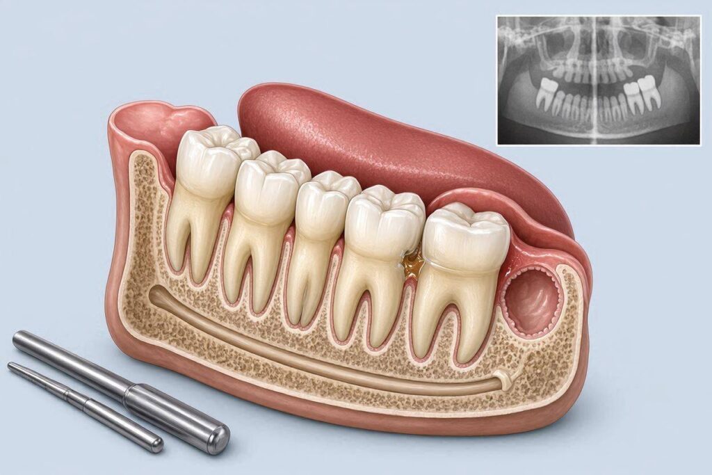
Dental impaction classifications describe the angulation of the third molar relative to the long axis of the adjacent second molar. A mesioangular impaction—the crown tilted toward the front of the mouth at roughly 45 degrees—is the most common. A horizontal impaction is a more extreme variant: the tooth is lying on its side, with the occlusal surface facing the roots or crown of the second molar. A distoangular impaction tilts toward the back of the mouth; a vertical impaction is upright but unable to erupt due to space constraints. Horizontal impactions represent roughly 5–15% of all impacted third molars, depending on the population studied.
The critical anatomical feature of a horizontal impaction is the direction of the eruptive force vector. A vertically impacted tooth pushes upward (or downward in the mandible) against the bone and soft tissue, causing pressure resorption of the overlying structures but not directly threatening the adjacent tooth. A horizontally impacted tooth pushes forward, directly into the distal root surface of the second molar. The eruptive force, even if it is weak and episodic, is applied to the periodontal ligament and cementum of the second molar’s distal root. This is the source of the most common and devastating pathology associated with horizontal impactions: external root resorption of the adjacent second molar.
External Root Resorption: The Silent Second Molar Killer
The second molar is a critical functional tooth. It is the largest molar in the arch, the primary grinding surface for mastication, and the anchor for the dental arch’s posterior support. When a horizontally impacted third molar’s crown presses against the distal root of the second molar, the pressure stimulates osteoclasts—not just in the bone, but on the root surface itself. The cementum and dentin of the second molar’s root are resorbed. This is external root resorption, and it is irreversible.
The process is asymptomatic. The patient feels nothing. The second molar is not infected, not decayed, and not mobile—until the resorption has destroyed so much root structure that the tooth becomes compromised. On a panoramic radiograph, the resorption appears as a dark, scooped-out defect on the distal root of the second molar, often with the crown of the horizontally impacted third molar nestled directly into the defect. By the time it is visible on a standard panoramic X-ray, the resorption is often extensive. A cone beam computed tomography (CBCT) scan will reveal the true three-dimensional volume of the lost root structure, which is frequently worse than the two-dimensional panoramic suggests.
The clinical consequence is tragic and preventable. The patient, now in their 30s or 40s, presents with a complaint of vague discomfort or food impaction in the back of the mouth. The X-ray reveals a horizontally impacted third molar that has been silently eroding the second molar’s root for a decade. The second molar may require extraction, root canal therapy with a heroic surgical attempt to save it, or a crown lengthening procedure to restore a deeply resorbed margin. The patient, who had no symptoms and no warning, loses a functionally critical tooth because the horizontal wisdom tooth was not removed when the patient was young, healthy, and the bone was forgiving. This is the most compelling argument for prophylactic removal of horizontally impacted third molars in young adulthood: the preservation of the adjacent second molar.
Cystic and Neoplastic Transformation: The Expanding Follicle
Every developing tooth is surrounded by a dental follicle, a sac of connective tissue that contains the enamel organ and the developing root. When a tooth erupts normally, the follicle is obliterated, and the tissue integrates into the gingiva. When a tooth is impacted and remains unerupted, the follicle persists around the crown. This follicular sac is lined with reduced enamel epithelium, which retains the potential to proliferate and undergo cystic degeneration.
A dentigerous cyst is a fluid-filled lesion that develops from the follicle of an impacted tooth. It is the second most common odontogenic cyst, after the radicular cyst associated with a non-vital tooth. A dentigerous cyst associated with a horizontally impacted third molar can expand slowly over years, resorbing bone and displacing the impacted tooth. The expansion is silent and painless until the cyst becomes large enough to cause visible facial swelling, displace adjacent teeth, or—in the mandible—thin the bone to the point of pathological fracture.
Even more concerning is the potential for neoplastic transformation. The epithelial lining of a dentigerous cyst can undergo dysplastic changes and develop into an ameloblastoma, a locally aggressive, benign but destructive tumor of odontogenic epithelium. Less commonly, a squamous cell carcinoma or a mucoepidermoid carcinoma can arise from the cyst lining. These are rare events, but they are catastrophic when they occur, requiring radical surgical resection with loss of a segment of the mandible, followed by complex reconstruction with a fibular free flap and dental implants. The horizontal impaction is the precursor lesion. The removal of the impacted tooth and its follicle in young adulthood eliminates the future risk of cystic and neoplastic degeneration.
Pericoronitis: The Acute Infection of the Partially Erupted
Not all horizontally impacted wisdom teeth remain completely buried. Some partially erupt through the gum, creating a communication between the oral cavity and the follicular sac. This communication is called an operculum—a flap of gum tissue that overlies the partially erupted crown. The space under the operculum is a bacterial incubator: warm, dark, anaerobic, and impossible to clean with a toothbrush or floss.
Pericoronitis is an acute or subacute infection of the soft tissue surrounding a partially erupted tooth, most commonly a mandibular third molar. It manifests as pain, swelling, a foul taste, trismus (limited jaw opening), and in severe cases, fever and lymphadenopathy. The infection can spread into the fascial spaces of the head and neck—the submandibular space, the buccal space, the pterygomandibular space—leading to cellulitis, abscess formation, and, in the most dangerous scenario, Ludwig’s angina, a rapidly spreading, life-threatening infection of the floor of the mouth that can compromise the airway.
A horizontally impacted tooth that has partially erupted is a persistent source of pericoronitis risk. The patient who has experienced one episode of pericoronitis will likely experience another. The definitive treatment is not antibiotics—those are a temporizing measure—but surgical removal of the impacted tooth. The question “Is this horizontal tooth harmful?” is answered in part by the presence of a communication with the oral cavity. A fully bony, completely submerged impaction may be biologically silent for decades. A partially erupted horizontal impaction is a ticking infection bomb.
Caries and Periodontal Disease of the Adjacent Tooth
The horizontal impaction is not just a structural threat to the second molar; it is a bacterial threat. The impacted tooth’s crown is positioned against the distal surface of the second molar, creating an interproximal area that cannot be cleaned. Food, plaque, and bacteria accumulate in the inaccessible space between the impacted crown and the second molar’s distal root. The second molar develops caries on its distal surface—decay that begins at the cementoenamel junction or on the root surface, below the gumline, invisible to the patient and often difficult to detect radiographically until it is advanced.
Simultaneously, the chronic bacterial load causes periodontal inflammation and bone loss on the distal aspect of the second molar. The periodontal pocket deepens. The attachment is lost. The second molar, even if it avoids decay and root resorption, may develop localized periodontitis that compromises its long-term prognosis. This is a direct, preventable consequence of the horizontal impaction. The removal of the impacted tooth eliminates the inaccessible bacterial niche and preserves the periodontal health of the second molar.
The Age-Dependent Risk Calculus: Why Youth Matters
The harmfulness of a horizontally impacted wisdom tooth is not a fixed property of the tooth itself; it is a function of time and the host’s healing capacity. The same horizontally impacted third molar that is a manageable surgical extraction in a healthy 18-year-old becomes a significantly higher-risk procedure in a 45-year-old.
In the young patient, the bone is elastic and forgiving. The follicular sac is wide, and the tooth can often be elevated out with minimal bone removal. The roots are incompletely formed—the apex is open, and the tooth is not fully ankylosed to the bone. The periodontal ligament is robust, and the tooth separates cleanly from the crypt. The risk to the inferior alveolar nerve, while never zero, is lower because the roots are shorter and further from the nerve canal. The post-operative healing is rapid, and the risk of complications—dry socket, infection, persistent numbness—is low.
In the older patient, the bone is dense and sclerotic. The follicular sac has atrophied, and the tooth may be partially ankylosed—fused directly to the bone. The roots are fully formed and often dilacerated (curved or hooked), wrapping around the nerve canal. The surgical extraction requires significant bone removal and tooth sectioning. The risk of nerve injury is higher. The post-operative healing is slower, and the risk of complications—prolonged swelling, trismus, infection, and mandibular fracture (in the extreme atrophic case)—is elevated. The horizontal impaction that could have been removed prophylactically in youth with low morbidity becomes a medically necessary, higher-risk procedure in middle age.
The “Observe, Don’t Operate” Criteria: When Leaving a Horizontal Tooth is Reasonable
Clinical guidelines, including those from the National Institute for Health and Care Excellence (NICE) in the UK and the American Association of Oral and Maxillofacial Surgeons, support the removal of impacted third molars when there is evidence of pathology—recurrent pericoronitis, caries, periodontal disease, cyst formation, or resorption of adjacent teeth. The guidelines generally do not support the prophylactic removal of asymptomatic, fully bony impacted third molars in patients over a certain age, particularly if there is no evidence of pathology and the surgical risk is high.
A horizontally impacted third molar that is completely encased in bone, with no communication with the oral cavity, no evidence of cyst formation or root resorption on serial radiographs, and no symptoms, in a patient over 35, may reasonably be managed with active surveillance. This involves periodic panoramic radiographs (every 2–3 years) to monitor for cystic changes, root resorption of the second molar, or positional changes. The patient must understand that the risk of cystic degeneration persists as long as the follicle is present, and that the risk is low but not zero. The patient and the surgeon together weigh the low-but-real long-term pathology risk against the immediate surgical risk of nerve injury and the morbidity of bone removal in an older patient.
This “watchful waiting” strategy is valid only for truly asymptomatic, fully bony impactions. A partially erupted horizontal impaction, an impaction with any radiographic evidence of follicle enlargement (a pericoronal radiolucency greater than 2.5–3 millimeters), or an impaction with any evidence of second molar root resorption should be removed regardless of age. The presence of pathology removes the option of observation.
The Inferior Alveolar Nerve: The Horizontal Impaction’s Most Feared Neighbor
The mandibular canal, which carries the inferior alveolar nerve (IAN), runs through the body of the mandible from the mandibular foramen to the mental foramen. The roots of the mandibular third molar often lie in close proximity to this canal. Horizontal impactions, because the tooth lies on its side, often have roots that point directly posteriorly toward the canal. The root apices can be intimately associated with the nerve—touching, grooving, or encircling the canal.
A cone beam CT scan is the standard of care for evaluating the relationship between the horizontally impacted third molar and the IAN when a panoramic X-ray shows radiographic signs of proximity: darkening of the root, narrowing of the canal, interruption of the lamina dura (the white line outlining the canal), or deviation of the canal around the root. The CBCT provides sub-millimeter resolution in three dimensions, allowing the surgeon to see whether the nerve is merely adjacent, grooved into the side of the root, or passing through the root (an inter-radicular course).
The surgical strategy is tailored to the nerve relationship. If the nerve is at high risk of injury, the surgeon may consider a coronectomy—removing the crown of the tooth and leaving the roots intentionally buried to move away from the nerve—or a staged extraction. The horizontal impaction’s harmfulness is not just in the pathology it causes; it is in the risk of iatrogenic injury during its removal. This risk must be quantified and disclosed to the patient as part of the informed consent process.
Comparative Table: Risks of Horizontal Impaction by Age
| Risk Factor | Young Adult (16–25) | Middle Age (35–50) | Older Adult (55+) |
|---|---|---|---|
| Root Resorption of 2nd Molar | Early, possibly reversible with removal. | Advanced; often irreversible without heroic measures. | Severe; second molar frequently lost. |
| Cyst Formation | Rare; prophylactic removal eliminates risk. | Follicle persists; low but real annual risk of degeneration. | Cumulative risk over decades; dentigerous cyst or ameloblastoma possible. |
| Pericoronitis | Common if partially erupted. | Recurrent episodes likely if partially erupted. | May present with severe spreading infection; compromised healing. |
| Surgical Morbidity | Low; elastic bone, incomplete roots. | Moderate; dense bone, complete roots. | High; sclerotic bone, ankylosis, poor healing. |
| IAN Nerve Injury Risk | Low. | Moderate; roots fully formed, close to canal. | Moderate to high; canal proximity often dictates coronectomy. |
| Mandibular Fracture Risk | Negligible. | Low. | Moderate if significant bone removal in atrophic mandible. |
Conclusion
Horizontally impacted wisdom teeth are almost always harmful over the long term, causing silent and irreversible external root resorption of the adjacent second molar, creating an inaccessible bacterial niche that fosters distal caries and periodontitis, and harboring a persistent follicular sac capable of cystic or neoplastic degeneration. The pathology is age-progressive: the damage accumulates silently, and the surgical risk of removal rises with age as bone density increases, roots mature, and nerve proximity tightens. Prophylactic removal in young adulthood is the evidence-based standard for preventing these predictable complications, while active surveillance may be considered only for the rare fully bony, completely asymptomatic impaction with no radiographic signs of pathology in a patient of advanced age.
FAQ
Q: Can a horizontally impacted wisdom tooth cause crowding of my other teeth?
A: The long-held belief that wisdom teeth push forward and cause anterior crowding has been largely debunked by longitudinal studies. Late mandibular incisor crowding occurs regardless of third molar presence and is attributed to normal mesial drift and arch narrowing with age. However, a horizontally impacted tooth exerts localized forward pressure on the second molar, which can cause that specific tooth to tip or shift, but it does not create a domino effect through the entire arch. The primary crowding risk is to the second molar’s position, not the anterior teeth.
Q: If my horizontal wisdom tooth doesn’t hurt, why can’t I just wait and see if it becomes a problem?
A: Because the most serious pathology—root resorption of the second molar—is completely painless until the damage is advanced and often irreversible. By the time you feel symptoms, you may have already lost a critical molar. The “wait and see” strategy is only appropriate for fully bony, deeply buried impactions with no radiographic signs of follicle enlargement or root resorption, monitored with periodic imaging. If your horizontal tooth is partially erupted or shows any pathology, the waiting period is actively allowing damage to accumulate.
Q: What is a coronectomy, and when is it performed for a horizontal impaction?
A: A coronectomy is a surgical procedure in which the crown of the impacted tooth is removed, but the roots are intentionally left in the jawbone. It is performed when the roots are intimately associated with the inferior alveolar nerve, and removing them would carry a high risk of permanent nerve damage and lip numbness. The roots, once separated from the crown, are no longer a source of pathology; they typically remain stable or are gradually encased in bone. Coronectomy is a valid, evidence-based alternative to complete extraction for high-risk horizontal impactions, and it dramatically reduces the risk of IAN injury.
Additional Resource
For clinical guidelines on the management of impacted third molars, including indications for removal versus observation, visit the American Association of Oral and Maxillofacial Surgeons at www.aaoms.org.