
When Dental Implants Go Wrong: Understanding Complications, Failures, and Solutions
A patient runs their tongue over a dental implant that has served them well for five years. Lately, the gum around it bleeds when they brush. There is a dull ache that was not there before. A small voice of worry whispers: Is something wrong with my implant? Dental implants enjoy remarkably high success rates, exceeding 95 percent in healthy patients over ten years. But no medical procedure is without risk. When implants go wrong, the consequences range from minor inconvenience to significant bone loss and the need for removal.
This comprehensive guide explores the ways dental implants can fail or develop complications. We examine the causes, the warning signs, the diagnostic process, the treatment options, and the preventive strategies that minimize the chances of problems. Knowledge empowers patients and clinicians alike to recognize trouble early and intervene effectively.
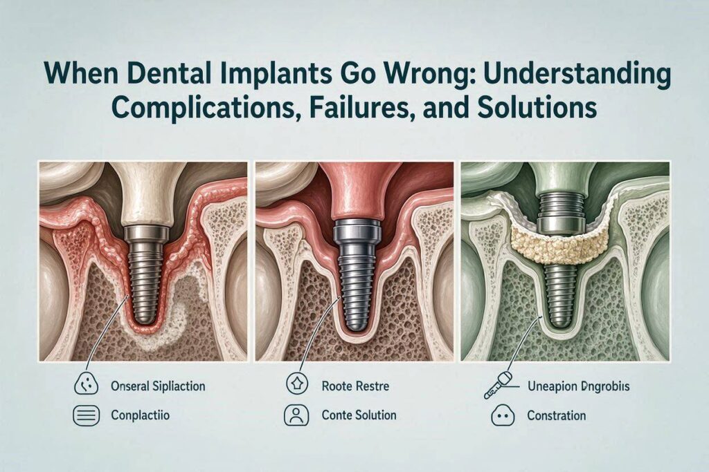
Defining Implant Complications Versus Implant Failure
The terms are related but distinct. Understanding the difference frames the discussion.
Implant Complications
A complication is an undesirable event that affects the implant or surrounding tissues but does not necessarily mean the implant will be lost. Complications can be mechanical, affecting the crown, abutment, or screw. They can be biological, affecting the gum or bone around the implant. They can be esthetic, resulting in an unacceptable smile. With proper management, many complicated implants can be salvaged and continue to function.
Implant Failure
Failure means the implant has lost osseointegration, is mobile, or has been removed. Failure is the endpoint where the implant can no longer serve its intended purpose. Some failures occur early, before the crown is attached. Others occur late, after years of function. Failed implants generally require removal and, if the site allows, eventual replacement.
The Spectrum of Problems
Between perfect health and outright failure lies a spectrum of ailing and failing implants. The goal is to identify problems while they are still manageable and reverse or halt the damaging process.
Early Implant Failure: When Healing Goes Wrong
Early failure occurs within the first few months after placement, before the implant has integrated or before it has been restored with a crown.
Failure to Achieve Osseointegration
The implant is placed, but instead of bone growing tightly against the titanium surface, a fibrous capsule forms. The implant remains mobile or develops mobility during the healing period. The surgeon removes it, often with minimal force because bone has not attached.
Causes of Early Failure
Surgical trauma: Overheating the bone during drilling, excessive pressure during implant insertion, or tearing of the soft tissue can compromise healing. Bone cells die if the temperature exceeds 47 degrees Celsius for more than a minute. Copious irrigation and sharp drills prevent this.
Infection at the surgical site: Bacteria contaminate the implant surface or the osteotomy site. This may result from poor sterile technique, contamination from adjacent teeth with periodontal disease, or patient noncompliance with postoperative instructions.
Compromised bone quality: Very soft, porous bone may not provide adequate primary stability. The implant moves slightly during healing. This micromotion prevents bone cells from attaching to the implant surface.
Systemic patient factors: Uncontrolled diabetes, heavy smoking, immunosuppression, or medications that impair bone healing, such as bisphosphonates, increase early failure risk.
Overloading during healing: If the patient chews on the implant before osseointegration, or if a poorly designed provisional restoration transmits forces, the implant can fail.
Signs of Early Failure
The patient may notice persistent pain beyond the expected postoperative discomfort. Swelling and redness may not resolve. The implant may feel loose. Radiographs show a radiolucency, a dark halo, around the implant instead of bone contact.
Late Implant Failure: The Sleeping Problem Awakens
Late failure occurs after the implant has successfully integrated and functioned for a year or more. This is more devastating psychologically because the patient has come to rely on the implant as a permanent tooth replacement.
Peri-Implantitis: The Leading Cause
Peri-implantitis is an inflammatory condition affecting the soft and hard tissues around an osseointegrated implant. It is characterized by bleeding on probing, suppuration, and progressive bone loss beyond the initial bone remodeling. It is the implant equivalent of advanced periodontitis.
The process begins with peri-implant mucositis, a reversible inflammation of the soft tissue only, analogous to gingivitis. If not treated, it progresses to peri-implantitis, where the inflammation extends to the bone, causing irreversible resorption.
The Microbial Culprit
The bacteria that cause peri-implantitis are similar to those in periodontitis: anaerobic gram-negative rods, spirochetes, and other pathogens. They form a biofilm on the implant surface below the gum line. The rough surface of modern implants, designed to promote bone attachment, unfortunately also provides an ideal substrate for bacterial colonization once exposed.
Risk Factors Specific to Peri-Implantitis
- History of periodontitis: Patients who lost teeth to gum disease are at significantly higher risk.
- Poor oral hygiene: Inadequate plaque removal around the implant restoration.
- Lack of regular professional maintenance: Implants require professional cleaning, just like natural teeth.
- Smoking: A powerful risk factor for both periodontitis and peri-implantitis.
- Diabetes, especially uncontrolled: Impaired host immune response.
- Excess cement: Cement retained crowns can leave subgingival cement remnants that act as a bacterial nidus.
- Ill-fitting prosthesis: Open margins, overcontoured crowns that impede hygiene.
- Genetic predisposition: Some patients are biologically more susceptible to inflammatory bone loss.
Biomechanical Overload
An implant can fail mechanically even if the biology is sound. Excessive occlusal forces cause microfractures in the bone around the implant, leading to loss of osseointegration. This is distinct from peri-implantitis, though the two can coexist.
Causes of Overload
- Bruxism: Clenching or grinding, especially during sleep.
- Poor occlusal design: The crown has heavy, off-axis contacts.
- Cantilevered prostheses: Extensions beyond the supporting implants create leverage forces.
- Insufficient number of implants to support the prosthesis: Too few implants for the load.
Signs of Late Failure
The patient may notice bleeding when brushing around the implant, a bad taste or pus, deepening pockets around the implant, gum recession exposing the implant threads, mobility of the implant itself, or radiographic bone loss.
Mechanical Complications: When Hardware Breaks
Not all implant problems are biological. Mechanical failures affect the components.
Abutment Screw Loosening or Fracture
The screw that connects the abutment to the implant can loosen over time. The patient feels a slight movement of the crown. The dentist can often simply retighten or replace the screw. If unrecognized, the loose screw can fracture, leaving a portion inside the implant that is challenging to retrieve.
Implant Fixture Fracture
The implant body itself breaks. This is rare but catastrophic. The fracture typically occurs at the neck of the implant, the point of greatest stress concentration where the implant meets the bone crest. It results from metal fatigue over time, often in the setting of heavy occlusal forces, bruxism, or an implant with a narrow diameter placed in a high-load posterior location. A fractured implant must be removed surgically.
Crown or Bridge Fracture
The ceramic crown or bridge on top of the implant can chip, crack, or fracture entirely. This is a restorative complication. The implant itself may be perfectly healthy. The crown is replaced.
Loss of Retention for Cemented Crowns
A cemented crown can debond from the abutment. The patient may feel the crown come loose. It can often be recemented if the abutment and implant are intact.
Esthetic Complications: The Smile That Does Not Look Right
An implant can be biologically integrated and mechanically sound but esthetically unacceptable.
Gum Recession and Implant Thread Exposure
The gum recedes, revealing the gray metal of the implant or abutment. This is particularly distressing in the anterior esthetic zone. It may result from thin gum biotype, bone loss on the buccal plate, or an implant placed too far facially.
Mismatched Crown Color or Shape
The crown does not match the adjacent natural teeth. This may be due to limitations of the ceramic material, the skill of the laboratory technician, or changes in the natural teeth over time as they darken or wear.
Black Triangles Between Teeth
Gaps between the implant crown and adjacent teeth, where the interdental papilla has been lost. This creates an unaesthetic, aged appearance. It results from bone loss between the implant and the natural tooth, often due to inadequate spacing.
Poor Emergence Profile
The crown emerges from the gum at an unnatural angle, looking bulky or flat compared to a natural tooth. This stems from implant position or abutment contour.
Diagnostic Process: When a Problem Is Suspected
The clinician systematically evaluates the troubled implant.
Patient History and Symptoms
The dentist asks about pain, swelling, bleeding, bad taste, looseness, and changes in bite or esthetics. The duration and progression of symptoms are noted.
Clinical Examination
Visual inspection: The dentist looks for redness, swelling, fistulas draining pus, gum recession exposing threads, and any visible fracture or chip.
Probing: A periodontal probe is gently passed around the implant. Bleeding on probing, probing depths greater than 5 or 6 millimeters, and suppuration are signs of peri-implant disease. Probing should be gentle to avoid damaging the fragile soft tissue seal.
Mobility testing: The implant is checked for any movement. An integrated implant should be absolutely immobile. Any perceptible mobility indicates loss of osseointegration.
Percussion: Tapping on the implant may produce a dull sound instead of the ringing sound of a solid bone-implant interface.
Occlusal analysis: The dentist checks for heavy contacts, fremitus during clenching, and excursive interferences.
Radiographic Evaluation
Periapical radiographs: High-resolution images show bone levels around the implant. Sequential radiographs over time are compared to determine the rate and pattern of bone loss.
Cone-beam computed tomography: CB-CT provides three-dimensional information about bone volume on all sides of the implant, the position relative to nerves and sinuses, and any bony defects.
Microbiological Testing
In some refractory cases, a sample of the subgingival biofilm is taken and analyzed to identify the specific pathogens present. This can guide targeted antibiotic therapy.
Treatment Strategies for Ailing Implants
The treatment depends on the diagnosis. Many implants can be saved if caught early.
Treatment of Peri-Implant Mucositis
This is fully reversible. Treatment involves professional debridement to remove plaque and calculus from the implant surface and abutment, oral hygiene instruction, and the use of antimicrobial mouth rinses such as chlorhexidine. The patient must improve home care. Resolution is the rule with good compliance.
Non-Surgical Treatment of Peri-Implantitis
For early to moderate peri-implantitis, non-surgical debridement is attempted. The clinician uses special instruments, such as plastic or titanium curettes, ultrasonic scalers with non-metal tips, or air abrasion devices, to clean the exposed implant surface without scratching it. The goal is to decontaminate the surface and reduce the bacterial load. Systemic or local antibiotics may be used as an adjunct.
Surgical Treatment of Peri-Implantitis
When non-surgical therapy fails or when bone loss is advanced, a surgical flap is elevated. The surgeon visualizes the contaminated implant surface directly. They decontaminate it mechanically and chemically using various protocols: titanium brushes, air powder abrasion, citric acid, hydrogen peroxide, or laser. Granulation tissue is removed. The bony defect may be grafted with bone substitute materials to try to regenerate lost bone. The flap is sutured closed.
Managing Mechanical Complications
A loose abutment screw is retightened to the specified torque, often with a new screw. A fractured screw is retrieved with specialized removal kits. A fractured implant fixture requires removal of the entire implant. A fractured crown is replaced.
Managing Esthetic Complications
Soft tissue grafting with connective tissue from the palate can thicken the gum and mask thread exposure. Crown replacement with better color matching or contour adjustment addresses restorative esthetic problems. In severe cases, the implant may need to be removed and the site reconstructed.
Implant Removal: When Salvage Fails
When bone loss is extensive, when the implant is mobile, or when the patient’s symptoms cannot be resolved, the implant must be removed.
The Explantation Procedure
Removing an integrated implant requires surgical techniques to break the osseointegration. The surgeon uses piezoelectric instruments or trephine burs to cut the bone-implant interface. The goal is to remove the implant with minimal additional bone loss. After removal, the socket is debrided, and bone graft material is often placed to preserve ridge dimensions for potential future implant placement.
The Patient’s Emotional Response
Removing an implant that the patient invested significant time, money, and hope in is emotionally difficult. The clinician must communicate with compassion, explain the reasons clearly, and present a plan for moving forward. Acknowledging the loss and the patient’s frustration is part of the care.
What Comes After Removal?
The site heals for three to six months. If adequate bone remains or has been regenerated, a new implant can be placed. In some cases, a different tooth replacement option is chosen, such as a bridge or a removable partial denture. The decision depends on the reason for the initial failure and the patient’s preferences and risk factors.
Table: Common Implant Problems, Causes, and Solutions
| Problem | Typical Cause | Treatment | Prognosis |
|---|---|---|---|
| Peri-implant mucositis | Poor oral hygiene, plaque accumulation | Professional cleaning, improved home care | Excellent, fully reversible |
| Early peri-implantitis | Progression of mucositis, risk factors | Non-surgical debridement, antibiotics | Good if caught early |
| Advanced peri-implantitis | Untreated infection, heavy bone loss | Surgical debridement, grafting, or implant removal | Guarded; may still lose implant |
| Abutment screw loosening | Insufficient torque, heavy occlusal forces | Retighten or replace screw, adjust occlusion | Excellent |
| Implant fracture | Metal fatigue, overload, narrow implant | Implant removal | Requires replacement |
| Crown fracture | Heavy biting, material failure | Crown replacement | Excellent for implant |
| Esthetic failure | Poor placement, tissue loss | Soft tissue graft, new crown, or removal | Variable |
| Early failure (no integration) | Surgical trauma, infection, patient factors | Removal, allow healing, reassess | Good for future implant if cause addressed |
Prevention: The Best Strategy Against Implant Problems
The most effective way to manage implant complications is to prevent them.
Thorough Preoperative Assessment
The clinician must evaluate the patient’s medical history, smoking status, periodontal health, and occlusal habits. A patient with untreated periodontitis should not receive implants until their periodontal condition is stabilized. A heavy smoker should be counseled about the increased failure risk.
Meticulous Surgical Technique
Atraumatic preparation, avoidance of overheating, achievement of primary stability, and placement in a prosthetically driven position all reduce the risk of future problems.
Prosthetically Sound Restoration
The crown or bridge must be designed with cleansable contours, passive fit, and an occlusion that avoids overload. Screw-retained restorations eliminate the risk of retained cement. If cemented, cement excess must be meticulously removed.
Patient Education and Engagement
The patient must understand that an implant is not a “set it and forget it” solution. It requires daily cleaning and professional maintenance. The clinician should demonstrate proper brushing, flossing, and use of interdental brushes or water flossers around the implant.
Regular Professional Maintenance
The patient should return to the dentist or periodontist at least every six months, and more frequently if they have a history of periodontitis. The professional maintenance visit includes probing around the implant, radiographs at appropriate intervals, and professional cleaning of any accessible biofilm.
Early Intervention When Problems Arise
The patient should be instructed to report any bleeding, swelling, bad taste, or looseness immediately. Early peri-implant mucositis is easily treated. Advanced peri-implantitis is not.
The Role of the Patient in Preventing Implant Problems
Implants demand a partnership. The dentist places and restores, but the patient maintains.
Daily Oral Hygiene Around Implants
Brushing twice daily with a soft toothbrush, focusing on the gum line around the implant. Interdental cleaning with floss designed for implants, soft picks, or an oral irrigator. The surfaces where the crown meets the gum require particular attention.
Lifestyle Modifications
Smoking cessation is the single most impactful action a smoking patient can take to protect their implants. Smoking impairs blood flow, immune response, and healing. Controlled diabetes management similarly protects implants.
Protecting Against Overload
If the patient grinds or clenches, a custom night guard should be fabricated and worn consistently. The guard protects the implant from excessive forces during sleep.
When to Seek a Second Opinion
A patient with a failing or complicated implant may benefit from a second opinion, particularly if the treating clinician seems uncertain or if the proposed treatment is implant removal.
Finding a Specialist
A periodontist or prosthodontist with experience in managing implant complications is the ideal consultant. The patient should bring all records, radiographs, and a timeline of the problem. A fresh set of eyes can sometimes identify a salvageable situation that was initially deemed hopeless.
Questions to Ask the Second Opinion Clinician
- What is the exact diagnosis? Is it peri-implantitis, overload, or a mechanical problem?
- Is the implant salvageable? What are the chances of success with treatment?
- If removal is recommended, why is it not salvageable?
- What are the alternatives to removal?
- If a new implant is placed later, what will be different to prevent recurrence?
Conclusion
Dental implants can go wrong through biological failures like peri-implantitis and loss of osseointegration, mechanical complications such as screw loosening or implant fracture, and esthetic problems that compromise the smile. Early recognition of warning signs such as bleeding, swelling, pus, or mobility allows for intervention while the implant is still salvageable. Treatment ranges from non-surgical debridement for early disease to surgical regenerative procedures for advanced bone loss, with implant removal reserved for failed cases. Prevention through patient selection, meticulous surgical and restorative technique, and lifelong maintenance is the most powerful strategy. When an implant fails, understanding the cause and learning from it is essential before moving forward with replacement.
Frequently Asked Questions
What are the early signs that a dental implant is failing?
Early signs include bleeding when brushing around the implant, persistent redness or swelling of the gum, a bad taste or pus coming from around the implant, and deepening pockets. Pain is not always present. Mobility of the implant is a late sign of failure.
Can a failing implant be saved?
Yes, if caught early. Peri-implant mucositis is fully reversible with professional cleaning and improved home care. Early peri-implantitis can often be arrested with non-surgical or surgical debridement. Advanced bone loss may still allow salvage with regenerative surgery, but the prognosis is more guarded.
How common is dental implant failure?
Success rates for dental implants in healthy patients are above 95 percent over ten years. The failure rate is low but real. Risk factors like smoking, uncontrolled diabetes, and a history of periodontitis increase the failure risk.
What happens if my implant needs to be removed?
The implant is surgically removed under local anesthesia. Bone graft is often placed to preserve the ridge. After a healing period of several months, a new implant may be placed, or an alternative tooth replacement can be chosen.
Does dental insurance cover the treatment of implant complications?
Coverage varies. The surgical removal of a failed implant may be covered. Treatment of peri-implantitis may or may not be covered, depending on the plan. The replacement implant and crown are typically subject to the same limitations and waiting periods as the original implant.
Additional Resources
American Academy of Periodontology – Peri-Implant Diseases
https://www.perio.org/for-patients/periodontal-treatments-and-procedures/dental-implant-procedures/
Patient education on peri-implant diseases, prevention, and treatment from the specialists in implant health.


