Does Dental Implants Cause Bone Loss?
A patient considering dental implants hears a confusing statement: “Implants prevent bone loss.” Another source claims implants can cause bone loss. Both statements contain truth, and both oversimplify a complex biological reality. The relationship between dental implants and the jawbone is not a simple binary of preservation versus destruction. It is a dynamic interaction influenced by surgical technique, prosthetic design, oral hygiene, patient health, and time.
This guide provides a thorough, evidence-based exploration of how dental implants affect jawbone. We examine the bone preservation benefits of implants, the circumstances under which bone loss occurs around implants, the distinction between normal remodeling and pathological resorption, and the strategies to maintain bone health for the long term.
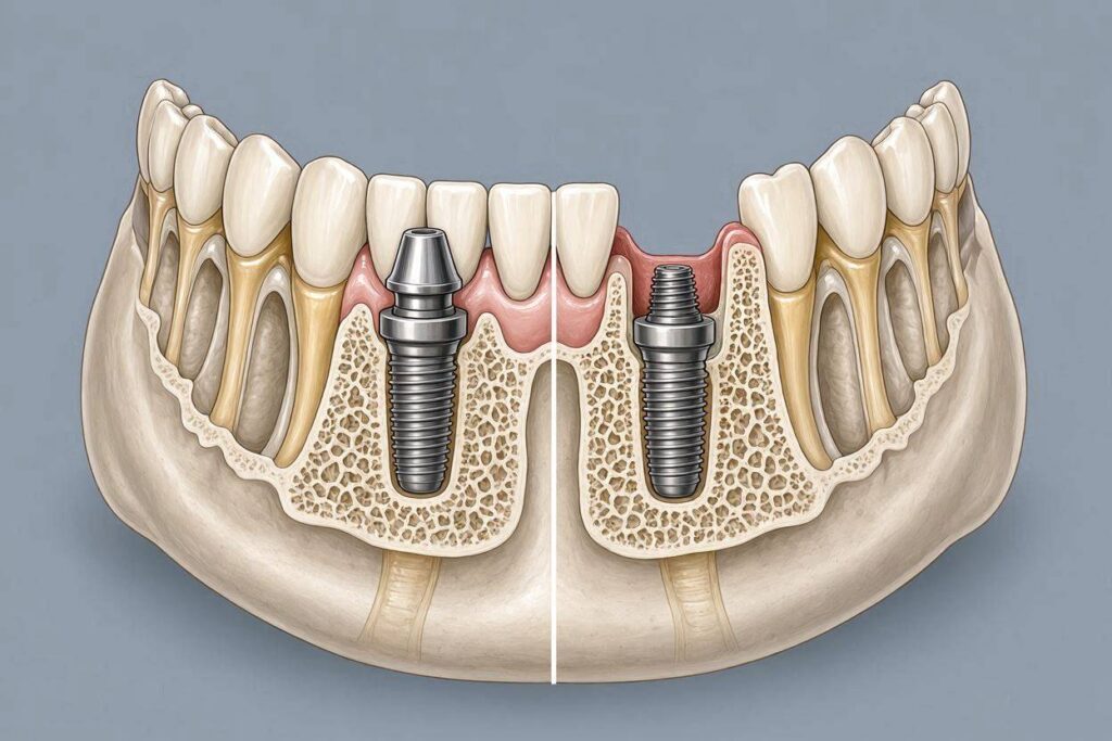
The Natural Consequence of Tooth Loss: Why Bone Disappears
To understand what implants do to bone, one must first understand what happens to bone when a tooth is lost.
The Function of the Alveolar Bone
The alveolar bone exists for one purpose: to support teeth. It forms in response to tooth eruption. It is maintained by the mechanical stimulation of chewing forces transmitted through the periodontal ligament. When a tooth is extracted, this stimulus disappears. The body, in its metabolic efficiency, resorbs bone that no longer serves a function. This is Wolff’s law in reverse: bone that is not loaded is removed.
The Pattern of Post-Extraction Resorption
Bone loss after extraction follows a predictable pattern. The buccal bone plate, which is thinner than the lingual or palatal plate, resorbs more rapidly and extensively. Within the first year after extraction, the alveolar ridge can lose up to 50 percent of its width, with the majority of this loss occurring in the first three to six months. Height is also lost, though to a lesser degree. This resorption is irreversible and progressive, continuing at a slower rate throughout life.
The Clinical Consequence
The edentulous mandible and maxilla become progressively more atrophic. Dentures become loose. The face develops a collapsed, aged appearance. Nerve foramina become superficial, making dentures painful. This relentless process is what implants seek to halt.
How Dental Implants Preserve Bone
The primary reason to place a dental implant is to replace the missing tooth root and restore the mechanical stimulus that preserves bone.
Direct Load Transfer Through Osseointegration
Unlike a bridge or a removable denture, which sits on top of the gum and transmits forces to the surface of the ridge, an implant transmits chewing forces directly into the jawbone through the osseointegrated interface. The bone experiences strain. This strain signals osteocytes to maintain bone density. The bone remodels in response to functional loading, just as it does around natural teeth.
Prevention of Disuse Atrophy
The implant occupies the space once filled by the tooth root. The surrounding bone remains loaded and therefore remains present. This is the most significant bone-related benefit of dental implants. Long-term studies show that bone loss in edentulous areas with implants is dramatically less than in edentulous areas without implants.
The Implant as a Bone Stimulator
Think of an implant as a bone stimulator. It does not grow bone in a deficient ridge. It preserves existing bone. The mechanical signals generated by chewing travel down the implant, through the implant-bone interface, and into the surrounding trabecular and cortical bone. This signal is what prevents the body from resorbing bone it deems unnecessary.
When Bone Loss Occurs Around Dental Implants
Bone preservation is not automatic or guaranteed. Bone loss around implants occurs under several circumstances, and understanding these is essential for prevention.
Initial Bone Remodeling After Implant Placement
Some bone loss around the implant neck is expected and normal. After the implant is uncovered and exposed to the oral environment, the bone remodels to establish a biological width, a soft tissue and bone architecture that allows a protective seal around the implant. This remodeling typically results in 0.5 to 1.5 millimeters of crestal bone loss in the first year after loading. This is considered physiological and does not jeopardize the implant.
Peri-Implantitis: The Pathological Bone Loss
Peri-implantitis is the primary cause of excessive, pathological bone loss around implants. It is an inflammatory condition triggered by bacterial biofilm on the implant surface. The host inflammatory response drives bone resorption. Without treatment, peri-implantitis progresses, causing crater-like bone defects around the implant. Over years, this bone loss can reach the apical portion of the implant, leading to mobility and failure.
The bone loss from peri-implantitis is not a consequence of the implant material itself. It is a consequence of bacterial infection in a susceptible host. The implant provides a surface for biofilm formation, just as a natural tooth root does.
Biomechanical Overload and Bone Microfracture
Excessive occlusal forces can cause microfractures in the bone around the implant. The bone attempts to repair these microfractures, but if overload is chronic, the repair cannot keep pace. The result is a radiographic radiolucency around the implant, often a circumferential saucerization, without the suppuration and bleeding typical of peri-implantitis. This is a biomechanical, not infectious, cause of bone loss. The two can coexist, making diagnosis challenging.
Surgical Trauma and Bone Necrosis
Overheating the bone during implant osteotomy preparation can cause bone cell death. If a significant volume of bone around the implant is devitalized, it will resorb and be replaced, potentially compromising initial stability. This is a surgical complication resulting in early bone loss and possible early implant failure. Copious irrigation, sharp drills, and a controlled drilling speed prevent this.
Bone Loss Due to Implant Malposition
An implant placed too close to an adjacent tooth, too close to another implant, or too far buccally may experience bone loss. If the implant is too buccal, the thin buccal bone plate resorbs, exposing threads. If implants are too close, the interimplant bone may resorb due to compromised blood supply and overlapping inflammatory zones.
Table: Types of Bone Loss Around Dental Implants
| Type of Bone Loss | Cause | Onset | Radiographic Appearance | Reversible? |
|---|---|---|---|---|
| Physiologic remodeling | Establishment of biological width | First year after loading | 0.5-1.5 mm crestal loss | No, stabilizes |
| Peri-implantitis | Bacterial biofilm, host inflammatory response | Any time, often after years | Crater-like or saucer-shaped defects, progressive | Can be arrested with treatment |
| Biomechanical overload | Excessive occlusal forces | Variable | Circumferential radiolucency, often no soft tissue inflammation | Possibly, if force reduced |
| Surgical trauma | Bone overheating during drilling | Early, before loading | Generalized radiolucency around implant | No, leads to early failure |
| Malposition | Implant placed too buccal or too close to adjacent | Early | Thread exposure, buccal bone loss | No, may require removal |
Radiographic Monitoring of Bone Levels
Bone loss around implants is typically asymptomatic in its early stages. Radiographs are the primary diagnostic tool.
Baseline and Sequential Radiographs
A baseline periapical radiograph is taken at the time of crown delivery. The bone level at this point becomes the reference against which future bone levels are compared. Follow-up radiographs at one year, then every one to three years, are compared to the baseline. A change in bone level of more than 0.2 millimeters per year after the first year is a cause for concern.
Cone-Beam CT for Advanced Assessment
When significant bone loss is detected on periapical films, or when the defect morphology must be understood for surgical treatment planning, a cone-beam CT scan provides three-dimensional visualization. It reveals buccal and lingual bone loss that is invisible on two-dimensional periapical films. It shows the relationship of the defect to vital structures.
The Probing Examination
Radiographs show bone loss that has already occurred. Periodontal probing around implants provides real-time information about the soft tissue attachment and, indirectly, the bone level. Probing depths, bleeding on probing, and suppuration are recorded at maintenance visits. Changes in probing depth over time, combined with bleeding, suggest active inflammation and potential bone loss.
How to Prevent Bone Loss Around Implants
Prevention is far more effective than treatment.
Meticulous Oral Hygiene
The patient must clean around the implant daily. A soft toothbrush, interdental brushes sized to fit the spaces, floss specifically designed for implants, and oral irrigators all play a role. The dentist or hygienist demonstrates proper technique and reinforces it at every visit.
Regular Professional Maintenance
Patients with implants should see the dental hygienist at intervals determined by their risk profile, typically every three to six months. The maintenance visit includes probing, assessment of oral hygiene, professional cleaning of the implant surfaces with non-metallic instruments, and review of home care. Radiographs are taken at appropriate intervals to monitor bone levels.
Occlusal Management
The occlusion on implant restorations must be carefully designed and monitored. The crown should have light contact in maximum intercuspation and no contact in excursive movements when possible. Night guards protect against the destructive forces of bruxism. If parafunctional habits are identified, the patient must be educated and protective measures implemented.
Smoking Cessation
Smoking is a major risk factor for peri-implantitis and bone loss. Patients who smoke must be counseled about the risk and offered cessation resources. Some clinicians decline to place implants in heavy smokers due to the high failure and complication rates.
Management of Systemic Conditions
Diabetes, when well-controlled, does not significantly increase the risk of bone loss around implants. Poorly controlled diabetes impairs wound healing and the immune response, increasing susceptibility to peri-implantitis. The patient’s physician may need to be involved in optimizing glycemic control before implant treatment and on an ongoing basis.
Implant Design and Its Effect on Bone Stability
The implant itself influences bone behavior.
Platform Switching
Platform switching is a design concept where the abutment is narrower than the implant platform. This inward shift of the implant-abutment junction moves the inflammatory cell infiltrate away from the crestal bone. Studies have shown that platform-switched implants exhibit less crestal bone remodeling than conventional platform-matched implants. The design contributes to bone preservation.
Implant Surface Characteristics
Moderately rough implant surfaces promote osseointegration and bone-to-implant contact. However, if the rough surface becomes exposed to the oral cavity due to bone loss, it retains more biofilm than a smooth surface. Some implant systems feature a smooth collar at the coronal portion designed to be at the bone crest or slightly supracrestal, minimizing biofilm retention if bone remodeling occurs.
Implant Geometry and Thread Design
Microthreads at the implant neck have been shown to distribute stress more favorably and preserve crestal bone. Tapered implant bodies can achieve higher primary stability in soft bone, reducing micromotion during healing. The surgical protocol matched to the implant design is essential for bone preservation.
Treatment of Bone Loss Around Implants
When bone loss is detected, intervention is required.
Non-Surgical Treatment for Early Peri-Implantitis
Debridement of the implant surface with specialized instruments, local or systemic antibiotics, and improved oral hygiene can arrest early peri-implant bone loss. The defect is cleaned, but lost bone does not typically regenerate without surgical intervention.
Surgical Regeneration of Peri-Implant Bone Defects
For more advanced defects, a surgical flap is elevated. The contaminated implant surface is decontaminated using mechanical, chemical, or laser methods. The bony defect is grafted with bone substitute material and covered with a membrane. The goal is to regenerate lost bone and re-establish osseointegration on the previously contaminated surface. Success is not guaranteed. The extent of regeneration is variable.
Explantation and Site Reconstruction
If the bone loss is so extensive that the implant is mobile or if treatment fails to arrest the disease, the implant must be removed. The site is debrided, grafted, and allowed to heal. A new implant may be placed after adequate healing if the cause of the original bone loss has been addressed.
Bone Loss with Different Types of Implant Prostheses
The prosthetic design affects bone loading and hygiene access, both of which influence bone stability.
Single Crowns
Single implant crowns allow the best oral hygiene access. Bone loss around single crowns is typically minimal if the patient maintains hygiene and the occlusion is sound.
Implant-Supported Fixed Bridges
Splinting multiple implants together changes the force distribution. The bone around each implant is loaded differently. Hygiene under the pontics and around the connectors is more challenging. Bone loss is more common around implant bridges than single crowns, driven by hygiene difficulties.
Full-Arch Fixed Prostheses
An All-on-Four or similar full-arch fixed prosthesis places the implant-bone interface under complex loads. The prosthesis itself, especially a hybrid design with acrylic denture teeth on a metal framework, can be difficult to clean thoroughly. Bone loss around these implants, particularly the tilted posterior implants, requires vigilant monitoring.
Implant-Retained Overdentures
The removable prosthesis allows excellent access for hygiene around the implants themselves. The locator attachments or bars require cleaning. Bone loss around overdenture implants tends to be less than around fixed full-arch prostheses, likely due to better hygiene access and the removable design allowing overnight removal to reduce load.
Conclusion
Dental implants do not inherently cause bone loss. On the contrary, their primary biological benefit is the preservation of jawbone by replacing the mechanical stimulation lost when a tooth is extracted. The bone loss that does occur around implants is largely attributable to bacterial peri-implantitis, biomechanical overload, surgical complications, or poor prosthetic design, not to the implant material itself. Physiologic crestal bone remodeling of 0.5 to 1.5 millimeters in the first year is normal. Progressive bone loss beyond this is pathological and demands intervention. With proper planning, meticulous surgical technique, patient-maintained oral hygiene, regular professional care, and attention to occlusal forces, an implant can preserve bone and function for decades.
Frequently Asked Questions
Will an implant prevent bone loss in my jaw?
Yes, an implant provides the mechanical stimulation that signals the body to maintain bone. It preserves existing bone far better than a bridge or removable partial denture. It does not, however, regenerate bone that was lost before implant placement.
Is it normal to lose some bone around an implant?
Yes. A small amount of crestal bone remodeling, typically 0.5 to 1.5 millimeters, occurs in the first year after the implant is loaded. This stabilizes and is considered physiological. Progressive bone loss after the first year is not normal.
What causes bone loss around a dental implant years later?
The most common cause is peri-implantitis, a bacterial infection of the tissues around the implant. Other causes include occlusal overload, especially in patients who grind their teeth, and systemic conditions such as uncontrolled diabetes.
Can bone loss around an implant be treated?
Early peri-implantitis can be treated non-surgically to arrest bone loss. More advanced defects may require surgical debridement and bone grafting. Severely compromised implants with extensive bone loss may need to be removed.
How often should I have my implants checked for bone loss?
You should have professional maintenance visits every three to six months, including periodontal probing. Bitewing or periapical radiographs are typically taken annually or as recommended by your dentist to monitor bone levels.
Additional Resources
American Academy of Periodontology – Dental Implant Maintenance
https://www.perio.org/for-patients
Patient information on maintaining implants and understanding the risks of peri-implant diseases and bone loss.