How Gums Are Built For Dental Implants?
You have seen the final photograph. A dental implant crown emerges from a collar of pink, healthy, perfectly contoured gum tissue. It looks as if the tooth has simply grown there, nature undisturbed. But you know that beneath the surface, a complex biological and surgical story has unfolded. The question you ask is not about the titanium post or the ceramic crown. It is about the living frame that holds it all together: how are gums built for dental implants?
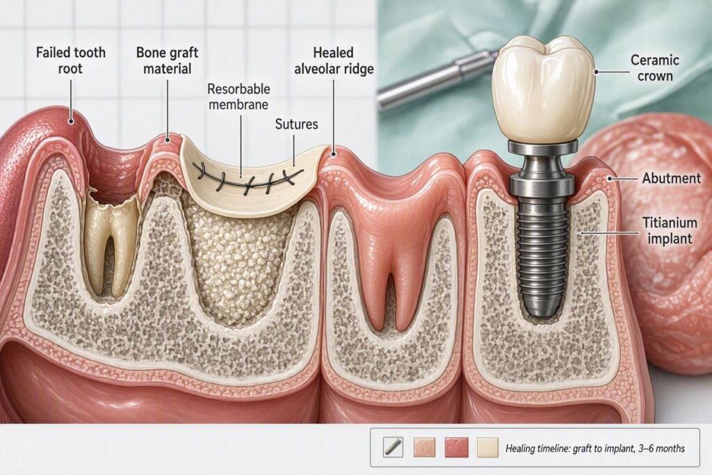
The answer is that gum tissue, or peri-implant mucosa, is not a passive bystander. It must be actively, thoughtfully, and sometimes surgically created. A successful dental implant is not just a bone-integrated post. It is a tooth that emerges through a healthy, immobile, attached band of keratinized gum tissue that forms a protective, cleansable seal. This article will explain the biological requirements for peri-implant health, the surgical techniques used to build and shape gum tissue, and the critical role of the healing abutment in sculpting the final emergence profile.
The Biological Requirement: Keratinized Tissue and the Biologic Width
Natural teeth are surrounded by a specialized type of gum tissue called keratinized or attached gingiva. This is the firm, pale pink, stippled tissue that wraps tightly around the neck of each tooth. It is rich in collagen fibers and resistant to the mechanical forces of chewing and brushing. Below this attached tissue, a delicate, non-keratinized lining mucosa moves freely. The attached gingiva acts as a protective cuff, a physical barrier that resists the invasion of bacteria.
Around a dental implant, this protective cuff is equally essential. The implant does not have a periodontal ligament with a rich blood supply from the bone. Its blood supply comes entirely from the overlying gum tissue. A zone of thick, attached, keratinized tissue around the implant is biologically critical for long-term health. It forms a stable, non-mobile seal against the abutment. It resists the pull of the cheek and lip muscles. It provides a firm, resilient surface that the patient can effectively clean with a toothbrush and floss.
If an implant is placed in an area with little or no attached gum tissue, surrounded only by thin, mobile lining mucosa, the prognosis is compromised. This type of tissue is fragile and easily inflamed. Muscle pull can tug on the margin, opening a pathway for bacteria. Oral hygiene becomes painful and ineffective. The patient is on a fast track to peri-implant mucositis and, eventually, the bone-destroying progression of peri-implantitis. Building an adequate zone of attached, keratinized gum is not an aesthetic luxury. It is a fundamental biological requirement for implant survival.
The Soft Tissue Graft: Building Gum from the Palate
When a patient has a deficiency of attached gum tissue at an implant site, the solution is a soft tissue graft. This is a surgical procedure, often performed by a periodontist, to harvest a piece of keratinized tissue from the patient’s own palate and transplant it to the implant site.
The palate, the roof of the mouth, is a reliable donor site. The tissue on the hard palate is firmly bound down to the underlying bone and is composed almost entirely of the dense, collagen-rich, keratinized connective tissue that the implant site needs. There are two primary graft techniques.
The Free Gingival Graft
This is the classic technique for building a zone of attached tissue. The surgeon delineates a rectangular piece of tissue on the palate, about the size of a small postage stamp, and carefully dissects it from the underlying connective tissue. This full-thickness graft is then sutured to the recipient site at the implant, which has been prepared by creating a bed of bleeding, vascular tissue. The graft is not placed over the implant. It is placed adjacent to it, to augment the width of the keratinized band.
The free gingival graft is a powerful, predictable procedure. The donor site on the palate heals by secondary intention, like a scrape, over a period of two to four weeks. The discomfort from the palatal wound is the main downside, often described as a pizza burn that lasts for a week. The graft itself initially looks white and dead, but it is quickly revascularized from the underlying bed and matures into a thick, healthy, keratinized collar around the implant.
The Subepithelial Connective Tissue Graft
This technique is used primarily for root coverage on natural teeth and for augmenting the thickness and contour of the gum around an implant, rather than just increasing the width of the attached zone. The surgeon makes an incision on the palate and opens a flap. Instead of removing the full-thickness surface tissue, they harvest only the subepithelial connective tissue from underneath the palatal flap. The palatal flap is then sutured closed, protecting the donor site and making the post-operative recovery significantly more comfortable.
The harvested connective tissue is then carefully placed under a pouch or flap created at the implant site. It acts as a biological filler, plumping up a deficient ridge, masking the grey shadow of a titanium abutment, and providing a thick, resilient bed of tissue for the implant crown to emerge from. This graft is often used in the aesthetic zone to create a natural, convex gum contour that mimics the anatomy around a natural tooth root.
The Healing Abutment: The Gum Sculptor
Gum tissue is not just built in bulk. It is sculpted, shaped, and trained to form a precise, natural-looking emergence profile for the implant crown. This is the role of the healing abutment, also called a healing cuff or permucosal extender.
After the implant is placed, and before the final crown is made, a small, cylindrical or anatomical healing abutment is screwed into the implant. This abutment protrudes through the gum into the oral cavity. Its purpose is to create and maintain a precisely shaped transmucosal channel—the tunnel through which the final crown will emerge.
The healing abutment acts as a mold. The gum tissue heals around it, forming a tight, circular collar with a defined sulcus depth. The restorative dentist can select different diameters and profiles of healing abutments to shape the gum in a way that mimics the emergence of a natural tooth. A molar site will get a wide, circular healing abutment. A central incisor site, particularly in a patient with a scalloped, thin gum phenotype, may get an anatomical healing abutment that is wider on the facial aspect to create the natural triangular gum contour.
The healing abutment is left in place for several weeks, allowing the gum to mature and form a stable, keratinized seal around it. When it is finally removed, the restorative dentist is presented with a perfect, circular, biologically healthy tunnel, ready to receive the final abutment and crown. Without this staged tissue shaping, the final crown would emerge from an amorphous, shapeless, and often inflamed gum hole.
The Provisional Restoration: The Ultimate Sculptor
In the most demanding aesthetic cases, particularly in the front of the mouth, the healing abutment is replaced by a custom-fabricated provisional (temporary) implant crown. This provisional is not just a placeholder. It is a sophisticated soft tissue sculpting tool.
The provisional crown is screwed onto the implant and is shaped by the restorative dentist to support the gum tissue in a way that exactly mimics the final desired emergence profile. Over weeks and months, the gum tissue remodels around the carefully contoured provisional, forming a perfectly anatomical, scalloped, and papillae-filled frame. The patient wears this provisional during the osseointegration period, allowing the gum to mature into the ideal aesthetic form before the final impression is even taken. The master ceramist then copies this beautifully sculpted emergence profile when fabricating the final crown. This technique, called “provisionalization for soft tissue sculpting,” is the gold standard for achieving truly invisible implant restorations in the aesthetic zone.
The Healing Period is Not Just for Bone: A common misconception is that the healing period after implant placement is solely for the bone to fuse to the implant. This is half the story. The simultaneous healing and maturation of the gum tissue are equally critical. A well-osseointegrated implant with poor, thin, inflamed gum tissue is a clinical failure waiting to happen. A successful implant requires both a solid bony foundation and a healthy, keratinized, well-contoured soft tissue seal.
Conclusion
Gums for dental implants are built through a combination of biological understanding and precise surgical technique. Soft tissue grafts, harvested from the patient’s own palate, augment deficient areas to create a thick, protective zone of keratinized attached tissue. Healing abutments and custom provisional restorations then sculpt this tissue into a natural, cleansable, and aesthetically beautiful emergence profile. The creation of healthy, stable peri-implant mucosa is not an afterthought; it is a central, defining element of a successful, long-lasting implant restoration.
Frequently Asked Questions
Is gum grafting for an implant painful?
The implant recipient site itself is typically not painful after grafting. The donor site on the palate is the source of post-operative discomfort. A free gingival graft, which leaves an open wound on the palate, is more uncomfortable than a connective tissue graft, where the palate is sutured closed. The discomfort is usually managed with over-the-counter pain medication and a protective plastic palatal stent, and it resolves significantly within one to two weeks.
Do I always need a gum graft before getting an implant?
No. Many patients have an adequate zone of attached, keratinized gum tissue at the proposed implant site. The need for a graft is determined by the periodontist or implant surgeon during the clinical examination. If you have a broad band of firm, pink gum, you may not need any soft tissue augmentation.
How long does it take for the gum to heal around a dental implant?
The initial soft tissue healing around an implant, where the gum closes and forms a seal around the healing abutment, takes about two to four weeks. The complete maturation of the peri-implant mucosa, where it becomes a stable, keratinized, and resilient collar, takes several months. This is why the final impression for the crown is often delayed for three to six months after implant placement, to allow the soft tissue to fully stabilize before the final restoration is fabricated.
Additional Resource:
For a detailed explanation of soft tissue grafting procedures and their importance in dental implant treatment, visit the American Academy of Periodontology’s patient resource on gum grafts: https://www.perio.org/consumer/gum-graft-surgery