How Dental Implants Stay in the Mouth?
You have a titanium post in your jaw. You chew. You talk. You bite into a crisp apple. And yet, your dental implant does not budge. It feels as solid as a natural tooth, perhaps even more so. How is this possible? What holds it in place? There is no glue, no suction, and no clasps. So what is the secret?
The answer lies in one of the most remarkable discoveries in modern medicine: osseointegration. This is the biological process that fuses living bone directly to the surface of a titanium implant. It is not a mechanical interlock. It is not a chemical adhesive. It is a living, dynamic union between your body and a biocompatible material. Understanding this process will give you a profound appreciation for the engineering and biology that keeps your implant secure for decades.
This guide will explain the science of implant retention, from the macroscopic threads that provide initial stability to the microscopic cellular bonds that lock the implant in place forever.
The Dual Mechanism of Retention: Mechanical and Biological
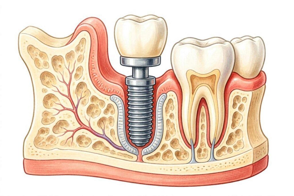
A dental implant stays in the mouth through two distinct mechanisms that work sequentially.
- Primary Stability (Mechanical): This is the immediate, physical lock achieved the moment the surgeon places the implant. It depends on the bone quality, the implant design, and the surgical technique. Think of it as the friction and mechanical interlock of a screw in wood.
- Secondary Stability (Biological): This is the long-term, living connection. Over weeks and months, new bone cells grow directly onto the implant surface, creating a biological weld. This is osseointegration. Primary stability fades as the bone remodels, but secondary stability rises to replace it.
If an implant lacks primary stability at placement, it will likely fail. If secondary stability never develops, it will also fail. But when both work together, the result is a tooth that can withstand hundreds of pounds of chewing force for a lifetime.
Primary Stability: The Immediate Mechanical Lock
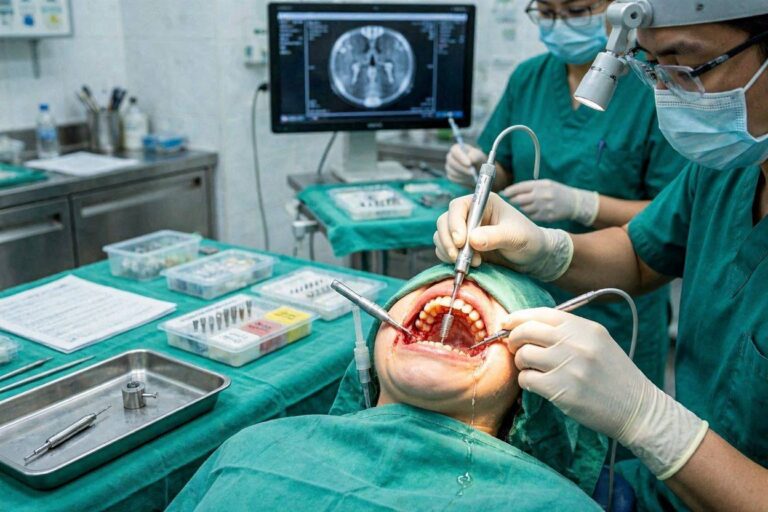
Imagine the surgeon drilling a precisely sized hole into your jawbone. The drill bit is slightly narrower than the implant. When the implant is screwed into this prepared site, it compresses the surrounding bone. This creates a tight, frictional grip. The threads of the implant engage the bone walls like a wood screw biting into a stud.
Factors That Determine Primary Stability
- Bone Density: Dense, cortical bone provides a stronger initial grip than soft, spongy cancellous bone. The lower front jaw (mandibular symphysis) has the densest bone in the oral cavity and gives the best primary stability. The upper back jaw (posterior maxilla) has thin, soft bone and often presents challenges.
- Implant Design: Modern implants are not simple cylinders. They have aggressive, self-tapping threads that cut a precise path into the bone. The thread pitch, depth, and shape are engineered to maximize surface contact and compression. Tapered implant bodies wedge into the bone, increasing stability.
- Surgical Technique: The surgeon uses a sequence of increasingly larger drills to prepare the site without overheating the bone. If the bone is soft, the surgeon may use a technique called “undersized drilling,” using a slightly smaller final drill to achieve more compression and grip. They then measure the insertion torque with a torque wrench. A high insertion torque (typically above 35 Ncm) correlates with good primary stability.
The Fragile Window
Primary stability is at its maximum on day one. Almost immediately, the bone at the microscopic interface begins to remodel in response to the surgical trauma. A thin layer of damaged bone cells dies off and must be resorbed. During this period (weeks 2-6), primary stability actually decreases. This is why secondary stability must ramp up in time to take over. This transition is the most vulnerable period in the life of an implant.
Secondary Stability: The Miracle of Osseointegration
This is the true answer to how implants stay in. Osseointegration is the direct structural and functional connection between living bone and the surface of a load-bearing implant. There is no intervening soft tissue, no periodontal ligament. Under a microscope, bone cells appear to be in intimate contact with the titanium oxide layer.
The Cellular Process Step-by-Step
- Blood Clot and Protein Adsorption: The moment the implant is placed, blood fills the gap between the implant and the bone. Plasma proteins, including fibrinogen and growth factors like TGF-β and BMPs, immediately adsorb onto the titanium surface. This protein coating makes the inert metal biologically recognizable.
- Platelet Activation and Clot Formation: Platelets aggregate and degranulate, releasing a cocktail of growth factors that signal the body to start healing. A fibrin clot forms, acting as a provisional matrix.
- Angiogenesis and Cell Migration: New blood vessels sprout into the clot. Mesenchymal stem cells migrate from the surrounding bone marrow and periosteum. The implant surface, now coated with a protein film, is an attractive scaffold for these cells.
- Osteoblast Attraction and Differentiation: The stem cells differentiate into osteoblasts, the bone-building cells. They migrate through the fibrin matrix and settle directly onto the implant surface. The rough, micro-textured surface of a modern implant is critical here. It provides microscopic undercuts and peaks that the cells can latch onto.
- Woven Bone Deposition: The osteoblasts begin secreting osteoid, an unmineralized collagen-rich matrix. This osteoid fills the microscopic roughness of the implant surface. Calcium phosphate crystals then deposit into this matrix, mineralizing it. This forms immature woven bone directly on the implant. This contact osteogenesis happens rapidly.
- Bone Remodeling and Maturation: Over months, the woven bone is remodeled into stronger, organized lamellar bone. The bone-implant interface is a dynamic structure. It is constantly being resorbed and rebuilt in response to mechanical loading. The bone adapts to the forces you place on it, becoming denser where stress is highest.
The Titanium Oxide Secret
Why does bone love titanium? Pure titanium is highly reactive with oxygen. The instant a titanium implant is machined and exposed to air, its surface spontaneously forms a thin, dense, highly adherent layer of titanium dioxide (TiO2). This layer is ceramic-like, incredibly stable, and biologically inert. It does not corrode in the body’s salty fluids.
Crucially, this oxide layer has properties that promote bone bonding. It has a high dielectric constant and can adsorb calcium and phosphate ions from the body fluids. Hydroxyl groups on the surface can form chemical bonds with the mineral phase of bone. There is evidence that a true chemical bond, a form of biointegration, occurs at the molecular level between the titanium oxide and the bone mineral hydroxyapatite.
“Osseointegration is not a scar tissue encapsulation. It is not a mechanical interlock. It is a dynamic biological union where living bone cells adhere to and mineralize directly against a stable, passive oxide surface.”
The Role of Implant Surface Technology
Not all implant surfaces are created equal. The evolution of surface technology is a major reason for today’s high success rates.
- Machined Surfaces (1960s-1990s): The original Brånemark implants had a relatively smooth, turned surface. Osseointegration was possible but slower and less predictable. These implants required a longer healing time and were less forgiving in poor-quality bone.
- Roughened Surfaces (2000s-Present): Modern implants undergo surface treatments to create controlled micro-roughness. This includes:
- Grit Blasting: The implant is blasted with particles of titanium oxide, alumina, or resorbable calcium phosphates, creating microscopic pits and peaks.
- Acid Etching: The surface is etched with strong acids (hydrochloric, sulfuric, hydrofluoric) to create a fine, intricate micro-porosity.
- SLA (Sandblasted, Large Grit, Acid-Etched): A common combination treatment that produces a moderately rough surface optimal for bone cell attachment.
- Anodization: An electrochemical process that thickens the oxide layer and creates a porous surface structure.
- Hydrophilic Surfaces: Some implants are stored in a solution or treated to maintain a highly hydrophilic (water-loving) surface. When placed, blood and proteins spread instantly across the surface, accelerating the initial healing cascade.
This surface roughness increases the surface area for bone contact by up to six times compared to a smooth surface. It also provides microscopic interlock. Bone cells grow into the tiny crevices. The result is a mechanical interdigitation at the micron scale that dramatically increases the strength of the interface.
The Biological Seal: The Soft Tissue Defense
The bone-implant interface keeps the implant anchored. But the mouth is a bacteria-filled environment. A second critical retention and defense mechanism exists: the soft tissue seal.
Where the implant abutment emerges through the gum, a collar of specialized tissue forms. This is the peri-implant mucosa. It consists of a sulcus lined with sulcular epithelium, which is permeable like the gum pocket around a tooth. Deeper down, the epithelium attaches to the abutment surface via a junctional epithelium, forming a seal with hemidesmosomes and a basement membrane.
Below this epithelial attachment, a zone of connective tissue rich in collagen fibers contacts the abutment. In natural teeth, these fibers insert perpendicularly into the cementum, creating a strong suspensory ligament. Around implants, because there is no cementum, the collagen fibers run in a parallel, circular orientation around the abutment. This is a weaker attachment, which is why implants are more vulnerable to bacterial penetration and peri-implantitis than natural teeth.
Nevertheless, this soft tissue cuff provides a critical biological seal. It prevents oral bacteria and their toxins from reaching the bone-implant interface. Maintaining this seal with gentle, meticulous oral hygiene is essential for long-term retention.
Mechanical Loading: How Chewing Forces Are Handled
Once the crown is placed, the implant enters a life of hard work. Every time you chew, forces are transmitted through the crown, down the abutment, through the implant body, and into the surrounding bone.
Force Transmission in a Natural Tooth vs. an Implant
A natural tooth has a periodontal ligament. This ligament is viscoelastic; it absorbs shock, distributes forces evenly, and provides proprioceptive feedback. The tooth can move slightly within the socket.
An osseointegrated implant has no ligament. It is rigidly fused. When force hits the crown, it is transmitted nearly 100% to the bone interface, with no shock absorption. The bone must handle the full brunt of the force. This is why implant occlusion is designed carefully. The dentist adjusts the crown to have slightly lighter contacts than natural teeth, especially in side-to-side movements. The goal is to protect the bone-implant interface from overload.
Wolff’s Law and Bone Remodeling
The good news is that bone is a dynamic tissue. Wolff’s Law states that bone remodels in response to the mechanical stresses placed upon it. When an implant transmits controlled, physiological forces into the bone, the bone around the implant threads maintains its density and may even increase in density. This is the principle behind immediate loading and early loading protocols.
However, if the forces are excessive (bruxism, a poorly designed bite), the bone cannot adapt. Microfractures accumulate, and bone resorption overtakes formation. This leads to peri-implant bone loss and eventual failure. The implant stays in the mouth as long as the mechanical forces remain within a physiological window.
The Long-Term Equilibrium
So, how do dental implants stay in the mouth for 30, 40, or even 50 years?
The answer is a state of dynamic equilibrium. The bone-implant interface is not a dead, static weld. It is a living tissue that is constantly being micro-resorbed and re-deposited. As long as the mechanical forces are appropriate, the oral hygiene is excellent, and the patient’s systemic health is stable, the bone will continue to embrace the implant.
The titanium post itself is essentially inert and indestructible in the oral environment. It will not decay. It will not degrade. The weak point is never the metal; it is the biological response of the host. Care for your body, care for your gums, and the osseointegration bond will endure.
Conclusion
Dental implants stay in the mouth through a two-phase retention mechanism. Primary stability is the immediate mechanical grip achieved by the screw threads compressing against the bone. Secondary stability, the true long-term answer, is the biological process of osseointegration, where living bone cells attach directly to the titanium oxide surface and mineralize onto it. Supported by a protective soft tissue seal and maintained by balanced mechanical forces, this living bone-to-implant bond can securely anchor a replacement tooth for a lifetime.
FAQ
1. Is the implant glued into the bone?
No, there is no glue or cement. The implant is held in place by osseointegration, a natural biological process where your bone cells attach directly to the titanium surface. The initial stability comes from the tight mechanical fit of the screw threads in the bone.
2. Can the bone ever let go of the implant?
Yes, unfortunately. The bone-implant bond can be destroyed by a chronic bacterial infection called peri-implantitis, which causes progressive bone loss. It can also fail due to excessive mechanical overload from bruxism or a poorly fitting crown.
3. How strong is the osseointegration bond?
Osseointegration is remarkably strong. The implant-bone interface can withstand hundreds of pounds of compressive force during chewing. It typically takes a very high reverse torque to break the bond, far exceeding normal physiological forces.
4. Does the implant feel different from a natural tooth?
Yes, slightly. A natural tooth has a periodontal ligament that provides a tiny bit of spring and fine tactile feedback. An implant is rigidly fused and lacks this ligament. You feel pressure and vibration, but the subtle “feel” of chewing is different. Most patients adapt to this difference quickly.
Additional Resource
For a visual exploration of osseointegration and implant surface science, visit the Academy of Osseointegration: https://www.osseo.org/