D0431 Dental Code: A Complete Guide to Adjunctive Pre-Diagnostic Testing
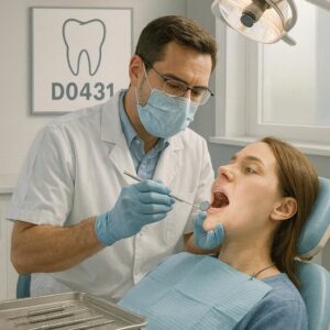
You schedule a periodic oral evaluation for a patient. During the conversation, they mention a sensitive area on the tongue or a spot that feels different. You look, and you see something that requires more than a visual inspection and palpation. You need a diagnostic aid. This is precisely the moment the D0431 dental code enters the clinical picture.
Many dental professionals struggle with this code. Insurance coordinators often put it aside for later review. Yet understanding it transforms how you practice risk assessment and early detection. This guide walks you through every detail of D0431. It offers clarity on its clinical application, coding nuances, reimbursement realities, and documentation essentials.
Let’s explore this diagnostic code without unnecessary complexity.
What Exactly Is the D0431 Dental Code?
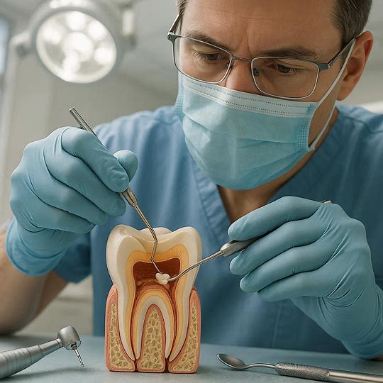
The American Dental Association maintains the Code on Dental Procedures and Nomenclature. Within this system, D0431 falls under the Diagnostic category. Its official descriptor reads: “Adjunctive pre-diagnostic test that aids in detection of mucosal abnormalities including premalignant and malignant lesions, not to include cytology or biopsy procedures.”
This is a screening aid. It does not give you a diagnosis. It helps you decide if a biopsy or referral is necessary. Think of it as a sophisticated triage instrument. You use an adjunctive device to visually highlight tissue changes that remain invisible to the naked eye under standard white light.
Dentists often confuse this test with a definitive diagnostic procedure. It is not. A biopsy provides the histological diagnosis. D0431 simply tells you, “Look closer here. This area needs more investigation.”
The Clinical Technology Behind the Code
Multiple devices and systems support the D0431 procedure. Each employs a different biological principle to distinguish between normal and potentially abnormal tissue.
Fluorescence-based systems use a specific wavelength of light. Normal mucosal tissue fluoresces a pale green color when exposed to this light. Dysplastic or suspicious tissue disrupts the collagen matrix and cellular metabolism. It appears as a dark, non-fluorescing area against the green background. The loss of fluorescence signals a loss of normal tissue architecture.
Chemiluminescence devices employ a different approach. A patient rinses with a dilute acetic acid solution. This solution desiccates the cell nuclei slightly and removes the glycoprotein layer. A specialized light source then illuminates the tissue. Abnormal cells, with their higher nuclear-to-cytoplasmic ratio, reflect light differently. They appear distinctly white, a phenomenon termed “aceto-whitening.”
Tissue reflectance systems analyze the spectral characteristics of reflected light. Hemoglobin absorption and nuclear scattering patterns differ between healthy and dysplastic tissue. The device captures this data and creates a color-coded map on a monitor.
The code covers the technical component and the professional interpretation. You bill D0431 once per encounter, regardless of the number of sites examined.
When Should You Use the D0431 Code?
Clinical judgment drives the appropriate application of D0431. Not every patient requires this test at every hygiene visit. Overuse dilutes its clinical value and attracts payer scrutiny.
High-Risk Patient Profiles
Certain patients benefit most from adjunctive screening. The decision to employ the test should correlate with recognized risk factors for oral cancer and potentially malignant disorders.
Tobacco users constitute a primary high-risk group. This includes cigarettes, cigars, pipes, and smokeless tobacco products. The duration and intensity of use increase the cumulative risk. Document the pack-year history or the years of smokeless tobacco use.
Heavy alcohol consumers face a synergistic risk when combined with tobacco use. The relative risk multiplies rather than adds. Establish a threshold in your practice protocol. Many clinicians define heavy consumption as more than 14 drinks per week for men and more than 7 for women.
Patients with prior oral cancer or premalignant lesion history require vigilant surveillance. A previous diagnosis of oral squamous cell carcinoma, verrucous carcinoma, or carcinoma in situ places them in an ongoing high-risk category. The same applies to a history of oral epithelial dysplasia, erythroplakia, or proliferative verrucous leukoplakia.
Immunocompromised individuals need consideration. Patients with HIV/AIDS, organ transplant recipients on immunosuppressive therapy, or those with chronic autoimmune conditions may face elevated risk. The immune surveillance system plays a crucial role in eliminating aberrant cells.
HPV-positive individuals represent an emerging risk group. Human papillomavirus, particularly HPV-16, is associated with oropharyngeal squamous cell carcinoma. These cancers often occur in the tonsillar pillars, base of tongue, and soft palate. Visual inspection alone easily misses these areas.
Age over 40 with additional risk factors serves as a general guideline. The incidence of oral cancer increases with age, although HPV-related cancers are shifting the demographic toward younger males.
Clinical Scenarios Triggering Use
Beyond risk profiles, specific clinical findings prompt the use of D0431.
A clinically suspicious lesion identified during the routine exam justifies the test. This includes leukoplakia that does not rub off, erythroplakia, mixed erythroleukoplakia, ulcerations without obvious traumatic etiology persisting beyond two weeks, or any exophytic growth.
A patient report of persistent soreness, numbness, or a sensation of something stuck in the throat may trigger the test. Unexplained symptoms without a visible corresponding lesion on conventional examination warrant deeper investigation.
Asymmetric tonsillar enlargement or a unilateral neck mass discovered during the extraoral exam should prompt consideration of adjunctive testing for the oropharyngeal region.
Follow-up monitoring of previously identified but not yet biopsied lesions represents a valid application. If you and the patient elected watchful waiting for a low-suspicion lesion, D0431 aids in tracking any change in character over time.
Step-by-Step Clinical Protocol for D0431
Consistency in execution ensures reliable results and supports billing compliance.
Pre-Procedure Preparation
Explain the procedure to the patient in simple terms. Obtain verbal consent. You do not typically need a separate written consent form for this non-invasive test, but your practice policy may vary.
A sample patient script reads: “Mrs. Johnson, based on your history of smoking and the rough white patch I see on your cheek, I want to use a special light that helps us see tissue changes we cannot see with the office lights. It is painless and takes about two minutes. It helps us decide if we need to take a small sample for the lab.”
Perform a comprehensive conventional oral cancer screening first. This includes visual inspection under white light and bimanual palpation of all oral soft tissues. The extraoral exam covers the head, neck, thyroid, and lymph nodes. The intraoral exam systematically evaluates the labial mucosa, buccal mucosa, floor of the mouth, ventral and lateral tongue, dorsum of the tongue, hard and soft palate, tonsillar pillars, and oropharynx.
The adjunctive test does not replace this conventional exam. It adds to it. Document the complete conventional exam findings before initiating D0431.
Performing the Test
Follow the manufacturer’s instructions precisely for your specific device. General principles apply across platforms.
Dim the ambient operatory lighting. Overhead lights and dental chair lights may interfere with the device’s light emission and your perception of tissue fluorescence or reflectance.
Position the patient appropriately. A semi-supine position works well for anterior structures. You may need to ask the patient to extend their neck or protrude their tongue for posterior visualization.
For fluorescence devices, shine the light onto the mucosal surface from a distance of approximately 20-30 centimeters. Systematically scan all mucosal surfaces in a predetermined sequence. A consistent sequence prevents omission of areas.
Observe the tissue response. Normal tissue appears pale green. Areas of loss of fluorescence appear dark brown or black. Inflammation, ulceration, traumatic keratosis, and vascular lesions can also cause loss of fluorescence. A positive finding does not equal cancer. It identifies an area requiring differential diagnostic consideration.
For chemiluminescence systems, after the acetic acid rinse, the tissue is examined under the specialized light. Normal tissue maintains a bluish hue. Abnormal tissue appears bright white with sharp margins.
For reflectance spectroscopy, place the probe tip gently on the tissue surface. The device captures spectral data. Some systems display a risk stratification score.
Photograph or diagram the positive findings. Note the location, size in millimeters, color, and margin characteristics. This baseline documentation proves invaluable for monitoring.
Post-Procedure Steps
Discuss the findings with the patient immediately. If the test is negative, offer reassurance that no suspicious areas were identified with the adjunctive light, but reinforce that this does not replace the biopsy as the gold standard if a visible lesion still concerns you.
If the test is positive, frame the discussion carefully. A statement like “The special light shows an area on the side of your tongue that looks different from the surrounding tissue. This does not mean it’s cancer. It means we need to take a closer look or possibly send a tiny sample to the pathologist for a definitive answer” provides honest information without unnecessary alarm.
Document the D0431 code, the device used, the findings, the interpretation, and the recommended next steps.
Documentation Requirements for D0431
Payers scrutinize claims for D0431. Inadequate documentation is the leading cause of claim denials for this code. A strong clinical note supports medical necessity and facilitates timely reimbursement.
Essential Elements of the Clinical Note
Your documentation should stand alone. Another clinician reading the note should understand precisely why you performed the test, what you found, and what it means.
Reason for the test. State the risk factors or clinical findings that prompted the adjunctive screening. Examples: “Patient is a 45-year smoker with a 40-pack-year history and daily alcohol consumption,” or “Visual exam revealed a 12mm x 8mm non-homogeneous leukoplakia on the right lateral tongue border, persisting despite elimination of possible traumatic etiology.”
Type of device and system used. Record the specific technology and device name. “OralID fluorescence device,” “VELscope Vx,” “ViziLite Plus with TBlue,” or “Identafi reflectance system” are appropriate entries.
Areas examined. Document that a full-mouth mucosal evaluation was performed. “All oral mucosal surfaces were examined, including the buccal mucosa, floor of the mouth, ventral and lateral tongue, hard and soft palate, and oropharyngeal tissues.”
Findings. Note the appearance under the adjunctive device. Use descriptive terminology. “Under fluorescence examination, a discrete area of loss of fluorescence was observed on the right lateral tongue border corresponding to the clinically noted leukoplakia. The area measured 10mm x 7mm with irregular margins. No other areas of fluorescence loss were identified.”
Interpretation. This is the clinical judgment piece. “The finding represents a suspicious lesion that requires further evaluation. The loss of fluorescence suggests disruption of the normal tissue architecture and warrants biopsy.”
Recommended next steps. State the plan clearly. “Referral to oral surgeon for incisional biopsy of the right lateral tongue lesion. Patient advised to schedule this within two weeks. Follow-up exam scheduled to discuss biopsy results.”
Images or diagrams. If your system captures images, include them in the patient record. A simple diagram depicting the lesion location adds value. Note: Some payers may require image submission with the claim.
Audit-Proofing Your Documentation
Auditors look for consistency. The risk factors documented at the initial exam should align with the decision to use D0431. The findings from the conventional exam should justify the use of the adjunctive test. The interpretation should logically connect to the next steps.
Document the medical necessity clearly. A note that states “D0431 performed” without context fails to establish medical necessity. A note that explains the patient’s significant risk profile and the clinical suspicion for malignancy does.
Reimbursement, Fees, and Payer Policies for D0431
Financial considerations play a role in clinical decision-making. Understanding the reimbursement landscape helps you set appropriate fees, communicate effectively with patients, and manage denial appeals.
Setting Your Fee
Calculate your fee based on a factor. Consider the cost of disposables (sheaths, rinse solutions, marking aids), the amortized cost of the device, the additional clinical time required (typically 2-5 minutes), and the professional interpretation time.
Fees in the United States typically range from $65 to $150 per test. Geographic location, practice overhead, and market demand influence the specific fee. Conduct a fee survey in your area or consult with your dental society.
Do not set the fee so low that it appears nominal. A very low fee suggests a lack of value. Do not set it so high that patients consistently decline the service. Transparency about the fee and potential insurance coverage before performing the test minimizes billing surprises.
Insurance Coverage Variability
D0431 faces inconsistent coverage across dental benefit plans. This reality frustrates many practitioners.
Medical insurance may cover the test under the medical plan when it is performed for the evaluation of a specific lesion or symptom. The link to a potential medical diagnosis (e.g., a lesion suspicious for malignancy) makes it a medical diagnostic test. You must use medical claim forms and appropriate medical diagnosis codes. A code from the ICD-10-CM system, such as K13.21 (Leukoplakia of oral mucosa, including tongue) or R19.8 (Other specified symptoms and signs involving the digestive system and abdomen), supports medical necessity.
Dental insurance policies vary widely. Some carriers consider D0431 an integral part of the periodic oral evaluation and bundle it with D0120 or D0150. Others consider it experimental or investigational. A growing number of carriers recognize the value and offer coverage as a separate benefit, particularly for high-risk patients.
Common Payer Policies:
| Payer Type | Typical Policy for D0431 | Patient Responsibility |
|---|---|---|
| Delta Dental (varies by state) | Often bundles with periodic exam; separate coverage increasing | Usually patient pays out-of-pocket |
| Aetna Dental | May consider medically necessary with risk factors; pre-determination recommended | Varies by plan, often covered with documentation |
| Cigna Dental | Coverage varies; some plans include once per year for high-risk patients | Co-pay or co-insurance may apply |
| MetLife Dental | Generally does not cover; considered adjunctive and not separately reimbursable | Patient responsible for full fee |
| Humana Dental | Increasingly covers with documented risk factors and suspicious lesion | Varies by specific plan |
| Medical Plans (BCBS, UHC, Aetna Medical) | May cover if billed as medical diagnostic test with appropriate medical diagnosis codes | Subject to medical deductible and co-insurance |
Important Note: Payer policies change frequently. Always verify coverage for the specific patient’s plan before providing the service. Use the payer’s online portal or call provider services. Document the name of the representative, the reference number, and the coverage details provided.
Billing Tips for Successful Claims
Submit with the appropriate diagnostic code. For medical claims, link D0431 to one or more ICD-10-CM codes. K13.21 (Leukoplakia), K13.22 (Minimal keratinized residual ridge mucosa), K13.29 (Other disturbances of oral epithelium), K13.70 (Unspecified lesions of oral mucosa), Z85.810 (Personal history of malignant neoplasm of tongue), and Z85.818 (Personal history of malignant neoplasm of other sites of lip, oral cavity, and pharynx) are commonly used.
Include a narrative report. When submitting on a medical claim form (CMS-1500), include a brief narrative or a copy of the procedure note in box 19 or as an attachment. This preempts many requests for additional information.
Pre-determination. For high-fee procedures or uncertain coverage situations, submit a pre-determination with supporting documentation. This obtains a coverage decision before you perform the procedure and allows the patient to make an informed financial decision.
Appeal denials. A denial is not final. Write a concise appeal letter. Include the clinical note, relevant literature supporting the use of adjunctive screening in high-risk patients, and a clear statement of why the test was medically necessary for this specific patient.
D0431 Versus Other Diagnostic Codes: A Comparative View
Understanding the differences between related codes prevents mis-coding and claim rejections.
D0431 vs. D0120 (Periodic Oral Evaluation)
D0120 is the routine comprehensive oral exam performed at regular intervals. D0431 is a separate, specific procedure that involves a specialized technology. If you perform an adjunctive test during a periodic evaluation, both codes are reported. Modifier -51 (Multiple Procedures) is generally not required for dental claims, but some payers may request it.
Document both services distinctly. The periodic exam note covers the overall oral health status. The D0431 note covers the specific findings of the mucosal screening.
D0431 vs. D7286 (Biopsy of Oral Tissue – Soft)
D7286 is the surgical removal of a tissue sample for pathological examination. It is a diagnostic surgical procedure. D0431 is an adjunctive aid used to decide if a biopsy (D7286) is necessary. They are not mutually exclusive. A patient may receive D0431 to identify a suspicious area, and if the clinical suspicion is high, D7286 to obtain the definitive diagnosis.
D0431 vs. D7288 (Brush Biopsy)
D7288, often used for transepithelial cytology sampling, involves the collection of cells for cytological analysis. It is not an adjunctive pre-diagnostic test per the D0431 definition. D0431 specifically excludes “cytology or biopsy procedures.” You use D0431 to screen and decide if D7288 or D7286 is needed.
Comparison Table: D0431 and Related Diagnostic Codes
| Code | Description | Invasiveness | Purpose | Technology Used |
|---|---|---|---|---|
| D0431 | Adjunctive pre-diagnostic test for mucosal abnormalities | Non-invasive | Screening aid to detect areas of concern | Fluorescence, chemiluminescence, reflectance spectroscopy |
| D0120 | Periodic oral evaluation | Non-invasive | Routine comprehensive oral assessment | Visual, tactile, radiographic |
| D7286 | Biopsy of oral tissue – soft | Minimally invasive | Definitive histological diagnosis | Scalpel or punch, histopathology |
| D7288 | Brush biopsy, transepithelial sample | Minimally invasive | Cytological sampling for atypical cells | Brush, cytology analysis |
| D7285 | Incisional biopsy of oral tissue – hard | Invasive | Definitive histological diagnosis of bone | Surgical access, trephine or chisel, histopathology |
Integrating D0431 Into a Comprehensive Oral Cancer Screening Protocol
Adjunctive testing works best within a structured practice protocol. A systematic approach ensures consistency across providers and maximizes patient safety.
The Multi-Tiered Screening Model
Many practices adopt a three-tiered model.
Tier 1: Conventional Screening. Every patient receives a comprehensive extraoral and intraoral exam at every periodic evaluation. This is the minimum standard of care. It requires only operatory lighting, gauze, and gloves.
Tier 2: Adjunctive Screening for Risk-Stratified Patients. High-risk patients receive D0431 at designated intervals, typically annually. The risk factors determine eligibility for Tier 2. This step identifies lesions not visible under white light.
Tier 3: Diagnostic Intervention. Suspicious findings from Tier 2, or highly suspicious findings even under conventional screening, proceed to biopsy or specialist referral. This tier establishes the definitive diagnosis.
Sample Practice Protocol
Develop a written protocol for your practice. Train all clinical staff. Consistency of application is key.
Step 1: Risk Assessment. At each patient visit, update the risk factor profile. Use a standardized form or digital health history module. Factors assessed include tobacco use, alcohol consumption, prior history of oral cancer or premalignant lesions, HPV status, and immunosuppression status.
Step 2: Conventional Exam. Perform the standard exam. Document all findings, even normal ones. Use a systematic descriptive approach or a standardized oral cavity diagram.
Step 3: Decision Point. If risk factors are present AND/OR a suspicious lesion is identified, proceed to D0431. If the patient is low-risk and the conventional exam is entirely normal, D0431 is generally not indicated.
Step 4: Adjunctive Exam. Perform D0431 using the practice’s chosen device. Document findings meticulously.
Step 5: Clinical Decision. If D0431 is negative but a visually suspicious lesion persists, consider biopsy or close follow-up. The adjunctive test reduces, but does not eliminate, the false-negative risk. If D0431 is positive, proceed to biopsy or referral.
Step 6: Communication and Follow-up. Discuss the findings and plan. Schedule the follow-up or referral. Document the discussion.
Potential Pitfalls and How to Avoid Them
Experience teaches valuable lessons. Learn from common missteps to refine your diagnostic process.
Clinical Pitfalls
Over-reliance on a negative test result. A negative D0431 test is not a guarantee of tissue health. Subtle changes, early neoplastic transformation, or lesions in difficult-to-visualize areas can be missed. Never allow a negative adjunctive test to override your clinical suspicion based on the visual and tactile exam. If you see a lesion that concerns you, pursue definitive diagnosis regardless of the D0431 result.
Misinterpreting inflammatory lesions. Inflammation causes loss of fluorescence. A traumatic ulcer, lichen planus, or simple cheek bite can appear as a dark area. This is a false positive. Correlate the D0431 findings with the clinical history and appearance. A history of trauma, an adjacent sharp cusp, or classic reticular Wickham striae helps differentiate inflammatory from dysplastic lesions.
Using the wrong code. Reporting D0431 when performing a brush biopsy (D7288) or an incisional biopsy (D7286) is incorrect. Each has its own code. The descriptor for D0431 explicitly excludes cytology and biopsy.
Performing the test without appropriate patient communication. A patient who sees a dark spot on a screen and is told “we need to biopsy this” without adequate explanation experiences unnecessary fear. Frame the test as an advanced screening tool, not a cancer detector.
Administrative Pitfalls
Inadequate documentation. As discussed, this is the most common reason for claim denial. Use a template. Ensure every note includes the required elements.
Billing without verifying coverage. This leads to patient dissatisfaction. Always check coverage beforehand for high-risk patients or when a suspicious lesion is present and D0431 is clearly indicated.
Failure to appeal denials. Many denials are overturned on appeal. A well-written appeal letter supported by clinical documentation and literature frequently results in payment. Do not leave reimbursement uncollected simply because the initial submission was denied.
The Future of Adjunctive Screening and D0431
The landscape of oral mucosal disease screening continues to evolve. Emerging technologies and an expanding evidence base are shaping the future.
Artificial Intelligence Integration
Machine learning algorithms are being developed to analyze fluorescence and reflectance images. These systems learn to differentiate between inflammatory and dysplastic patterns based on thousands of annotated images. A computer-aided diagnostic system may soon provide an objective risk score, reducing operator-dependent variability in interpretation.
Biomarker-Based Screening
Salivary diagnostics and tissue-based biomarker testing are advancing. Future adjunctive tests may combine the optical screening of D0431 with a rapid point-of-care biomarker panel. Imagine a device that shines a light and simultaneously analyzes the tissue’s protein expression profile through a sensor.
Expanded Coverage
As the evidence base linking early detection to improved survival and reduced treatment costs solidifies, payer coverage for D0431 is expected to broaden. Professional organizations continue to advocate for recognition of the test as a valuable component of comprehensive oral healthcare.
Teledentistry Integration
Remote monitoring of lesions using patient-captured images or store-and-forward technology may extend the reach of screening. A D0431 test performed in a primary care office or a community health setting could be interpreted by a remotely located oral medicine specialist or oral and maxillofacial pathologist.
Frequently Asked Questions About D0431
Is D0431 a cancer screening test?
D0431 is an adjunctive screening aid that helps detect mucosal abnormalities that may include premalignant and malignant lesions. It is not a standalone cancer diagnostic test. A definitive diagnosis requires tissue biopsy and histopathological examination.
Does dental insurance cover D0431?
Coverage varies. Some plans cover it as a separate benefit, particularly for high-risk patients. Others bundle it with the periodic oral evaluation. Medical insurance may cover it when billed with appropriate medical diagnosis codes for a suspicious lesion. Always verify coverage before providing the service.
How often can D0431 be performed?
For high-risk patients, such as those with a history of oral cancer or heavy tobacco and alcohol use, an annual D0431 screening is often considered appropriate. For patients with suspicious lesions requiring monitoring, a shorter interval may be indicated. Payer policies may limit frequency.
Can I bill D0431 and D0120 on the same day?
Yes. The D0431 adjunctive test is a separate procedure from the D0120 periodic oral evaluation. Document both services distinctly. Report both codes on the claim.
What is the difference between D0431 and a VELscope exam?
VELscope is a brand of fluorescence visualization device used to perform the procedure described by code D0431. The code describes the procedure; VELscope is one of several technologies that can be used to perform it. Other devices include OralID, Identafi, and ViziLite.
Does a positive D0431 result mean the patient has cancer?
No. A positive result indicates an area of tissue that appears different under the adjunctive light. Inflammation, trauma, benign keratosis, and other non-malignant conditions frequently cause positive findings. The positive result means further investigation, such as monitoring, cytology, or biopsy, is indicated.
Conclusion
The D0431 dental code represents an adjunctive pre-diagnostic test that enhances conventional oral mucosal examination by highlighting tissue changes not visible to the naked eye. Clinicians use this technology for high-risk patients and to further evaluate suspicious lesions, guided by careful documentation and appropriate payer communication. When integrated thoughtfully into a structured screening protocol, D0431 supports earlier intervention and clearer clinical decision-making without replacing the gold standard of tissue biopsy.
Additional Resource
For current ADA coding guidance and any revisions to the Code on Dental Procedures and Nomenclature, visit the American Dental Association’s official coding resource at:
https://www.ada.org/publications/cdt
Disclaimer: This article provides general information about the D0431 dental code as of the date of writing. It does not constitute legal, coding, or billing advice. Dental codes and payer policies change. Always verify current coding information with the American Dental Association and verify coverage with the specific payer before providing services. Consult with a qualified dental billing specialist or healthcare attorney for practice-specific guidance.