What Causes Dental Implants to Fail?
A dental implant is the most predictable and durable tooth replacement in modern medicine, with a documented long-term success rate exceeding 95%. That statistic is a triumph of engineering and biology. But it also means that in a small percentage of cases, the implant does not survive. The failure of a dental implant is a complex, often silent event that unfolds over weeks or years. It is rarely a sudden, catastrophic event. It is more often a slow, progressive loss of the bond between the titanium surface and the living bone, or a mechanical fracture of the components under years of cyclic loading. Understanding why implants fail is not a cause for fear. It is the most powerful tool a patient has to partner with their dentist in prevention. This guide is a complete taxonomy of dental implant failure. It dissects the biological, mechanical, and iatrogenic causes, explains the difference between early and late failure, places peri-implantitis under a microscope, and gives you the specific warning signs that demand immediate professional attention.
The Two Categories: Early Failure vs. Late Failure
Implant failure is classified by time. The distinction is clinically essential because the causes are entirely different.
Early Failure (Before the Crown is Placed)
An early failure occurs before osseointegration is complete, typically within the first 3 to 6 months. The implant fails to fuse with the bone. It is a failure of biology. The implant either never achieves primary stability, or the healing process is disrupted. The implant becomes mobile, or a fibrous, non-integrated scar tissue capsule forms around it instead of rigid bone. Early failure is often painless. The dentist discovers mobility at the time of the uncovery appointment, or the patient notices a loose implant.
Late Failure (After the Crown is in Function)
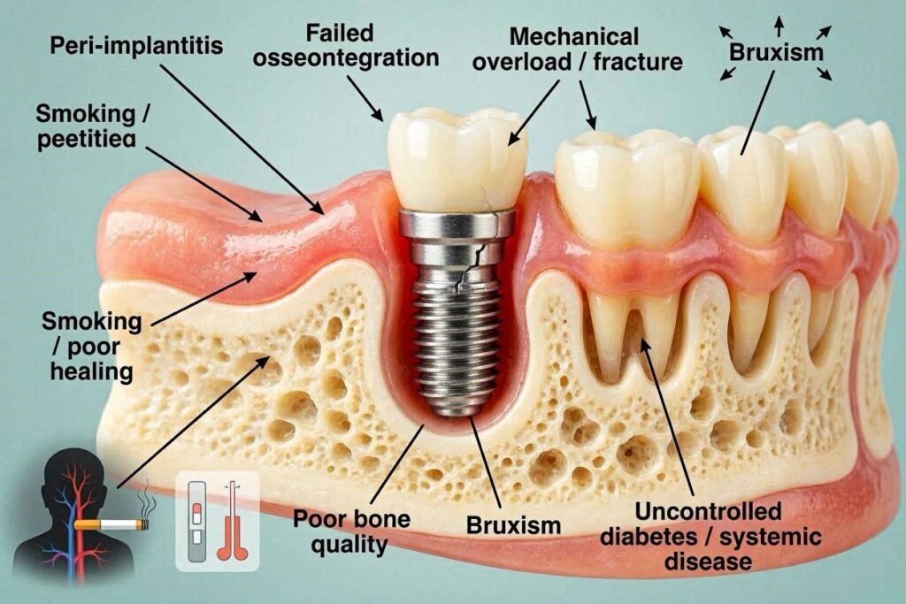
A late failure occurs after the implant has successfully osseointegrated and has been restored with a crown and loaded with chewing forces. It is a failure of the established bone-implant interface. The two primary drivers of late failure are peri-implantitis, a bacterial infection that destroys the bone, and mechanical overload, where excessive forces fracture the implant or cause the bone to fatigue and resorb. Late failure can occur years or decades after the implant was placed.
The Deep Dive: The Causes of Early Failure
The seeds of early failure are sown during the surgery or in the immediate healing phase.
Failure of Primary Stability
When the surgeon places the implant, they measure the insertion torque. A value of 35-50 Ncm is the target. If the bone is too soft (type D4 in the posterior maxilla), the implant may not achieve a rigid mechanical grip. A loose implant at placement will not osseointegrate. Micro-movement in the first weeks of healing is a death sentence for an implant. The surgeon’s decision to bury the implant and wait, rather than load it immediately, is a protective measure when primary stability is borderline.
Thermal Necrosis of the Bone
This is the most devastating and entirely preventable cause of early failure. The drilling of the osteotomy generates frictional heat. If the bone temperature exceeds 47 degrees Celsius (116.6 degrees Fahrenheit) for more than one minute, the osteocytes, the living bone cells, die. The implant is placed into a socket of dead bone. The body will then slowly resorb this necrotic bone, and the implant will lose its support. The cause is inadequate irrigation with chilled saline during the drilling sequence, a dull or worn drill, or drilling with excessive pressure. This is a surgical error. A skilled surgeon uses copious, constant irrigation and sharp, new drills, and drills with a light, pumping motion.
Bacterial Contamination at the Surgical Site
The implant must be placed into a sterile site. If bacteria are introduced during the surgery—from a breach in sterile technique, from the saliva of the patient, or from an adjacent infected tooth socket—the bacteria can colonize the implant surface before the bone cells can. This results in a low-grade, smoldering infection that prevents osseointegration. Pre-operative chlorhexidine rinsing and strict sterile surgical protocol are the defenses.
Over-Compression of the Bone
If the surgeon uses an implant that is too large for the osteotomy, or compresses the bone too aggressively during placement, the excessive pressure can crush the delicate trabecular bone in the threads, causing localized bone necrosis. This is micro-fracture and cell death at the implant interface.
Systemic and Lifestyle Patient Factors
- Smoking: Nicotine is a potent vasoconstrictor. It strangles the capillary blood flow to the oral tissues, reducing the oxygen tension and the delivery of immune cells and healing factors. Smokers have a documented early implant failure rate 2 to 3 times higher than non-smokers.
- Uncontrolled Diabetes: Elevated blood glucose impairs wound healing, reduces white blood cell function, and alters collagen metabolism. Poorly controlled diabetes is a significant risk factor for early failure.
- Medications: Bisphosphonates (used for osteoporosis) and other anti-resorptive drugs can severely impair bone turnover and healing. Patients on these drugs are at risk of medication-related osteonecrosis of the jaw, a catastrophic, non-healing bone exposure.
- Radiation Therapy: Patients who have received high-dose radiation to the jaws have a permanently compromised blood supply. Implants placed in irradiated bone have a significantly higher failure rate.
The Deep Dive: The Causes of Late Failure
Once the implant is successfully functioning, a new set of long-term threats emerges.
Peri-Implantitis: The Chronic Bacterial Infection
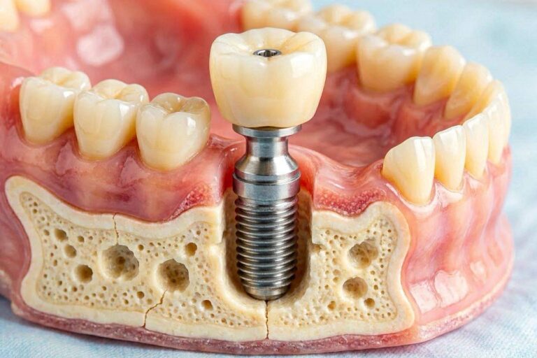
This is the leading cause of late implant failure. It is the analog of periodontitis around a natural tooth, but it is more aggressive. The bacterial biofilm builds up on the implant abutment surface, particularly if a patient’s home care is inadequate. The bacteria trigger an inflammatory host response in the peri-implant mucosa. The inflammation, if unchecked, migrates down the implant surface. The bone begins to resorb. A crater-shaped defect forms around the implant.
The danger is that an implant lacks the defensive periodontal ligament of a natural tooth. The blood supply is limited, and the inflammatory infiltrate can spread directly and unimpeded. The infection often progresses painlessly. The patient may notice bleeding, pus, a bad taste, or gum recession, but often the bone loss is a silent radiographic finding. Advanced peri-implantitis results in a deep, bleeding pocket, a mobile implant, and a radiographic saucer of bone loss. Treatment is surgical and difficult. Prevention through daily, meticulous cleaning of the abutment surface is the only true defense.
Mechanical Overload and Occlusal Trauma
An implant does not have a shock-absorbing periodontal ligament. The forces of chewing are transmitted directly to the bone. If the implant crown is in a hyper-occlusion, meaning it hits harder than the natural teeth, or if the patient is a severe bruxer who grinds and clenches with immense force, the bone around the implant can suffer fatigue micro-fractures and slowly resorb. The implant components themselves can fracture. This is mechanical overload. The failure is not biological but physical. A properly adjusted bite and a custom-fitted night guard are the protections.
Implant Fracture
This is rare but catastrophic. The titanium body of the implant can fracture, usually at the neck where the bending stress is highest. The cause is almost always a combination of metal fatigue over many years, an excessively narrow implant diameter placed in a high-load molar position, and a patient with a heavy bite or bruxism. Once fractured, the implant must be surgically removed.
Abutment Screw Loosening and Fracture
The tiny titanium screw that connects the abutment to the implant can loosen over time. This is the most common mechanical complication. The patient feels a clicking or a slight wobble in the crown. If ignored, a loose screw can fracture, leaving a broken fragment deep in the implant. This is a manageable complication if addressed immediately.
Cement-Induced Peri-Implantitis
If a crown is cemented onto the abutment, excess cement can be squeezed under the gum and left there, invisible and inaccessible. Dental cement is a rough, plaque-retentive foreign body. The gum will inflame around it, and the bone will resorb. This is a purely iatrogenic cause of late implant failure. The dentist’s meticulous technique in cement removal is the critical factor.
The Warning Signs You Must Not Ignore
Early detection is the difference between a salvageable complication and a terminal failure.
- Bleeding on gentle brushing or flossing around the implant.
- Swollen, red, or tender gums around the implant.
- A bad taste or persistent bad breath emanating from the implant area.
- Pus or a small pimple on the gum (a fistula).
- Receding gum line exposing the abutment surface.
- A feeling of looseness or movement in the implant tooth.
- A change in how the implant tooth feels when you bite.
- Radiographic bone loss visible on an X-ray.
Any one of these signs demands an immediate visit to your implant dentist.
Conclusion
Dental implant failure is divided into early failure, occurring before the crown is placed due to a failure of osseointegration from surgical trauma, infection, or systemic factors like smoking and uncontrolled diabetes, and late failure, occurring after years of function, primarily caused by the bacterial bone infection peri-implantitis or by mechanical overload from a poorly adjusted bite or bruxism. The silent, progressive nature of peri-implantitis makes daily biofilm disruption around the abutment, professional maintenance visits, and immediate attention to bleeding or a loose crown the non-negotiable pillars of long-term implant survival.
Frequently Asked Questions
Can a failed implant be replaced?
Yes, in the vast majority of cases. The failed implant is atraumatically removed, the infected or necrotic tissue is debrided, and the site is grafted with bone. After a healing period of 3 to 6 months, the bone is reassessed, and a new implant can be placed. A replaced implant can heal successfully.
Is implant failure painful?
Early failure is often painless. Late failure from peri-implantitis is typically painless until the infection becomes acute and an abscess forms. Mechanical failure from a loose abutment screw causes a clicking, not pain. The silent nature of implant failure is why regular professional monitoring is essential.
What is the failure rate of dental implants?
The long-term success rate for a single implant in a healthy, non-smoking patient is over 95%. The failure rate is approximately 2-5%. In smokers, the failure rate can rise to 10-15%. In patients with a history of severe periodontitis, the risk of peri-implantitis and late failure is significantly higher.
Can a failing implant be saved?
An implant with early peri-implant mucositis, where the inflammation is confined to the soft tissue with no bone loss, is completely reversible with a professional cleaning and improved hygiene. An implant with early, moderate peri-implantitis can be surgically treated with flap surgery, debridement, and decontamination of the implant surface. A mobile implant with advanced bone loss cannot be saved and must be removed.
Additional Resource:
For further scientific information on peri-implant disease and implant complications, visit the American Academy of Periodontology at www.perio.org.
Disclaimer: This article provides a comprehensive educational overview of dental implant failure. It is not a diagnostic tool. If you suspect a problem with your dental implant, you must seek an immediate clinical examination and radiographic evaluation from your implant surgeon or dentist.