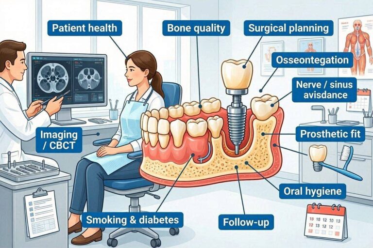
What Is the Working Principle of Dental Implants?
You have probably heard that dental implants work by fusing to the jawbone, but that simple explanation barely scratches the surface of what is arguably the most significant innovation in restorative dentistry. A dental implant does not merely sit in the bone like a screw in wood. It becomes biologically integrated with living tissue in a way that no other medical device achieves. The implant tricks your body into treating titanium as if it belongs there, recruiting the very cells that build your skeleton to construct a custom connection between metal and bone. Understanding how this works transforms the way you think about implant treatment, moving it from a purely mechanical solution to an elegant collaboration between surgical precision and biological intelligence.
The Fundamental Principle: Osseointegration
The working principle of dental implants rests on a single biological phenomenon discovered in the 1950s by a Swedish orthopedic surgeon who was studying blood flow in rabbit bone. His accidental finding would eventually change dentistry forever.
The Discovery That Changed Dentistry
Per-Ingvar Brånemark was conducting research on bone healing at Lund University in Sweden when he inserted titanium optical chambers into rabbit leg bones to observe blood flow. When he attempted to remove these chambers at the conclusion of his experiments, he discovered that the bone had fused to the titanium so completely that the chambers could not be removed without cutting them from the bone. The titanium had not been rejected. It had not been walled off with scar tissue. The bone had accepted it as if it belonged.
Brånemark recognized the potential significance of this finding for medicine and dentistry. He named the phenomenon osseointegration and spent the next decades studying its mechanisms and developing clinical applications. The first dental implant patient received treatment in 1965, and that patient lived with functioning implants until death decades later. The implants were still integrated and functional.
This discovery challenged the prevailing understanding that foreign materials placed in the body would inevitably be encapsulated by fibrous tissue and eventually rejected. Titanium, under the right conditions, provoked a different response entirely. The body did not reject it. The body incorporated it.
The Biological Definition of Implant Success
Osseointegration is formally defined as the direct structural and functional connection between ordered living bone and the surface of a load-bearing implant. Every word in this definition matters. The connection is direct, meaning no intervening soft tissue layer exists between bone and implant. It is structural, meaning the connection can transmit mechanical forces. It is functional, meaning the connection persists under the loading conditions of normal use.
This connection occurs at the microscopic level. Under an electron microscope, bone is seen in direct contact with the titanium oxide surface. The collagen fibers of the bone matrix approach within nanometers of the oxide layer. There is no gap, no fluid space, no fibrous membrane. The bone and the implant are in intimate apposition.
The clinical manifestation of osseointegration is implant stability. An integrated implant cannot be moved. It produces a high-pitched ring when percussed, indicating solid contact with bone. It transmits forces efficiently to the surrounding skeleton. It feels, to the patient, exactly like a natural tooth root buried in bone, because functionally and biologically, it has become one.
How Osseointegration Differs from Other Body-Implant Interactions
Most materials placed in the body do not osseointegrate. Stainless steel screws used in orthopedic fracture fixation develop a fibrous capsule. Silicone breast implants are walled off by scar tissue. Artificial joints made of cobalt-chromium alloys develop a fibrous membrane at the bone-cement or bone-implant interface. This fibrous encapsulation is the body’s default response to foreign materials.
The fibrous capsule serves to isolate the foreign material from the body. It creates a biological barrier that walls off the implant from living tissue. For many medical devices, this encapsulation is acceptable. The device functions mechanically despite the fibrous layer. For dental implants, however, fibrous encapsulation would be failure. The implant would be mobile, unable to support a functional restoration, and vulnerable to bacterial invasion along the fibrous interface.
Titanium’s unique ability to avoid fibrous encapsulation and instead achieve direct bone contact is the foundation of implant dentistry. Without osseointegration, dental implants as we know them would not exist. The entire field depends on this specific biological response to this specific material.
The Implant as an Artificial Tooth Root
A dental implant functions as an artificial tooth root, replacing both the physical presence of the natural root and its mechanical function of transmitting forces to the jawbone. This root replacement principle distinguishes implants from all other forms of tooth replacement.
How Natural Tooth Roots Function
Natural tooth roots serve several essential functions that implants must replicate. The most obvious is anchorage, holding the tooth securely in the jaw so that it can withstand the forces of chewing without being dislodged. A healthy natural tooth can withstand forces of 70 to 150 pounds per square inch during normal function, and higher forces during parafunctional activities.
Natural roots also transmit mechanical forces to the surrounding bone. This force transmission is not simply a passive consequence of anchorage. It is an active biological signal that maintains bone density. When a tooth is loaded during chewing, the force is transmitted through the root to the periodontal ligament and then to the bone. This mechanical stimulation signals the bone cells to maintain bone density. Without this signal, the bone resorbs.
The periodontal ligament, a specialized connective tissue between the root surface and the bone, provides additional functions that implants cannot replicate. It acts as a shock absorber, dampening the forces transmitted to the bone. It provides proprioceptive feedback, the ability to sense the position and loading of the tooth, which allows precise control of chewing forces. It contains cells that can repair root surface damage and remodel the surrounding bone in response to changing forces.
How Implants Replicate Root Functions
The dental implant replicates the anchorage function of the natural root through osseointegration. The implant is rigidly fixed in the bone and resists displacement under functional loading. Unlike the natural root, which is suspended by the periodontal ligament, the implant is in direct contact with the bone. This direct contact provides even greater primary stability than the natural root, which has some physiological mobility.
The implant replicates the force transmission function of the natural root, but in a different way. Forces applied to the implant crown are transmitted directly to the bone through the implant body, without the dampening effect of the periodontal ligament. This direct transmission means that the bone around implants experiences higher peak stresses than the bone around natural teeth. The implant and restoration must be designed to manage these stresses appropriately.
The implant cannot replicate the periodontal ligament functions. There is no shock absorption, no proprioceptive feedback from the ligament, and no ligament-mediated remodeling. The implant patient relies on feedback from the opposing natural teeth, the jaw muscles, and the temporomandibular joint to regulate chewing forces. This lack of proprioceptive feedback is one reason that occlusal management is so critical for implant restorations.
The Role of the Abutment and Crown
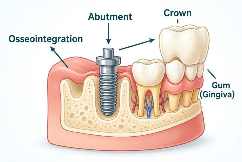
The implant body buried in the bone is only one component of the complete tooth replacement. The abutment and crown are essential for translating the implant’s root function into a visible, functional tooth.
The abutment connects the implant to the crown. It emerges from the implant platform through the gum tissue, providing the structural link between the buried implant and the visible restoration. The abutment may be a separate component screwed into the implant, or it may be integral with the crown in some designs. The abutment transmits forces from the crown to the implant body.
The crown replicates the visible portion of the natural tooth. It is shaped, sized, and colored to match the adjacent natural teeth. It restores the ability to chew food, speak clearly, and smile confidently. The crown is the only component visible in the mouth, but it depends entirely on the implant root for its stability and function.
The complete implant system, from implant body through abutment to crown, functions as a mechanical and biological unit. Forces applied to the crown travel through the abutment to the implant body, then to the surrounding bone. The health of each component depends on the health of the others. A problem at any level affects the entire system.
The Titanium-Bone Interface
The interface between the titanium implant surface and the living bone is where the working principle of implants actually operates. Understanding this interface at the cellular and molecular level reveals the elegance of the biological solution.
The Titanium Oxide Layer
The surface of a titanium implant is not actually titanium metal. It is titanium dioxide, an oxide ceramic that forms spontaneously when titanium is exposed to air or water. This oxide layer, only 3 to 10 nanometers thick, is the surface that the body’s cells and proteins actually encounter.
The oxide layer is chemically stable and electrically insulating. It prevents the underlying titanium metal from reacting with the biological environment. Unlike iron oxide, which flakes away and exposes fresh metal to continued corrosion, titanium oxide is adherent and self-healing. If the oxide layer is scratched, it reforms instantly in the presence of oxygen.
The oxide layer’s surface properties determine the biological response. The surface is negatively charged at physiological pH, which influences protein adsorption. The oxide is hydrophilic, meaning it attracts water, which promotes protein adsorption in the conformations that favor cell attachment. The surface energy of the oxide layer is high, which enhances cell spreading and differentiation.
Protein Adsorption and Cell Attachment
The first event when the implant contacts blood and tissue fluids is protein adsorption. Within milliseconds to seconds, plasma proteins coat the oxide surface. The types of proteins that adsorb, and their three-dimensional conformation on the surface, determine how cells will respond.
Fibronectin, vitronectin, and other cell adhesion proteins adsorb to the titanium dioxide surface in orientations that expose their cell-binding domains. These domains contain the amino acid sequence arginine-glycine-aspartic acid, commonly called the RGD sequence, which is recognized by integrin receptors on the surface of osteoblasts and other cells.
When an osteoblast precursor cell approaches the implant surface, its integrin receptors bind to the RGD sequences on the adsorbed proteins. This binding triggers intracellular signaling cascades that tell the cell it has found a suitable surface for attachment. The cell spreads out on the surface, establishes additional attachments, and begins the process of differentiation into a mature, bone-forming osteoblast.
Without this specific protein adsorption and cell attachment, osseointegration would not occur. The titanium oxide surface has evolved through decades of research to optimize this interaction. Modern implant surfaces are modified through sandblasting, acid etching, anodization, and other treatments to enhance protein adsorption and cell attachment beyond what occurs on a smooth titanium surface.
Bone Formation Directly on the Implant Surface
Once osteoblasts have attached to the implant surface and differentiated, they begin the process of bone formation. They synthesize and secrete type I collagen, the primary organic component of bone matrix. The collagen molecules self-assemble into fibrils that form the scaffold for mineralization.
The osteoblasts then mineralize the collagen matrix by depositing calcium and phosphate ions, which crystallize into hydroxyapatite, the mineral phase of bone. This mineralization occurs directly on the titanium oxide surface. The first bone formed is woven bone, with randomly oriented collagen fibers. Over time, this woven bone is remodeled into lamellar bone with highly organized collagen architecture.
The bone that forms on the implant surface is living tissue, complete with osteocytes embedded in the mineralized matrix and a blood supply delivered through the haversian canal system. This bone is constantly remodeling throughout the implant’s lifetime, responding to mechanical forces and repairing microdamage. The implant has become part of the body’s skeletal system.
Mechanical Force Distribution
The working principle of dental implants involves not only the biological integration but also the mechanical function of force transmission. How the implant manages the forces of chewing determines its long-term survival.
How Chewing Forces Are Managed
When you bite down on an implant crown, the force is transmitted through the crown to the abutment, through the abutment to the implant body, and through the implant body to the surrounding bone. At each interface, the force must be transferred efficiently without creating stress concentrations that could cause failure.
The implant body is designed to distribute forces favorably. The threaded design increases the surface area available for force transfer. The threads convert some of the vertical biting force into compressive forces along the thread faces, which bone tolerates well. The implant diameter and length determine the total bone-implant contact area, which influences the stress distribution.
The bone surrounding the implant responds to the transmitted forces according to Wolff’s law, which states that bone remodels in response to the mechanical demands placed on it. Areas of high stress stimulate bone formation. Areas of low stress experience bone resorption. Over time, the bone architecture adapts to optimize force distribution around the implant.
This adaptive capacity has limits. Excessive forces can exceed the bone’s ability to respond, leading to microfractures, bone resorption, and eventual implant failure. This is why occlusal management, including careful design of the implant restoration and protection against parafunctional forces, is essential for long-term implant success.
The Absence of the Periodontal Ligament
The most significant mechanical difference between implants and natural teeth is the absence of the periodontal ligament. This ligament, approximately 0.2 millimeters wide around natural teeth, provides several functions that implants must manage differently.
The periodontal ligament acts as a viscoelastic shock absorber. When force is applied to a natural tooth, the ligament compresses slightly, distributing the force over time and reducing peak stresses on the bone. The implant, lacking this ligament, transmits forces instantaneously and at higher peak stress to the surrounding bone. The bone around implants must be able to withstand these higher peak stresses.
The periodontal ligament provides proprioceptive feedback through specialized nerve endings that detect tension in the ligament fibers. This feedback allows precise control of chewing forces, with reflex inhibition protecting the teeth from overload. Implant patients lack this feedback and must rely on other sensory inputs, including feedback from remaining natural teeth and muscle proprioception.
The absence of the periodontal ligament has clinical implications. Implant restorations must be designed with lighter occlusal contacts than natural teeth. Patients with implants should avoid using their implant crowns to test the hardness of foods, as they would with natural teeth. Night guards are recommended for patients with bruxism to protect implants from uncontrolled parafunctional forces.
Primary Stability and Its Importance
Primary stability refers to the mechanical engagement of the implant with the bone at the time of surgical placement. It is achieved by preparing the implant site slightly smaller than the implant diameter, so that the implant compresses the bone as it is inserted. Primary stability is the mechanical foundation on which biological integration builds.
High primary stability is essential for successful osseointegration. It prevents micromovement of the implant during the healing period, which would disrupt the delicate cellular processes of bone formation. The threshold for damaging micromovement is approximately 100 to 150 microns. Movement above this threshold directs healing toward fibrous encapsulation rather than osseointegration.
The surgeon measures primary stability at the time of placement using insertion torque, the force required to seat the implant. Values above 35 Newton-centimeters generally indicate adequate primary stability. Resonance frequency analysis provides an additional measure of implant stability that can be tracked over time to monitor the transition from primary mechanical stability to secondary biological stability as osseointegration occurs.
The Healing Sequence That Enables Function
The working principle of dental implants is not instantaneous. It requires a carefully orchestrated healing sequence that transforms the implant from a mechanically retained foreign body into a biologically integrated part of the skeleton.
The Blood Clot Phase
Immediately after implant placement, the space between the implant threads and the prepared bone walls fills with blood. This blood clot is not an incidental occurrence. It is the provisional matrix that orchestrates everything that follows.
Platelets within the clot degranulate, releasing growth factors that initiate the healing cascade. These growth factors include platelet-derived growth factor, which attracts mesenchymal stem cells to the site, and transforming growth factor-beta, which stimulates matrix production. Vascular endothelial growth factor stimulates new blood vessel formation.
The fibrin mesh of the clot provides the scaffold for cell migration. Mesenchymal stem cells from the surrounding bone marrow and periosteum crawl along the fibrin strands toward the implant surface. The clot is gradually replaced by granulation tissue, then by woven bone, as the healing process advances.
The Cellular Cleanup and Recruitment Phase
Within hours of implant placement, inflammatory cells arrive at the surgical site. Neutrophils and macrophages enter the wound, cleaning it of bacteria, debris, and damaged tissue. This inflammatory phase is essential for healing, not a complication to be avoided. The macrophages release additional growth factors that amplify the healing response.
Mesenchymal stem cells, attracted by the growth factor gradients established by platelets and macrophages, migrate toward the implant surface. These cells have the potential to differentiate into several cell types, including osteoblasts, fibroblasts, and chondrocytes. The local environment determines their fate. On the implant surface, under the influence of the adsorbed proteins and the mechanical environment, they differentiate into osteoblasts.
This is the critical decision point for implant success. If the implant is stable and the biological environment is favorable, the stem cells become osteoblasts and begin forming bone on the implant surface. If the implant is mobile or the environment is inflamed, the stem cells may become fibroblasts and form fibrous tissue instead. This is why primary stability and infection control are so critical during the early healing period.
Bone Remodeling and Long-Term Maintenance
The woven bone that initially forms on the implant surface is temporary. Over weeks to months, it is remodeled into mature lamellar bone through the coupled activities of osteoclasts, which resorb bone, and osteoblasts, which form new bone.
This remodeling process adapts the bone architecture to the mechanical forces transmitted through the implant. Bone is added where forces are highest, along the implant threads and at the implant neck. Bone is removed where forces are lowest. The result is an optimized structure that efficiently transfers forces from the implant to the skeleton.
The remodeling process continues throughout the implant’s lifetime. Approximately 10 percent of the bone surrounding an implant is replaced each year through normal skeletal maintenance. This ongoing remodeling allows the bone to repair microdamage, adapt to changing force patterns, and maintain its mechanical integrity. It also means that the implant-bone interface is a living, dynamic structure, not a static connection.
The Implant-Abutment-Crown System
The working principle of dental implants extends beyond the bone-implant interface to include the mechanical system that delivers the replacement tooth to the oral cavity.
The Sealing Function of the Soft Tissue
The implant body is buried in bone, but it connects to the oral environment through the abutment that penetrates the gum tissue. This penetration creates a potential pathway for bacteria to reach the underlying bone. The soft tissue seal around the implant and abutment is the barrier that prevents this bacterial invasion.
The junctional epithelium, a specialized epithelial tissue, attaches to the abutment surface through hemidesmosomes and a basal lamina, similar to its attachment to natural tooth surfaces. This epithelial attachment forms the most superficial component of the biological seal.
Deep to the junctional epithelium, connective tissue forms a cuff around the abutment. Unlike around natural teeth, where connective tissue fibers insert perpendicularly into the cementum, the fibers around implants run parallel to the abutment surface. This parallel orientation provides a less effective mechanical barrier than the perpendicular insertion around teeth, making the peri-implant soft tissue seal inherently more vulnerable to bacterial penetration.
Maintaining this soft tissue seal through good oral hygiene and regular professional maintenance is essential for long-term implant health. Breakdown of the seal leads to peri-implant mucositis and potentially peri-implantitis, the inflammatory conditions that threaten implant survival.
Force Transmission Through the Restoration
The implant crown receives the forces of chewing and transmits them through the abutment to the implant body. Each interface in this chain must be secure and stable to prevent mechanical complications.
The abutment-implant connection is a precision interface. The abutment fits into the implant with a tolerance of a few microns, creating a stable connection that resists rotational and lateral forces. The abutment screw that secures the abutment is tightened to a specific torque, typically 25 to 35 Newton-centimeters, creating a clamping force that prevents micromovement.
The crown-abutment connection, whether cemented or screw-retained, must resist functional forces without loosening or fracturing. Cement-retained crowns rely on the cement bond for retention. Screw-retained crowns use a screw through an access hole in the crown to secure it directly to the implant or abutment.
Any loosening or failure at these interfaces compromises the entire restoration. Loose abutment screws cause the crown to move, potentially damaging the implant internal connection and creating a pathway for bacterial invasion. Loose crowns allow food and bacteria to accumulate, causing inflammation and tissue damage. Regular professional maintenance checks the integrity of these connections.
Why Implants Preserve Jawbone
One of the most significant working principles of dental implants is their ability to preserve jawbone, a function that no other tooth replacement option provides. This bone preservation is a direct consequence of the force transmission through the implant.
The Mechanism of Bone Maintenance
Bone is a dynamic tissue that requires mechanical stimulation to maintain its density. Osteocytes, the cells embedded within the bone matrix, act as mechanosensors, detecting the strains produced when force is applied to the bone. When strains are adequate, the osteocytes signal that bone maintenance should continue. When strains are inadequate, the osteocytes signal for bone resorption.
When a tooth is extracted, the mechanical stimulation that the tooth root provided to the surrounding bone is lost. The osteocytes in that bone detect the reduced strain and signal for increased osteoclast activity. Bone resorption begins within weeks of extraction and continues indefinitely. The alveolar ridge, the specific bone that supported the teeth, resorbs both vertically and horizontally.
An implant replaces the mechanical stimulation that was lost with the natural tooth. Forces applied to the implant crown are transmitted through the implant body to the surrounding bone, creating the strains that osteocytes require to signal bone maintenance. The bone around a functioning implant remains dense and stable, while the bone in edentulous areas without implants continues to resorb.
This bone preservation has significant clinical implications. Implants maintain facial contours that collapse when teeth are lost. They preserve the bone needed for future implant placement if the original implant should ever fail. They prevent the progressive bone loss that makes denture wearing increasingly difficult over time.
The Consequences of Missing Teeth Without Implants
When teeth are lost and not replaced with implants, the resulting bone loss creates a cascade of problems. The alveolar ridge narrows and shortens, reducing the bone available for any form of tooth replacement. Adjacent teeth lose some of their supporting bone as the ridge resorbs. The loss of vertical bone height brings critical anatomical structures, including the inferior alveolar nerve and the maxillary sinus, closer to the ridge surface.
Facial appearance changes as the bone supporting the lips and cheeks resorbs. The face develops a collapsed, aged appearance. The distance between the nose and chin decreases as vertical bone height is lost. These changes are particularly dramatic when all teeth are missing, but they occur to some degree with any tooth loss.
Denture retention becomes progressively more difficult as the ridge resorbs. The denture base, which relies on ridge contour for stability, loses its foundation. The denture rocks, shifts, and causes sore spots. Implants provide the only means of preserving the bone needed for stable, comfortable dentures.
Conclusion
The working principle of dental implants is osseointegration, the direct structural and functional connection between living bone and the titanium implant surface, discovered accidentally by Per-Ingvar Brånemark and refined over decades into the most successful tooth replacement technology available. The titanium oxide layer that forms spontaneously on the implant surface adsorbs specific proteins that trigger osteoblast attachment and differentiation, leading to bone formation directly on the implant surface without the fibrous encapsulation that occurs with most other materials. The implant functions as an artificial tooth root, transmitting chewing forces to the surrounding bone and providing the mechanical stimulation necessary to maintain bone density, preventing the progressive resorption that follows tooth loss. The complete implant system, from the bone-integrated implant body through the soft tissue-penetrating abutment to the visible crown, replicates the functions of the natural tooth while requiring meticulous surgical and restorative technique, adequate healing time, and ongoing maintenance to achieve the decades of service that well-executed implant treatment can provide.
Frequently Asked Questions
How does a dental implant stay in place?
A dental implant stays in place through osseointegration, the direct fusion of living bone to the titanium implant surface. Bone cells attach to the titanium oxide layer and deposit mineralized bone matrix directly onto the implant. This creates a connection so secure that the implant cannot move and can withstand normal chewing forces.
Is the implant as strong as a natural tooth?
An integrated dental implant can withstand forces comparable to or exceeding those of a natural tooth. The implant is made of titanium or zirconia, materials with high strength. The limiting factor is typically the supporting bone rather than the implant itself. Properly placed and restored implants function successfully for decades under normal chewing forces.
Why can’t the body reject a titanium implant?
The body does not reject titanium because the titanium oxide surface is biologically inert and does not trigger the immune response that causes rejection of transplanted organs. The immune system does not recognize titanium as foreign in the way it recognizes transplanted tissue. Instead of attacking the implant, the body builds bone directly on its surface.
How does an implant feel different from a natural tooth?
An implant lacks the periodontal ligament that provides natural teeth with slight mobility and sensory feedback. The implant feels more rigid, with no give when pressed. You cannot feel pressure on the implant itself the way you can feel pressure on a natural tooth. Sensation comes from the surrounding gum tissue and the opposing teeth.
Can an implant set off metal detectors?
Dental implants are small and made of titanium, which is non-magnetic. They typically do not set off metal detectors at airports or security checkpoints. The amount of metal is too small and the material too non-reactive to trigger most detection systems.
How does the implant connect to the crown?
The crown connects to the implant through an abutment, a connector that screws into the implant body and extends through the gum. The crown is either cemented onto the abutment or secured with a screw through an access hole in the crown. This connection transfers chewing forces from the crown to the implant to the bone.
Do dental implants conduct electricity?
Titanium implants can conduct electricity, and the titanium oxide surface layer is a semiconductor. This conductivity is why patients with implants should avoid using electronic oral devices that pass current through the mouth unless specifically designed for use with implants. Normal oral function does not generate electrical currents of concern.
How do upper jaw implants avoid the sinuses?
When the sinus floor is too low for standard implant placement, a sinus lift procedure raises the sinus membrane and places bone graft material beneath it, creating additional bone height. Alternatively, short implants may be used, or angled implants may be placed to engage bone adjacent to the sinus. Preoperative CBCT imaging maps the sinus position for precise planning.
Additional Resource
Academy of Osseointegration: Understanding Dental Implants
https://osseo.org/patient-resources/