What Is A Bone Graft In Dental Work?
A bone graft in dental work is a surgical procedure that places natural or synthetic bone material into a jawbone defect. The goal is to stimulate the body’s own bone cells to grow into the graft site, replacing the graft material over time with living, vascularized bone. This regenerated bone then provides the necessary volume and density to support a dental implant, stabilize a loose tooth, or restore the anatomical contour of the jaw. A dental bone graft is foundational regenerative surgery; it rebuilds the biological scaffold that disease or tooth loss has destroyed.
The Biological Rationale: Why the Jawbone Resorbs
To understand why a bone graft is necessary, you must understand the fate of the jawbone after a tooth is lost. Natural teeth transmit the forces of chewing through their roots into the surrounding alveolar bone. This mechanical loading stimulates the bone to constantly remodel and maintain its density. When a tooth is extracted, this physiological stimulus disappears. The body interprets the unloaded bone as metabolically unnecessary and begins to resorb it.
The resorption process is predictable and relentless. The most dramatic bone loss occurs in the first year after extraction, where the width of the ridge can decrease by up to 50%. The outer, buccal plate of bone, which is thinner and receives less blood supply, resorbs much faster than the inner, lingual or palatal plate. The bone also resorbs in height. Over time, an edentulous jaw can become so atrophic that it cannot house an implant, and a conventional denture loses its supporting ridge and becomes loose and uncomfortable. A bone graft at the time of extraction, a procedure called ridge preservation, is a preemptive intervention to minimize this inevitable resorption.
The Grafting Materials: A Spectrum of Sources
The term “bone graft” covers a range of materials with different origins and biological properties. The choice of material depends on the size of the defect, the surgeon’s preference, and the ultimate prosthetic goal. The materials are classified by their source.
Autografts: The Patient’s Own Bone
An autograft is bone harvested from the patient’s own body and transplanted to the defect site. It is the biological gold standard because it is the only graft material that is osteogenic, meaning it contains living bone cells that can directly form new bone. It also has osteoinductive properties, with natural growth factors that recruit stem cells to the area, and it provides a purely biocompatible scaffold.
The cost of this superiority is a second surgical site. For a dental bone graft, the most common donor site is the mandibular symphysis (the chin area) or the external oblique ridge at the back of the lower jaw. For very large defects, the iliac crest of the hip or the tibial plateau may be used. The donor site surgery adds post-operative pain and morbidity. For this reason, autografts are usually reserved for significant reconstructive cases where their unparalleled regenerative capacity is essential.
Allografts: Donor Human Bone
An allograft is human bone harvested from carefully screened deceased donors and processed by a tissue bank. The processing removes all living cells to eliminate any immune rejection risk, leaving behind the mineralized collagen matrix of the bone. This processed allograft is osteoconductive, meaning it acts as a perfect, biocompatible scaffold into which the patient’s own bone cells can migrate and slowly remodel. Allograft is the most widely used graft material in routine dental implant site development. It eliminates the need for a second donor site surgery entirely.
Xenografts: Animal-Derived Bone Mineral
A xenograft is bone mineral sourced from a different species, most commonly bovine or porcine. Bovine bone, derived from cows, is processed to completely remove all organic protein, leaving behind a pure, crystalline hydroxyapatite scaffold that is structurally almost identical to human bone mineral. This material is very slowly resorbed, and some particles may remain for years, embedded in the newly formed bone. This slow resorption is actually a clinical advantage for preserving ridge volume long-term, making xenografts excellent for ridge preservation and large sinus lift procedures.
Alloplasts: Synthetic Bone Substitutes
An alloplast is a laboratory-synthesized, completely synthetic grafting material. These include bioactive glasses, calcium phosphate ceramics like hydroxyapatite and beta-tricalcium phosphate, and calcium sulfate. Alloplasts carry zero risk of disease transmission and have an unlimited supply. Their resorption rate can be engineered by the manufacturer. Beta-tricalcium phosphate, for example, resorbs relatively quickly and is replaced by new bone. Synthetic materials are popular for smaller, well-contained defects and are a reliable, safe option when the surgeon wants to avoid human or animal-derived materials.
The Major Clinical Applications
Bone grafting in dentistry is not a single procedure. It is a suite of techniques applied to different clinical scenarios, each with a specific surgical objective and prosthetic timeline.
Socket Preservation (Ridge Preservation)
This is the most common and most preventive bone grafting procedure. Immediately after a tooth is extracted, the empty socket is packed with a particulate graft material, often an allograft or xenograft. The graft is contained by a resorbable collagen membrane placed over the socket opening, which is then sutured closed. The graft material occupies the space and prevents the buccal and lingual walls of the socket from collapsing inward as the blood clot organizes. This simple, low-morbidity procedure preserves the width and height of the ridge, creating a much more favorable implant site four to six months later.
Sinus Lift (Sinus Augmentation)
The maxillary sinus is an air-filled cavity located above the roots of the upper posterior teeth. After these teeth are lost, the sinus floor often expands downward, leaving only a paper-thin layer of bone between the sinus membrane and the oral cavity. This bone is insufficient to stabilize an implant. A sinus lift procedure gently elevates the Schneiderian membrane lining the sinus floor and packs bone graft material into the newly created space. Over six to nine months, the graft matures into a solid layer of bone, increasing the vertical bone height to safely accommodate a standard-length implant.
Guided Bone Regeneration for Horizontal and Vertical Defects
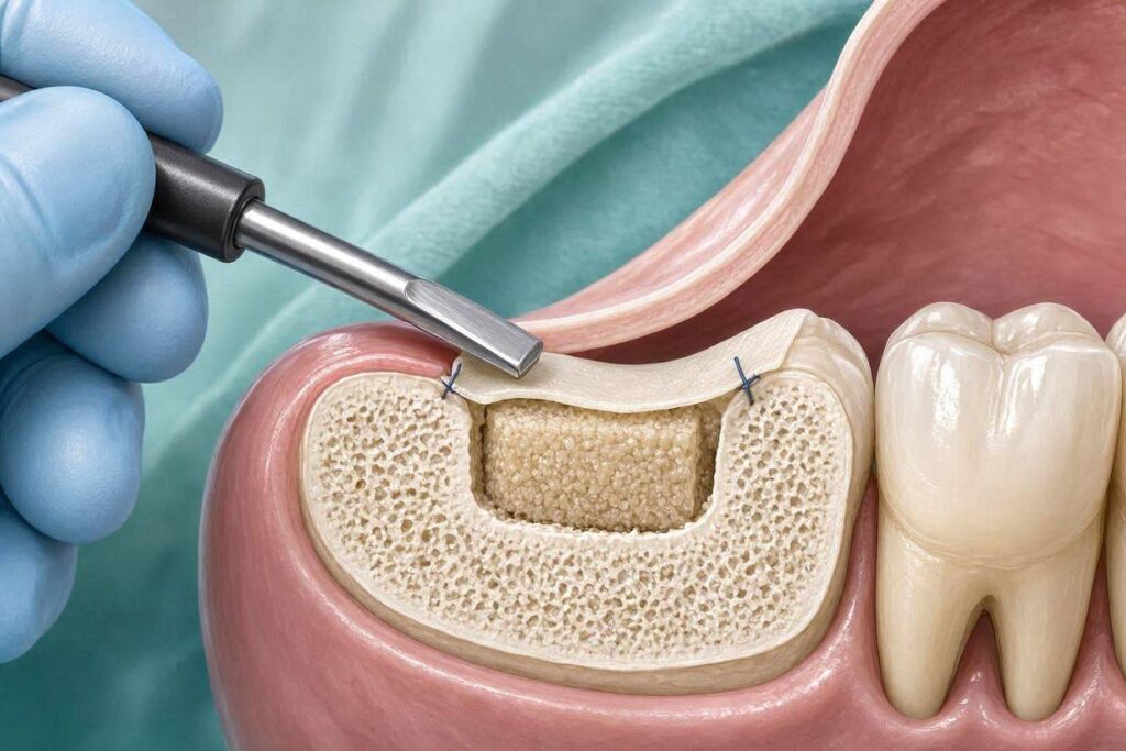
When a ridge defect is too wide or too deep to be treated with simple socket grafting, a more complex technique called guided bone regeneration is used. The surgeon places the particulate graft into the defect and then covers it with a barrier membrane. This membrane, which can be resorbable or a non-resorbable titanium-reinforced sheet, serves a critical biological purpose. It blocks the fast-growing soft tissue cells from the gum and cheek from growing into the graft site, while allowing the slower-migrating bone-forming cells to populate the area undisturbed. Titanium tacks or screws secure the membrane. This technique can successfully regenerate bone in even severely atrophic jaws.
Block Bone Grafts
For a large, non-contained horizontal or vertical defect, particulate graft alone may not provide enough structural integrity. A block bone graft, which is a solid piece of cortical and cancellous bone, is secured to the recipient jaw with titanium fixation screws. An autogenous block from the patient’s chin or ramus is the classic material, although processed allograft blocks are also used. After four to six months of healing, the block incorporates into the jaw, creating a solid, dense osseous platform for implant placement.
The Healing and Integration Process
The placement of a bone graft initiates a complex, staged biological cascade. The graft is not a static filler. In the first few days, a blood clot forms within the graft particles, bringing platelets, inflammatory cells, and growth factors. Tiny blood vessels from the surrounding native bone begin to sprout into the graft, a process called angiogenesis. The graft scaffold provides a surface for these new vessels and the circulating mesenchymal stem cells to adhere.
Over the next few months, the stem cells differentiate into osteoblasts, the bone-building cells. These cells lay down new organic osteoid matrix, which then mineralizes into woven bone. Simultaneously, osteoclasts, the bone-resorbing cells, begin to slowly remove the graft material. The rate of graft resorption versus new bone formation depends on the material type. Over six to nine months, the woven bone remodels into mature, lamellar bone, and the graft site becomes a vascularized, living part of the patient’s jaw, ready to receive a dental implant.
Post-Operative Care and Risks
A bone graft is a surgical procedure, and the patient must follow specific post-operative instructions. The primary risk is infection of the graft site. A course of prophylactic oral antibiotics and a chlorhexidine mouth rinse are standard. The patient must avoid disturbing the surgical site with a toothbrush or aggressive rinsing for the first few days. Smoking is an absolute contraindication; the vasoconstrictive and toxic effects of nicotine drastically reduce blood flow and oxygen delivery to the healing graft, leading to a high failure rate.
The most common complication is exposure of the barrier membrane through the gum incision during the first few weeks. A small exposure can sometimes be managed with meticulous hygiene and more frequent chlorhexidine rinses. A large, progressing exposure risks contamination of the graft and partial loss of the grafted bone. The patient must attend all follow-up appointments so the surgeon can monitor the soft tissue healing. Post-operative swelling, bruising, and mild discomfort are expected and managed with ice packs, anti-inflammatory medication, and prescribed analgesics.
Conclusion
A dental bone graft is a regenerative surgical procedure that implants a scaffold material—autograft, allograft, xenograft, or alloplast—into a jawbone defect to stimulate the body’s own bone to rebuild lost volume and density. The procedure ranges from a simple, preventive socket preservation at the time of extraction to complex sinus lifts and guided bone regeneration for severe atrophy. A successful bone graft creates the essential biological foundation that transforms a compromised jaw into a viable site for a stable, long-lasting dental implant, thereby restoring both function and facial structure.
Frequently Asked Questions
Q: Is a dental bone graft painful?
A: The procedure itself is performed under local anesthesia and is painless. Post-operative discomfort is typical for a few days, managed well with prescribed or over-the-counter pain medication and ice. The level of discomfort correlates with the size of the graft; a socket preservation is much easier than a block bone graft.
Q: Can my body reject the bone graft?
A: True immunological rejection, like an organ transplant rejection, does not occur with processed allograft, xenograft, or alloplast materials because the cells and immunogenic proteins have been removed. An autograft is from your own body and is perfectly compatible. Graft failure usually results from infection, smoking, or poor blood supply, not rejection.
Q: How long after a bone graft can I get my implant?
A: For a simple socket preservation graft, the healing period is typically three to four months. For a sinus lift or a larger guided bone regeneration procedure, the graft usually needs six to nine months to mature and fully integrate before implant placement is safe.
Additional Resource
For comprehensive patient education on bone grafting and dental implant preparatory procedures, this specialty organization is a definitive, science-based resource.
- American Academy of Periodontology Patient Resources: perio.org/for-patients