Will Dental Implants Stop Bone Loss?
A patient researching tooth replacement encounters a compelling claim: dental implants stop bone loss. The statement sounds definitive, almost miraculous. But is it accurate? Does the placement of a titanium fixture in the jaw truly halt the relentless process of bone resorption that follows tooth loss?
The answer is nuanced. Dental implants significantly reduce and, in many cases, effectively arrest the bone loss that occurs after tooth extraction. They do this by replacing the mechanical stimulus that natural teeth provide. However, the protection is not absolute, is localized to the bone immediately surrounding the implant, and depends on numerous factors including surgical technique, prosthetic design, patient health, and maintenance.
This guide provides a thorough, evidence-based exploration of how implants affect jawbone, the mechanisms of bone preservation, the limitations of that preservation, and the clinical factors that determine long-term bone stability.
The Biology of Bone Loss After Tooth Extraction
To understand how implants stop bone loss, one must first understand why bone disappears when a tooth is removed.
The Alveolar Bone: A Tooth-Dependent Tissue
The alveolar bone is the portion of the jaw that surrounds and supports the teeth. It is a unique tissue. Unlike the basal bone of the mandible and maxilla, which exists independently of the teeth, the alveolar bone forms in response to tooth eruption and is maintained by the presence of teeth. The periodontal ligament, which connects the tooth root to the alveolar bone, transmits mechanical forces generated during chewing. These forces create strain within the bone. Osteocytes, the mechanosensory cells embedded in the bone matrix, detect this strain and orchestrate a balance of bone formation and resorption that maintains bone density.
The Resorption Cascade Following Extraction
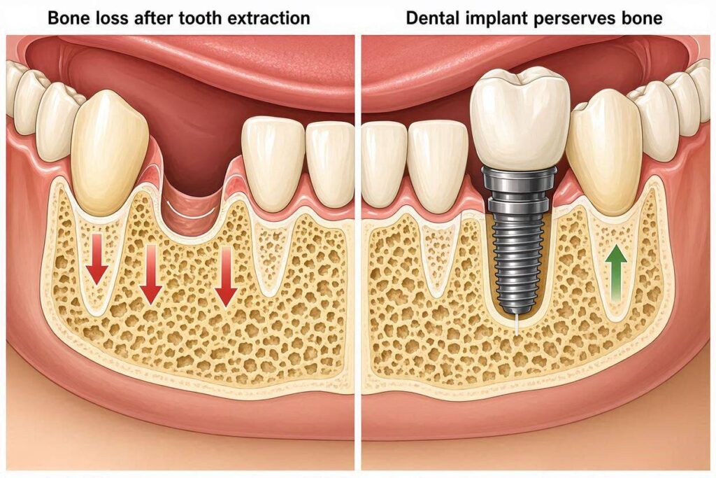
When a tooth is extracted, the periodontal ligament is lost along with the tooth. The mechanical stimulus disappears. The osteocytes no longer detect the chewing forces in that region. The balance shifts dramatically toward resorption. Osteoclasts, the bone-resorbing cells, become dominant. The alveolar bone, no longer needed by the body, is removed.
This resorption follows a predictable pattern. The buccal bone plate, which is thinner and receives less blood supply than the lingual or palatal plate, resorbs more rapidly and extensively. Within the first year, an extraction site can lose 40 to 60 percent of its buccolingual width. Height loss also occurs, though to a lesser degree. The process is most rapid in the first three to six months but continues at a slower, persistent rate throughout life.
The Cumulative Effect
In a fully edentulous patient, this resorption continues for decades. The mandibular ridge, which once supported teeth, flattens. The mental nerve, originally positioned mid-body, becomes superficial and can be compressed by a denture, causing pain. The maxillary ridge resorbs superiorly and palatally, reducing the space available for a denture and altering the support for the nose and upper lip.
The Mechanism of Implant-Mediated Bone Preservation
Dental implants interrupt this resorption cascade by restoring mechanical loading.
Direct Load Transfer Through Osseointegration
A dental implant is a titanium fixture that achieves osseointegration, a direct structural and functional connection between living bone and the implant surface. There is no periodontal ligament. The interface is rigid. When the patient chews, the forces travel from the crown, through the abutment, through the implant fixture, and directly into the surrounding bone at the implant-bone interface.
This load transfer creates strain in the bone surrounding the implant. The osteocytes detect this strain. The signal that bone is needed is restored. The osteoclast-osteoblast balance shifts back toward maintenance and formation. Bone is preserved.
The “Bone Stimulator” Analogy
Think of an implant as a bone stimulator. It does not create bone where none exists. It does not regenerate bone that was lost before placement. It preserves the bone that is present at the time of placement by providing the mechanical signal that the body requires to maintain bone. Without an implant, the bone receives no signal and is resorbed. With an implant, the bone receives functional loading signals and is maintained.
What the Evidence Shows: Implants and Bone Preservation
Decades of clinical research support the bone-preserving effect of implants.
Radiographic Studies of Crestal Bone Levels
Long-term studies measuring bone levels on standardized periapical radiographs show that after an initial remodeling period, bone levels around healthy implants remain remarkably stable. The typical pattern is 0.5 to 1.5 millimeters of crestal bone loss in the first year after loading, followed by annual bone loss of less than 0.2 millimeters per year, a rate that is clinically negligible and consistent with long-term stability.
Comparison to Edentulous Ridges Without Implants
Studies comparing bone levels in edentulous areas with and without implants demonstrate dramatic differences. The edentulous ridge without an implant continues to resorb. The ridge with an implant maintains its height and width, provided the implant remains healthy.
The Implant Overdenture Evidence
The two-implant overdenture in the mandible is one of the most studied implant treatments. Research shows that placing two implants in the anterior mandible preserves bone in that region. However, the posterior mandible, which does not have implants, continues to resorb. This demonstrates the localized nature of implant bone preservation.
The Limitations: What Implants Do Not Stop
The bone-preserving effect of implants has boundaries.
Bone Loss Beyond the Immediate Implant Vicinity
An implant preserves bone within its immediate zone of influence, typically a few millimeters around the fixture. Bone elsewhere in the jaw, in areas without implants, continues to resorb according to the natural history of edentulism. A single implant replacing a molar will preserve the bone around that implant. The bone in an edentulous area several teeth away will continue to resorb if it has no implant.
Bone Loss Due to Peri-Implantitis
An implant that develops peri-implantitis loses bone pathologically. The inflammatory process drives bone resorption that can exceed the rate of resorption in an edentulous ridge without an implant. An implant does not stop bone loss if it is diseased. In fact, a failing implant can accelerate local bone loss.
Initial Crestal Bone Remodeling
Some bone loss around the implant neck is physiological and expected. This remodeling establishes the biological width, the soft tissue and bone architecture that seals the oral environment from the underlying bone. This initial loss of 0.5 to 1.5 millimeters is not a failure of the implant to preserve bone. It is a normal adaptation.
Bone That Was Already Lost Before Implant Placement
The implant preserves bone from the time of placement forward. It does not regenerate bone that was lost in the months or years between tooth extraction and implant placement. If a patient waits five years after extraction to receive an implant, the bone loss that occurred during those five years is permanent unless bone grafting is performed.
Factors That Influence the Bone-Preserving Effect
Not all implants preserve bone equally. Several factors modulate the effect.
Implant Position and Angulation
An implant placed too far buccally, with insufficient bone thickness on the facial aspect, is at risk for buccal bone resorption. The thin buccal plate resorbs, exposing threads. An implant placed in the correct prosthetically driven position, with adequate surrounding bone, preserves that bone predictably.
Implant Diameter Relative to the Ridge
An implant that is too wide for the ridge leaves thin buccal and lingual bone walls. These thin walls resorb, leading to bone loss. The implant should be surrounded by at least 1.0 to 1.5 millimeters of bone on all sides.
The Prosthetic Design
The crown or prosthesis on the implant affects bone loading. Overcontoured restorations that impede oral hygiene contribute to inflammation and bone loss. Poorly designed occlusion that creates off-axis loading causes excessive stress on the bone and can lead to microfracture and resorption. A well-designed prosthesis with cleansable contours and a balanced occlusion supports bone health.
Patient Maintenance and Hygiene
The patient who maintains meticulous oral hygiene and attends regular professional cleanings preserves bone. The patient who neglects hygiene develops peri-implant inflammation and bone loss. The implant itself is not the sole determinant of bone stability. The host response to bacterial biofilm is equally important.
Systemic Health Factors
Well-controlled diabetes, non-smoking status, and good overall health support bone stability. Uncontrolled diabetes, heavy smoking, and systemic conditions that impair healing and immune response undermine the bone-preserving effect.
Table: Bone Preservation by Tooth Replacement Method
| Replacement Method | Bone Preservation Effect | Mechanism |
|---|---|---|
| No replacement | None; progressive resorption | No loading stimulus |
| Removable partial denture | Minimal; some surface loading | Clasps and rests transmit forces to ridge surface |
| Complete denture | None; may accelerate resorption | Pressure on ridge surface stimulates resorption |
| Fixed bridge (tooth-supported) | None at edentulous site | No loading of edentulous ridge; pontic rests on gum |
| Dental implant | Significant; localized to implant site | Direct load transfer to bone through osseointegration |
| Multiple implants with fixed prosthesis | Significant across implant sites | Distributed loading preserves bone at each implant |
The Clinical Implication: Timing Matters
The timing of implant placement relative to tooth extraction influences bone preservation.
Immediate Implant Placement
Placing the implant immediately into the extraction socket is the most effective strategy for preserving bone. The implant occupies the socket, supports the buccal plate, and provides immediate loading stimulus. However, immediate placement is not always possible. Active infection, inadequate bone beyond the socket apex, or inability to achieve primary stability precludes immediate placement.
Early Implant Placement
Placing the implant four to eight weeks after extraction allows soft tissue healing while minimizing bone resorption. The amount of bone lost during this short window is limited. Early placement is a reasonable compromise when immediate placement is contraindicated.
Delayed Implant Placement
Waiting three to six months or longer allows significant bone resorption to occur. The implant is placed into a healed ridge that is narrower and shorter than the original socket. Bone grafting is often required. The implant will preserve the bone that remains from that point forward, but the bone lost during the waiting period is gone.
The Use of Socket Preservation Grafting
When immediate implant placement is not possible, placing a bone graft into the extraction socket, a procedure called socket preservation or ridge preservation, reduces the bone loss that occurs during the healing period. The graft material acts as a scaffold, slowing resorption and maintaining ridge dimensions for future implant placement.
Bone Preservation in Full-Arch Implant Treatment
Full-arch treatment presents special considerations.
The All-on-Four Concept and Bone Preservation
Four to six implants placed in an edentulous arch preserve bone at those implant sites. The remaining edentulous ridge, particularly in the posterior areas where implants may not be placed, continues to resorb. Over years, this can create a discrepancy between the implant-supported anterior region and the resorbed posterior ridge. This does not affect the function of the prosthesis but can have esthetic consequences if the resorption alters facial support.
Zygomatic Implants and Maxillary Bone
Zygomatic implants anchor in the zygomatic bone rather than the maxillary alveolar bone. They do not preserve maxillary alveolar bone because they bypass it. The maxillary ridge continues to resorb. The prosthesis provides facial support prosthetically, not biologically.
Conclusion
Dental implants effectively stop or dramatically slow the bone loss that naturally follows tooth extraction by restoring the mechanical loading that signals the body to maintain bone. This preservation is localized to the bone immediately surrounding the implant, is dependent on the implant remaining healthy and free of peri-implantitis, and does not reverse bone loss that occurred before implant placement. The protective effect is maximized when the implant is placed soon after extraction, positioned correctly within adequate bone, restored with a well-designed prosthesis, and maintained with diligent oral hygiene and professional care. An implant is the only tooth replacement option that addresses the biological cause of post-extraction bone resorption rather than merely compensating for its consequences.
Frequently Asked Questions
Can dental implants reverse bone loss that has already occurred?
No. Implants preserve existing bone from the time they are placed. They do not regenerate bone that was lost before placement. Bone grafting is required to restore lost bone volume.
How much bone loss is normal after getting an implant?
Some crestal bone remodeling of 0.5 to 1.5 millimeters in the first year after loading is normal and expected. After this initial remodeling, annual bone loss should be less than 0.2 millimeters.
Will I still lose bone in areas of my mouth without implants?
Yes. The bone-preserving effect is localized to the immediate vicinity of the implant. Edentulous areas without implants will continue to resorb over time.
Does the type of implant affect bone preservation?
Standard-diameter, root-form implants with moderately rough surfaces placed with adequate surrounding bone provide the most predictable bone preservation. Mini implants preserve bone at their sites but have less surface area for osseointegration.
Can peri-implantitis cause bone loss despite having an implant?
Yes. Peri-implantitis is an inflammatory condition that causes progressive bone loss around an implant. An implant with untreated peri-implantitis can lose bone more rapidly than an edentulous ridge without an implant.
Additional Resources
American Academy of Periodontology – Dental Implant Information
https://www.perio.org/for-patients
Information on the relationship between dental implants and jawbone health, including the prevention of bone loss and management of peri-implant diseases.