Can You Get A Dental Implant If No Bone?
You sit in the dental chair, and the dentist reviews your X-rays. The news is not what you hoped for. You have been missing teeth for years, and the jawbone in those areas has resorbed. There is not enough bone to place a dental implant. You hear the words “insufficient bone volume” or “severe atrophy,” and you assume that implants are no longer an option for you. You resign yourself to a lifetime of dentures or bridges.
That resignation is premature. The question of whether you can get a dental implant when you have no bone, or very little bone, has an answer that has changed dramatically over the past two decades. Modern dentistry offers multiple techniques to rebuild lost bone, to bypass the areas where bone is missing, and to use alternative implant designs that require less bone. This guide will explain every option available to you, in plain language, so you can have an informed conversation with your dentist or seek a second opinion if necessary.
You should not accept “you have no bone” as the final word without understanding what can be done. The vast majority of patients who have been told they lack sufficient bone can, in fact, receive implants successfully.
Why Bone Disappears After Tooth Loss
To understand the solutions, you must first understand the problem. Bone loss after tooth extraction is a predictable biological process.
The Process of Resorption
The jawbone exists to support teeth. When a tooth is extracted, the body senses that the bone in that area is no longer needed for its primary function. Over time, the bone resorbs, meaning it dissolves and is absorbed back into the body. This process begins immediately after extraction and accelerates during the first year. Without the stimulation provided by a tooth root or an implant, the bone continues to shrink gradually over decades.
The rate of resorption varies by individual and by location in the mouth. The lower back jaw tends to resorb faster and more completely than the upper jaw. The front of the lower jaw resorbs more slowly. Patients who have worn dentures for many years often have severe resorption because the denture rests on the gum and transmits pressure to the bone rather than stimulating it.
The Anatomical Consequences
As the bone resorbs, several things happen. The height of the jawbone decreases, bringing critical structures closer to the surface. In the lower back jaw, the inferior alveolar nerve, which provides sensation to the lower lip and chin, moves closer to the top of the ridge. In the upper back jaw, the maxillary sinus, an air-filled cavity above the molar teeth, expands downward into the space where bone used to be.
The width of the jawbone also decreases. The bone becomes a narrow ridge that cannot accommodate a standard-diameter implant. The overall shape of the jaw changes, affecting facial support and creating the sunken appearance associated with long-term tooth loss.
Bone Grafting: Rebuilding What Was Lost
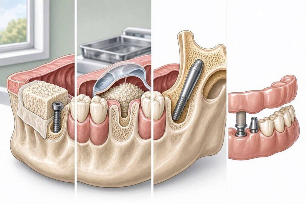
Bone grafting is the most common and well-established solution for insufficient bone volume. It involves placing bone material into the deficient area and allowing the body to remodel it into living bone capable of supporting an implant.
Types of Bone Graft Materials
Several types of graft material are available, each with advantages and disadvantages.
Autogenous bone is harvested from the patient’s own body, typically from the chin, the back of the lower jaw, or the hip for larger grafts. Autogenous bone is the gold standard because it contains living bone cells and growth factors that promote healing. The disadvantage is that it requires a second surgical site, which means additional pain, swelling, and recovery time.
Allograft bone is processed human bone from a tissue bank. It is thoroughly screened and sterilized, and it is safe. Allograft provides a scaffold into which the patient’s own bone cells migrate and eventually replace the graft material. It eliminates the need for a donor site surgery.
Xenograft bone is derived from an animal source, typically bovine bone from cows. Like allograft, it provides a scaffold for the patient’s own bone to grow into. Xenograft material is processed to remove all organic components, leaving a mineral structure that is very similar to human bone. It resorbs very slowly, which can be an advantage in maintaining graft volume over time.
Synthetic graft materials are manufactured from substances like calcium phosphate, hydroxyapatite, or bioactive glass. These materials are entirely synthetic, eliminating any theoretical concern about disease transmission. They are designed to be osteoconductive, meaning they support bone growth into the graft.
The Bone Grafting Procedure
Bone grafting is a surgical procedure typically performed in the dental office under local anesthesia, with or without sedation. The dentist reflects the gum tissue to expose the deficient bone, places the graft material into the defect, and covers it with a barrier membrane to protect it during healing. The gum is then sutured back into place.
The graft must heal and integrate with the surrounding bone before an implant can be placed. This healing period ranges from three to nine months, depending on the size and location of the graft. Larger grafts require longer healing. During this time, the patient wears a temporary replacement to maintain function and aesthetics.
After the graft has healed, new imaging confirms the bone volume, and the implant is placed. The implant then must integrate for an additional period before the crown is placed. The total timeline from grafting to final restoration can be 9 to 18 months.
Success Rates and Limitations
Bone grafting has high success rates when performed by an experienced surgeon on a healthy patient. The graft integrates predictably, and the resulting bone supports implants with success rates similar to those placed in native bone.
The limitations of bone grafting are the extended treatment timeline, the need for additional surgery, the cost, and the fact that some patients are not good candidates for extensive grafting due to medical conditions that impair healing. Patients who smoke, have uncontrolled diabetes, or take certain medications may have reduced graft success rates.
The Sinus Lift Procedure
The maxillary sinus presents a unique challenge for implant placement in the upper back jaw. As teeth are lost and bone resorbs, the sinus expands downward, leaving a thin layer of bone between the sinus membrane and the oral cavity. A sinus lift, also called a sinus augmentation, creates bone in this area.
How a Sinus Lift Works
The surgeon accesses the sinus through a small window created in the side of the maxilla. The sinus membrane is carefully elevated, and graft material is placed into the space created beneath it. The membrane is then allowed to drape back over the graft, and the surgical site is closed.
Over several months, the graft material matures into bone, increasing the height of available bone beneath the sinus. Implants can then be placed into this newly created bone. In some cases, if sufficient bone exists to stabilize the implant, the implant can be placed at the same time as the sinus lift, shortening the overall treatment timeline.
Sinus lifts are routine procedures with high success rates. They are typically performed by oral and maxillofacial surgeons or periodontists with training in the technique. Post-operative discomfort is moderate, and most patients return to normal activities within a few days.
Short and Narrow Implants
Not every case of limited bone requires grafting. In some situations, shorter or narrower implants can be placed in the available bone, avoiding the need for augmentation.
Short Implants
Standard implants are typically 10 to 14 millimeters in length. Short implants, usually defined as those less than 8 millimeters, can be placed in areas of reduced bone height. They are particularly useful in the back of the lower jaw where the inferior alveolar nerve limits implant length, and in the back of the upper jaw where the maxillary sinus limits height.
Short implants were once considered experimental, but research over the past two decades has demonstrated that they can be as successful as standard-length implants when used in appropriate cases. The key is adequate implant width and good bone quality. Short implants in dense bone perform well. Short implants in soft, low-density bone are less predictable.
The advantage of short implants is that they avoid the need for bone grafting, sinus lifts, or nerve transposition. The treatment is faster, less invasive, and less expensive. The disadvantage is that they may not be suitable for all situations, particularly when the bone is not only short but also of poor quality.
Narrow Implants
When the jawbone is too thin to accommodate a standard-diameter implant, which is typically 3.5 to 4.5 millimeters, a narrow implant, sometimes called a mini implant, may be an option. Narrow implants range from 1.8 to 3.3 millimeters in diameter.
Narrow implants can often be placed without grafting in thin ridges. They are commonly used to stabilize lower dentures, where multiple narrow implants can provide adequate retention without the need for bone augmentation. For single-tooth replacement, narrow implants require careful case selection. They are not as strong as standard-diameter implants and are more susceptible to fracture under heavy chewing forces.
Zygomatic Implants: Bypassing the Jawbone Entirely
For patients with severe bone loss in the upper jaw who cannot or do not want to undergo extensive bone grafting, zygomatic implants offer a remarkable alternative. These implants bypass the maxillary bone entirely and anchor into the zygomatic bone, also known as the cheekbone.
What Are Zygomatic Implants?
Zygomatic implants are much longer than standard implants, typically 30 to 50 millimeters. They are placed through the maxillary alveolar bone, through or alongside the maxillary sinus, and into the dense zygomatic bone. The zygomatic bone is not subject to the resorption that affects the maxilla, so it provides a stable, permanent anchor.
Zygomatic implants are typically used for full-arch restorations, not single teeth. One or two zygomatic implants on each side, combined with standard implants in the front of the jaw, can support a full fixed bridge. The procedure, called the zygomatic approach or zygomatic fixture technique, allows patients with severe maxillary atrophy to receive fixed teeth without bone grafting.
The Procedure and Its Demands
Zygomatic implant surgery is technically demanding and should only be performed by an oral and maxillofacial surgeon with specific training and experience in the technique. It is typically performed under general anesthesia. The surgeon must navigate the sinus and precisely place the implant into the zygomatic bone without injuring adjacent structures.
The success rates for zygomatic implants are high in experienced hands, above 95 percent. The recovery is similar to other major oral surgery. The result is a fixed set of teeth for a patient who previously had no options other than a removable denture.
Distraction Osteogenesis
Distraction osteogenesis is a technique borrowed from orthopedic surgery that allows the body to grow new bone in a controlled manner. It is used when a significant amount of bone height is needed and conventional grafting is insufficient.
How Distraction Works
The surgeon cuts the bone and places a distraction device across the cut. After a healing period, the patient or the surgeon activates the device, gradually separating the bone segments. The body responds by filling the gap with new bone. Over days to weeks, the bone is lengthened to the desired height. After a consolidation period, the distraction device is removed, and implants are placed into the newly formed bone.
Distraction osteogenesis is a specialized procedure used for severe vertical bone deficiencies. It is more invasive and time-consuming than grafting but can create bone in situations where grafting alone would fail.
Ridge Expansion and Split Crest Techniques
When the jawbone is wide enough in its deeper portion but too narrow at the top, a ridge expansion or split crest procedure can widen the bone without the need for a separate graft harvesting surgery.
The Split Crest Technique
The surgeon makes a cut along the top of the narrow ridge and gently separates the bone plates, creating a gap. Implants may be placed into the gap immediately, with the gap filled by graft material or left to fill with the patient’s own healing tissue. The technique uses the body’s natural healing response to generate new bone width.
Ridge expansion works well in the upper jaw, where the bone is more pliable. It is less predictable in the dense lower jaw bone. The advantage is that it can often be performed at the same time as implant placement, reducing the overall treatment time compared to a separate grafting procedure.
The All-on-Four and Tilted Implant Concepts
In some cases, the available bone is not where standard implants would typically be placed, but it is present in adjacent areas. The All-on-Four concept and related tilted implant techniques take advantage of this.
Angled Implants to Avoid Critical Structures
By tilting the posterior implants at an angle of 30 to 45 degrees, the surgeon can place longer implants that engage better-quality bone while avoiding the maxillary sinuses or the inferior alveolar nerve. The angled implants also increase the distance between the front and back implants, improving the biomechanical support for a full-arch prosthesis.
This approach often allows a fixed full-arch restoration without bone grafting, even in patients with moderate bone atrophy. The tilted implant technique is a cornerstone of the All-on-Four treatment concept.
Evaluating Your Candidacy
If you have been told you have insufficient bone for implants, the next step is a comprehensive evaluation by a clinician experienced in advanced implant procedures.
The Importance of Cone Beam CT Imaging
A cone beam CT scan is essential. It provides a three-dimensional view of your jaw anatomy, showing exactly how much bone is present, where the sinuses and nerves are located, and the bone density in different areas. The scan allows the clinician to plan implant placement virtually before any surgery is performed.
Two-dimensional panoramic X-rays do not provide enough information for complex cases. If a dentist has told you implants are not possible based only on a panoramic X-ray, seek a second opinion with cone beam CT imaging.
Seeking a Specialist Opinion
If your general dentist has told you that you lack sufficient bone, consider consulting a specialist. Oral and maxillofacial surgeons and periodontists have training in advanced bone grafting and are more likely to be familiar with the full range of options. A prosthodontist can provide a comprehensive treatment plan that coordinates surgical and restorative phases.
A multi-specialty implant center where surgeons and restorative dentists work together offers the advantage of collaborative treatment planning. Each specialist contributes their expertise, and the team can develop a plan that addresses even severe bone deficiencies.
Medical Considerations
Certain medical conditions and medications can affect your ability to undergo bone grafting and implant surgery. Uncontrolled diabetes impairs healing. Smoking dramatically reduces graft and implant success. Bisphosphonate medications and other antiresorptive drugs used for osteoporosis or cancer treatment increase the risk of osteonecrosis, a condition in which the jawbone fails to heal after surgery. Radiation therapy to the head and neck reduces blood supply and healing capacity.
These conditions do not necessarily preclude implant treatment, but they require careful management. Your implant team should communicate with your physician and develop a plan that minimizes risk.
The Realistic Outlook
The vast majority of patients who have been told they lack sufficient bone for dental implants can receive implants successfully. The solution may involve bone grafting, a sinus lift, short or narrow implants, zygomatic implants, or a combination of techniques. The treatment timeline will be longer and the cost higher than for a straightforward implant case, but the outcome, a fixed, functional, and aesthetic replacement for missing teeth, is achievable.
The key is to not accept the first “no” without exploring your options. Get a second opinion from a specialist who regularly handles complex implant cases. Ask specifically about the techniques described in this guide. You may find that the bone you need can be rebuilt, bypassed, or compensated for with an alternative implant approach.
Conclusion
You can get dental implants even if you currently have little or no bone, through bone grafting that rebuilds the jaw, sinus lift procedures that create bone beneath the maxillary sinus, short or narrow implants that work with available bone, zygomatic implants that anchor into the cheekbone, and distraction osteogenesis that grows new bone. The specific approach depends on the location and severity of the bone loss, your overall health, and the expertise of your treatment team. A comprehensive evaluation with cone beam CT imaging and consultation with an experienced implant surgeon or team is the essential first step.
Frequently Asked Questions
How long after bone grafting can I get an implant?
Healing typically requires 3 to 9 months depending on the graft size and location. Smaller grafts heal faster. Larger grafts and sinus lifts require longer. Your dentist will confirm readiness with imaging before scheduling implant placement.
Is bone grafting painful?
The procedure is performed under local anesthesia and should not be painful. Post-operative discomfort is similar to having a tooth extracted and is managed with pain medication and ice. Most patients return to normal activities within a few days.
How much does bone grafting cost?
Bone grafting costs vary by the size and type of graft. Minor grafts may cost a few hundred dollars. Major grafts, including sinus lifts and block grafts, can cost several thousand dollars. The fee includes the graft material, the surgical procedure, and follow-up care.
Can I wear my denture while the graft heals?
In most cases, yes, but the denture may need to be adjusted to avoid putting pressure on the graft site. Your surgeon will provide specific instructions. A soft reline of the denture can cushion the surgical area during healing.
What if bone grafting fails?
Graft failure is uncommon in healthy patients but can occur, particularly in smokers or those with healing impairments. If a graft fails, it can often be repeated. Alternative approaches, such as zygomatic implants, may be considered if repeated grafting is not successful.
Additional Resource:
For information on bone grafting and advanced implant procedures, visit the American Association of Oral and Maxillofacial Surgeons at https://www.aaoms.org.