Can A Dental Implant Become Infected?
You invested in a dental implant to replace a missing tooth. You followed the post-operative instructions. You healed well. The crown was placed, and for a time, everything felt perfect. Then you notice something. The gum around the implant looks redder than the surrounding tissue. It bleeds a little when you brush. There is a dull ache that was not there before. You wonder, can a dental implant get infected? Is that even possible, given that it is made of metal and porcelain? The answer is yes. A dental implant can become infected, and this infection is the most common cause of late implant failure. Understanding how it happens, how to recognize it, and how to treat it is essential for every implant patient.
This comprehensive guide will explain the two main types of implant infections: peri-implant mucositis and peri-implantitis. We will explore the causes, the risk factors, the diagnostic process, and the treatment options ranging from conservative therapy to surgical intervention. By the end of this article, you will know how to protect your implant from infection and what to do if an infection develops.
The Biological Difference: Why Implant Infections Matter
Before we delve into the infections themselves, you must understand why an infection around an implant is more dangerous than an infection around a natural tooth. A natural tooth has a defense system. The periodontal ligament, the connective tissue that attaches the tooth root to the bone, has a rich blood supply and an immune cell network that can fight off bacterial invasion. When bacteria accumulate in the sulcus, the crevice between the tooth and the gum, the body mounts an inflammatory response. Gingivitis develops. If the infection progresses to periodontitis, the bone around the tooth resorbs, but the body continues to fight.
A dental implant has no periodontal ligament. The implant is in direct contact with the bone. The blood supply to the interface between the implant and the bone is limited compared to a natural tooth. When bacteria invade the peri-implant sulcus, the body’s immune response is less effective at walling off the infection. The inflammation spreads more easily along the implant surface. The bone resorbs rapidly, and there are fewer biological barriers to halt the progression. This is why peri-implant infections can progress faster and be more destructive than periodontal disease around natural teeth.
Peri-Implant Mucositis: The Reversible Stage
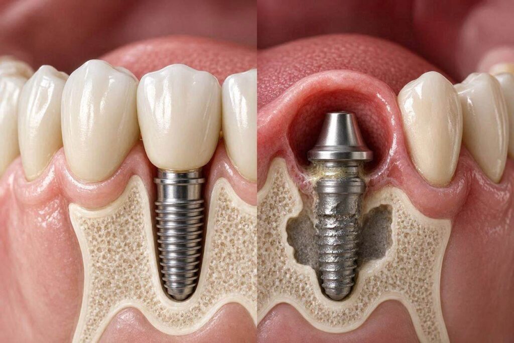
Peri-implant mucositis is the earliest stage of implant infection. It is an inflammation of the soft tissue surrounding the implant, analogous to gingivitis around a natural tooth. The key feature is that the inflammation is confined to the soft tissue. There is no bone loss. The implant remains stable and fully integrated.
The signs of peri-implant mucositis include redness and swelling of the gum margin around the implant, bleeding upon gentle probing or brushing, and possibly a slight tenderness. There is no pus, no deep pocketing beyond normal sulcus depth, and no radiographic evidence of bone loss. The condition is caused by the accumulation of bacterial plaque and biofilm on the implant surface and the abutment. Poor oral hygiene is the primary cause. Excess cement from a cemented crown can also trap bacteria and cause a localized mucositis.
The critical feature of peri-implant mucositis is that it is reversible. If the bacterial biofilm is removed and the patient improves their oral hygiene, the inflammation resolves completely. The bone is not permanently damaged. The implant can return to full health. This is why early detection and intervention are so important. A simple professional cleaning and a renewed commitment to home care can resolve the problem.
Peri-Implantitis: The Destructive Stage
Peri-implantitis is the progression of infection from the soft tissue to the supporting bone. It is characterized by inflammation of the peri-implant mucosa and progressive loss of the bone that supports the implant. The implant is under attack. The bone that holds it in place is being destroyed.
The signs of peri-implantitis include all the signs of mucositis, redness, swelling, and bleeding, plus additional, more ominous findings. There is often increased probing depth around the implant. A normal peri-implant sulcus is 3 to 4 millimeters deep. A probe that sinks 6, 8, or 10 millimeters indicates that the attachment between the gum and the implant has been destroyed, and the probe is traveling down into a deep, infected pocket. Pus, or suppuration, may be expressed from the pocket when pressure is applied. A bad taste or halitosis may be present. The patient may notice a dull ache or a feeling of pressure. Most definitively, a radiograph will show a crater-shaped or saucer-shaped bone defect around the implant. The bone level is lower than it was on previous X-rays or lower than expected.
Peri-implantitis is not reversible in the sense that the lost bone will grow back spontaneously. The goal of treatment is to stop the progression of the infection, arrest the bone loss, and maintain the implant in a healthy, functional state. Some bone regeneration procedures can restore some of the lost bone, but full regeneration is unpredictable.
The Causes of Implant Infections
The primary cause of peri-implant infections is bacterial biofilm. The same bacteria that cause periodontitis around natural teeth, particularly gram-negative anaerobes like Porphyromonas gingivalis, Tannerella forsythia, and Treponema denticola, are implicated in peri-implantitis. These bacteria colonize the rough surfaces of the implant, the abutment, and the crown margin. They form a sticky, protective matrix called biofilm that is resistant to antibiotics and host defenses.
Risk factors that predispose a patient to peri-implant infections include poor oral hygiene, which is the most significant modifiable risk factor. A history of periodontitis is a major risk factor. Patients who lost their natural teeth to gum disease are at higher risk of developing peri-implantitis because they harbor the pathogenic bacteria and have a susceptible immune response. Smoking is a powerful risk factor. Smokers have impaired blood flow, impaired immune function, and higher rates of implant failure and peri-implantitis. Diabetes, particularly uncontrolled diabetes with high blood sugar levels, impairs wound healing and immune function. Excess cement retained in the tissues from a cemented crown is an iatrogenic cause. A loose abutment or a poorly fitting prosthesis can create gaps where bacteria can harbor. Occlusal overload, excessive biting forces on the implant, may not directly cause infection but can contribute to bone loss that creates a niche for bacteria.
Diagnosis: How the Dentist Identifies an Infection
Diagnosing a peri-implant infection involves a systematic clinical and radiographic examination. The dentist will visually inspect the soft tissue for signs of erythema, edema, and alteration in contour. They will gently probe around the implant using a plastic or titanium probe to avoid scratching the implant surface. The probe measures the depth of the sulcus and detects bleeding upon probing, a key sign of inflammation. The dentist will palpate the tissue to see if pus is expressed. They will take a periapical radiograph, a small X-ray of the implant, to assess the bone level around the implant and compare it to previous radiographs. A baseline radiograph taken at the time of crown delivery is essential for this comparison. The dentist will also check the occlusion to ensure the implant is not being overloaded and will examine the integrity of the restoration, checking for loose screws or open margins.
Treatment of Peri-Implant Mucositis
The treatment for peri-implant mucositis is straightforward and highly effective if caught early.
Mechanical Debridement. The dentist or dental hygienist will use instruments to remove the plaque and calculus from the implant surface and the abutment. These instruments are often made of plastic, carbon fiber, or titanium, materials that are softer than the implant surface to avoid scratching it. A scratched implant surface provides more niches for bacteria. Ultrasonic scalers with non-metal tips can also be used.
Oral Hygiene Instruction. The patient must be re-educated on how to clean around the implant. Special tools are required. Interdental brushes with a nylon-coated wire can clean between the implant and the adjacent teeth. Water flossers with a non-metal tip can flush out the sulcus. Super floss, with a stiff end and a fuzzy middle, can be threaded under the prosthesis. The patient must commit to meticulous daily cleaning.
Antimicrobial Rinse. The dentist may prescribe a chlorhexidine gluconate mouth rinse for a short period to reduce the bacterial load. Chlorhexidine is a potent antiseptic but should not be used long-term due to staining and taste alteration.
Re-evaluation. The patient returns in four to six weeks for re-evaluation. The inflammation should be resolved. The bleeding on probing should be absent. If the mucositis has not resolved, a deeper cause, such as retained cement or a loose abutment, must be investigated.
Treatment of Peri-Implantitis
Treating peri-implantitis is more complex and requires surgical intervention in most cases.
Non-Surgical Therapy
For early, mild peri-implantitis with minimal bone loss, non-surgical debridement may be attempted. This involves scaling and root planing of the implant surface, often with the aid of local delivery of antibiotics. Small, biodegradable microspheres containing minocycline or doxycycline can be placed into the pocket after debridement. These release a high concentration of antibiotic locally over several weeks. The goal is to reduce inflammation and stabilize the bone level. Non-surgical therapy can halt the disease progression but rarely regenerates lost bone.
Surgical Therapy
For moderate to severe peri-implantitis, where significant bone loss has occurred, surgical access is required.
Open Flap Debridement. The gum is reflected surgically to expose the infected implant surface. Granulation tissue, the infected, inflamed soft tissue, is removed. The implant surface is meticulously cleaned. Various methods are used to decontaminate the implant surface, including titanium brushes, air powder abrasion with glycine powder, citric acid etching, and laser therapy. The goal is to remove all biofilm and endotoxins from the implant surface without damaging it. The area is then flushed with sterile saline, and the gum is sutured back into place. This procedure arrests the infection but does not replace lost bone.
Resective Surgery. If the bone defect is shallow and wide, the surgeon may reshape the bone, smoothing it and eliminating the defect. This reduces the pocket depth and creates a more cleansable environment. Some of the exposed implant surface may be removed, a procedure called implantoplasty, where the rough surface of the implant threads is smoothed and polished to reduce bacterial adherence.
Regenerative Surgery. If the bone defect is a contained, three-wall or four-wall defect, the surgeon may attempt to regenerate the lost bone. After debridement and decontamination, bone graft material is packed into the defect. A barrier membrane is placed over the graft to protect it and allow the bone cells to populate the graft without interference from fast-growing soft tissue cells. The gum is closed. Over several months, the graft may be replaced by new bone. The success of regeneration around implants is less predictable than around natural teeth, but significant improvement is possible in selected cases.
Implant Removal. If the infection has caused such extensive bone loss that the implant is mobile, or if the infection cannot be controlled, the implant must be removed. This is the definition of implant failure. After removal, the site is debrided, and bone grafting may be performed to rebuild the ridge for a future implant placement.
The Importance of Maintenance and Prevention
The best treatment for peri-implantitis is prevention. Once an implant is placed and restored, the patient enters a lifelong maintenance phase. Professional maintenance visits, typically every three to six months, are essential. During these visits, the dental hygienist cleans the implant, evaluates the tissue health, and reinforces home care techniques. Radiographs are taken periodically, usually annually, to monitor the bone level.
The patient must practice meticulous daily home care. Brushing twice a day with a soft toothbrush, focusing on the gumline around the implant. Interdental cleaning with specialized brushes, floss, or a water flosser. For full-arch fixed prostheses, cleaning under the bridge with super floss or a water flosser is critical. Patients who smoke must understand that smoking dramatically increases their risk of implant infection and failure. Smoking cessation is one of the most impactful actions an implant patient can take.
Conclusion
Dental implants can become infected, and the two forms of infection are peri-implant mucositis, a reversible inflammation of the soft tissue, and peri-implantitis, a destructive process involving bone loss. The primary cause is bacterial biofilm accumulating around the implant, and the major risk factors are poor oral hygiene, a history of gum disease, smoking, and uncontrolled diabetes. Peri-implant mucositis is treatable and reversible with professional cleaning and improved home care. Peri-implantitis requires more aggressive intervention, often surgical, to arrest the disease and save the implant. The key to a healthy implant is prevention through meticulous daily hygiene and regular professional maintenance. An implant is not a “set it and forget it” solution. It requires the same, if not greater, commitment to cleanliness as natural teeth.
Frequently Asked Questions (FAQ)
1. How do I know if my dental implant is infected?
Signs include red, swollen, or bleeding gums around the implant, a bad taste, pus, a dull ache, or a feeling that the implant is loose. If you notice any of these, see your dentist immediately.
2. Can an infected dental implant be saved?
Yes, especially if the infection is caught early. Peri-implant mucositis is fully reversible. Early peri-implantitis can often be stabilized with treatment. Advanced peri-implantitis with severe bone loss may require implant removal.
3. What causes a dental implant to become infected years later?
Late infections are usually caused by poor oral hygiene, the development of a loose abutment or crown, retained cement from the crown delivery, or the progression of the patient’s susceptibility to periodontal disease.
4. Is peri-implantitis painful?
Not always. It is often a silent, painless disease in its early stages, which is why regular dental checkups with X-rays are critical. Pain, swelling, or pus usually appear in more advanced stages.
5. Can antibiotics cure a dental implant infection?
Antibiotics alone cannot cure peri-implantitis because the bacteria are protected within a biofilm. Mechanical removal of the biofilm is essential. Antibiotics may be used as an adjunct to mechanical therapy.
6. How can I clean under my fixed implant bridge?
Use a water flosser with a non-metal tip, super floss, or interdental brushes specifically designed for implant care. Your dentist or hygienist will show you the proper technique.
7. Does a dental implant infection spread to the rest of the body?
A localized peri-implant infection is usually contained. However, the chronic inflammation associated with peri-implantitis may contribute to systemic inflammation. Rarely, an acute abscess can spread to adjacent structures.
Additional Resource
For more information on maintaining oral health with dental implants, visit the American College of Prosthodontists:
American College of Prosthodontists – Dental Implant Care