Why Are My Dental Implants Failing?
Experiencing a failing dental implant after investing time, money, and hope into restoring your smile is deeply distressing. Patients often blame themselves or their dentist, but the reality is that implant failure typically results from a combination of factors, some within the patient’s control and others not. Understanding why implants fail is the first step toward addressing the problem and preventing future failures.
Dental implants fail for several interconnected reasons: peri-implantitis (bacterial infection causing bone loss), failed osseointegration (the implant never bonds to bone), mechanical overload from excessive biting forces, surgical errors in placement or planning, systemic health conditions that impair healing, and lifestyle factors like smoking. The most common cause of late implant failure is peri-implantitis, an inflammatory condition that destroys the bone supporting a previously successful implant.
Identifying Why Your Specific Implant Is Failing
The cause of failure determines what can be done about it. A systematic approach to diagnosis is essential.
When Did the Failure Occur?
The timing of implant failure provides important diagnostic clues.
Early Failure (Within First Year, Before or Shortly After Restoration):
The implant never successfully integrated with bone. The problem likely relates to surgical factors, healing capacity, or early infection. The implant may have been mobile from the beginning or developed problems during the healing period.
Late Failure (After Successful Function):
The implant integrated and functioned well for months or years before problems developed. Peri-implantitis is the most common cause, though mechanical overload also contributes. Progressive bone loss around the implant eventually compromises support.
What Symptoms Are You Experiencing?
Different failure mechanisms produce different warning signs.
Mobility:
An implant that moves is failing or has already failed. Integrated implants are rigidly fixed in bone. Mobility indicates either failed osseointegration (early failure) or such advanced bone loss that all support is gone (late failure).
Bleeding and Swelling:
Gum tissue around a failing implant often bleeds when brushed or probed. Swelling and redness indicate active inflammation. These are hallmark signs of peri-implant disease.
Pus or Drainage:
Infection around an implant produces pus. This is a serious sign requiring immediate professional attention. The infection may be localized to soft tissue or may involve the bone supporting the implant.
Pain or Discomfort:
Healthy implants are comfortable. Pain when chewing or spontaneous discomfort suggests a problem. Pain that worsens over time rather than improving is particularly concerning.
Gum Recession:
Receding gum tissue around an implant may expose the implant surface, which appears metallic or dark. Recession often accompanies underlying bone loss.
Radiographic Bone Loss:
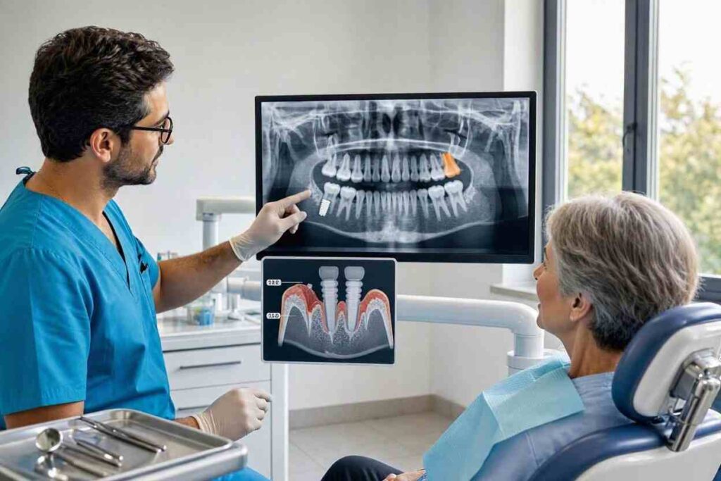
X-rays showing progressive bone loss around an implant confirm peri-implantitis or mechanical overload. Comparison with previous X-rays is essential for determining the rate and pattern of bone loss.
Common Causes of Implant Failure in Detail
Understanding each potential cause helps patients and clinicians develop appropriate treatment strategies.
Peri-Implantitis
This is the leading cause of late implant failure, affecting 10-20% of patients and 5-15% of implants over 10 years.
What It Is:
Peri-implantitis is a bacterially driven inflammatory condition that destroys the bone supporting a functioning implant. It begins as peri-implant mucositis (inflammation confined to soft tissue) and, if untreated, progresses to involve the underlying bone.
Why It Happens:
Plaque accumulation around the implant triggers an inflammatory response. In susceptible patients, this inflammation extends deeper and activates bone-resorbing cells. Unlike natural teeth, implants lack a periodontal ligament with its protective blood supply and defensive cells, potentially allowing more rapid disease progression.
Risk Factors Specific to You:
History of periodontitis (strongest risk factor). Inadequate oral hygiene. Irregular professional maintenance. Smoking. Poorly controlled diabetes. Excess cement from crown cementation. Ill-fitting restorations that trap plaque. Genetic susceptibility to inflammatory disease.
Failed Osseointegration
The implant never successfully bonds to bone. Fibrous scar tissue forms between the implant and bone instead of direct bone contact.
Why It Happens:
Surgical Factors:
Bone overheating during drilling. Implant contamination at placement. Inadequate primary stability. Poor surgical technique.
Patient Factors:
Inadequate bone quality or quantity. Smoking during the healing period. Uncontrolled diabetes. Medications affecting bone healing (bisphosphonates, corticosteroids). Radiation therapy history. Nutritional deficiencies.
Post-Operative Factors:
Premature loading of the implant. Infection at the surgical site. Excessive micromotion during healing.
Mechanical Overload
Excessive forces on the implant cause bone loss or component failure.
Why It Happens:
Bruxism (teeth grinding or clenching) generates forces far exceeding normal chewing loads. These forces concentrate at the implant-bone interface. The bone responds to excessive load by resorbing, similar to how orthodontic forces move teeth.
Contributing Factors:
Inadequate number of implants supporting the restoration. Cantilevered restorations that magnify forces. Poor occlusal design that concentrates rather than distributes loads. Large restorations on minimal implant support. Parafunctional habits like clenching or grinding.
Surgical and Prosthetic Errors
Errors in treatment planning or execution can doom an implant from the start.
Placement Errors:
Implant positioned too close to adjacent tooth roots. Implant placed at an angle that precludes proper restoration. Implant impinging on nerves, sinuses, or other anatomical structures. Inadequate bone surrounding the implant (less than 1-1.5mm on all sides).
Prosthetic Errors:
Ill-fitting restorations that trap plaque. Excess cement left beneath the gum tissue. Over-contoured crowns that prevent effective cleaning. Poor occlusion creating excessive or misdirected forces.
Systemic and Lifestyle Factors
Your overall health and habits significantly influence implant success.
Smoking:
Smoking increases implant failure risk 2-3 times. Nicotine reduces blood flow, impairs healing, and suppresses immune function. Smokers face dramatically higher peri-implantitis risk.
Diabetes:
Poorly controlled diabetes impairs wound healing and increases infection risk. Well-controlled diabetes (HbA1c below 7%) does not significantly increase failure risk.
Medications:
Bisphosphonates and other antiresorptives increase risk of medication-related osteonecrosis. SSRIs have been associated with increased implant failure in some studies. Corticosteroids impair healing and immune function.
Oral Hygiene:
The single most important patient-controlled factor. Inadequate plaque removal leads inevitably to peri-implant inflammation and disease.
Diagnosing the Cause of Your Implant Failure
Determining why your implant is failing requires professional evaluation.
Clinical Examination
Your dentist or specialist will perform several assessments.
Probing around the implant measures pocket depths and detects bleeding. Healthy peri-implant tissues exhibit shallow probing depths without bleeding. Increasing probing depths and bleeding indicate disease.
Mobility testing is critical. Any mobility in an implant that was previously stable indicates severe bone loss or failed osseointegration.
Evaluation of the restoration checks for loose screws, fractured components, poor fit, or occlusal problems that could contribute to failure.
Assessment of oral hygiene and plaque levels around the implant and remaining natural teeth provides insight into the role of home care in the failure.
Radiographic Evaluation
X-rays are essential for evaluating bone levels around the implant. Comparison of current X-rays with previous images reveals the rate and pattern of bone loss.
Cone beam CT provides three-dimensional visualization of bone defects when surgical treatment is planned. The scan reveals the true extent of bone loss, which is often underestimated on two-dimensional X-rays, particularly on the facial and lingual aspects of the implant.
Microbiological Testing
In some cases, bacterial sampling identifies the specific pathogens responsible for peri-implantitis. This information guides antibiotic selection if antimicrobial therapy is planned. Microbiological testing is not routine but may be valuable for refractory or aggressive cases.
Treatment Options Based on Cause of Failure
The appropriate treatment depends entirely on why the implant is failing.
Treating Peri-Implant Mucositis
Peri-implant mucositis is inflammation without bone loss. It is completely reversible.
Treatment involves professional debridement to remove plaque and calculus. Oral hygiene instruction and reinforcement. Correction of plaque-retentive factors. Short-term follow-up to verify resolution. This is the critical window for preventing progression to peri-implantitis.
Treating Early Peri-Implantitis
When bone loss is present but limited, non-surgical treatment may arrest disease progression.
Treatment involves mechanical debridement of the implant surface. Local or systemic antimicrobial therapy. Correction of modifiable risk factors. Frequent professional maintenance.
Surgical Treatment of Advanced Peri-Implantitis
When bone loss is significant or non-surgical treatment has failed, surgical intervention is necessary.
Surgical access and debridement expose the contaminated implant surface for thorough cleaning. Regenerative procedures attempt to rebuild lost bone using graft materials and barrier membranes. Implantoplasty smooths exposed implant surfaces to reduce bacterial adherence. Resective procedures reduce pocket depths by recontouring bone and soft tissue.
Managing Mechanical Complications
Loose screws are retightened or replaced. Fractured abutments are replaced. Fractured implant bodies require removal and replacement. Occlusal adjustment reduces excessive forces.
Removing a Failed Implant
When the implant cannot be saved, removal is necessary. Failed non-integrated implants are relatively easy to remove. Integrated implants require more surgical intervention. After removal and healing, a replacement implant can often be placed successfully.
Preventing Future Implant Failures
Whether you are trying to save a failing implant or planning for replacement, prevention strategies are essential.
Address modifiable risk factors:
Smoking cessation is the single most impactful change a smoking patient can make. Improved diabetes control. Better oral hygiene. Regular professional maintenance.
Treat underlying disease:
Active periodontitis must be treated before implant placement or replacement. Any remaining natural teeth should be periodontally healthy.
Choose providers carefully:
Specialist involvement for complex cases. Proper treatment planning with three-dimensional imaging. Meticulous surgical and restorative technique.
Commit to long-term maintenance:
Professional cleanings every 3-6 months. Annual radiographic evaluation. Prompt attention to any changes or symptoms.
Frequently Asked Questions
Why is my dental implant loose?
A loose implant indicates either failed osseointegration (early failure) or advanced bone loss from peri-implantitis (late failure). Both require immediate professional evaluation. Do not ignore implant mobility.
Can a failing implant be saved?
Often, yes. Early peri-implantitis is treatable. Advanced disease with mobility usually requires removal. Prompt treatment when symptoms first appear dramatically improves salvage potential.
Why is my implant failing if I take good care of it?
Implant failure sometimes occurs despite excellent home care. Possible explanations include undiagnosed occlusal overload, subclinical cement residue, genetic susceptibility, systemic factors, or surgical errors.
Is implant failure my fault?
Usually, multiple factors contribute to implant failure, some within your control and some not. Blame is less important than identifying the cause and addressing it for future treatment.
Why did my implant fail years after it was placed?
Late failure is most commonly caused by peri-implantitis, a progressive bacterial infection that destroys bone around the implant. Mechanical overload and systemic factors also contribute.
Can I get another implant after one fails?
In most cases, yes. The failed implant is removed, the site heals, and a replacement implant is placed after addressing the factors that caused the initial failure.
Will my dentist guarantee the replacement implant?
Policies vary. Some practices offer warranties or reduced fees for replacement implants. Discuss expectations with your provider before proceeding.
How can I prevent my other implants from failing?
Focus on meticulous oral hygiene, regular professional maintenance, smoking cessation, and managing systemic conditions. Early detection and treatment of problems around remaining implants is essential.
Additional Resources
For more information about dental implant failure:
- American Academy of Periodontology: www.perio.org
- American Academy of Implant Dentistry: www.aaid.com
- International Congress of Oral Implantologists: www.icoi.org
- American College of Prosthodontists: www.gotoapro.org