Are Dental Implants Susceptible To Plaque Growth?
The relationship between dental implants and plaque is a critical factor determining long-term implant success. While the materials that compose implants cannot decay like natural teeth, they are far from immune to the consequences of plaque accumulation. Understanding how plaque interacts with implant surfaces helps patients appreciate why meticulous oral hygiene remains essential after implant treatment.
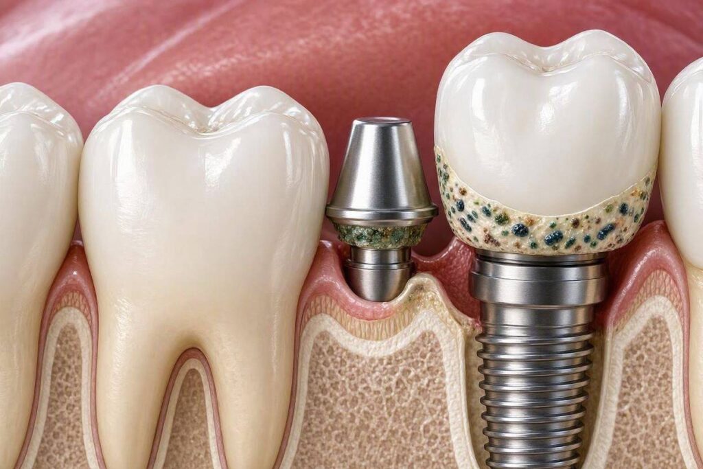
Yes, dental implants are highly susceptible to plaque growth. The same bacterial biofilm that forms on natural teeth readily accumulates on implant surfaces, abutments, and restorations. In fact, some implant surface characteristics may facilitate plaque retention more than natural tooth surfaces. The critical difference is not plaque susceptibility but the consequences: while plaque on natural teeth causes cavities and gum disease, plaque on implants causes peri-implant diseases that can lead to rapid bone loss and implant failure.
Plaque Formation on Implant Surfaces
The process by which plaque develops on implants follows the same biological principles as plaque formation on natural teeth, with some important differences.
The Biofilm Development Process
Plaque is not simply food debris stuck to teeth. It is a complex, organized community of bacteria embedded in a protective matrix, what microbiologists call a biofilm. Biofilm formation follows predictable stages on any surface placed in the oral environment, including dental implants.
Stage 1: Pellicle Formation
Within minutes of cleaning, salivary proteins adsorb onto all oral surfaces, including implant components. This thin protein layer, called the acquired pellicle, provides receptors to which bacteria can attach. The pellicle forms on titanium, zirconia, porcelain, acrylic, and all other materials used in implant restorations.
Stage 2: Initial Bacterial Colonization
Early bacterial colonizers, primarily gram-positive cocci and rods, attach to the pellicle receptors within hours. These pioneer species are generally non-pathogenic and prepare the surface for later colonizers.
Stage 3: Biofilm Maturation
Over days, additional bacterial species join the community. The bacteria multiply and produce an extracellular matrix that protects them from host defenses and antimicrobial agents. The biofilm community becomes increasingly complex and pathogenic.
Stage 4: Mature Biofilm
A mature, stable biofilm community develops over approximately 1-3 weeks if undisturbed. This mature plaque contains the anaerobic gram-negative bacteria most strongly associated with peri-implant diseases.
How Implant Surfaces Differ From Natural Teeth
While the plaque formation process is similar, implant surfaces present unique characteristics that affect plaque behavior.
Surface Roughness:
Modern implant surfaces are intentionally roughened to promote bone integration. This microroughness that benefits osseointegration also provides more surface area for plaque accumulation. Rough surfaces retain more plaque and are more difficult to clean than smooth surfaces.
When bone loss exposes this rough implant surface to the oral environment, plaque accumulation increases dramatically. The exposed rough surface acts as a plaque reservoir that is essentially impossible for patients to clean effectively.
Lack of Epithelial Attachment:
Natural teeth benefit from a specialized epithelial attachment where gum tissue cells adhere directly to the tooth surface via hemidesmosomes. This biological seal resists bacterial penetration. Around implants, the epithelial attachment is less robust, consisting primarily of a cuff of epithelium that contacts the implant surface without the specialized attachment structures. This weaker seal allows easier bacterial access to the underlying tissues.
Absence of Periodontal Ligament:
The periodontal ligament around natural teeth provides a vascular supply that delivers immune cells and tissue fluid that continuously flushes the sulcus. Implants lack this ligament entirely, removing an important host defense mechanism. The implant sulcus is more vulnerable to bacterial colonization and less capable of mounting an effective immune response.
Restoration Contours:
Implant crowns and bridges often have different contours than natural teeth. The emergence profile where the crown meets the implant may create ledges or concave areas that trap plaque. Multi-unit restorations create additional interfaces where plaque can accumulate.
| Characteristic | Natural Tooth | Dental Implant | Impact on Plaque |
|---|---|---|---|
| Surface Roughness (subgingival) | Smooth cementum | Microrough titanium | Rough surface retains more plaque |
| Epithelial Attachment | Strong hemidesmosomal attachment | Weaker epithelial cuff | Reduced barrier to bacterial invasion |
| Connective Tissue Attachment | Perpendicular fiber insertion | Parallel fiber orientation | Weaker seal; easier bacterial access |
| Vascular Supply | Periodontal ligament provides rich blood supply | No ligament; limited vascularity | Reduced immune surveillance |
| Sulcular Fluid Flow | Continuous outward flow flushes bacteria | Reduced or absent flow | Less natural cleansing |
Where Plaque Accumulates on Implants
Identifying the specific sites of plaque accumulation guides effective cleaning strategies.
The Implant-Abutment Junction
The microscopic gap where the implant meets the abutment represents a critical plaque accumulation site. No matter how precisely machined, this junction harbors a space of 1-10 micrometers that bacteria can colonize. Plaque accumulation at this junction triggers inflammation in the surrounding tissues.
Microleakage of bacteria and bacterial products through this junction contributes to peri-implant tissue inflammation. Some implant designs incorporate features to minimize this effect, such as platform switching, where the abutment is narrower than the implant platform, moving the junction inward and away from the bone.
The Transmucosal Surface
The portion of the implant or abutment that passes through the gum tissue is exposed to the oral environment. Smooth, polished surfaces in this zone resist plaque accumulation better than rough surfaces. When rough implant surfaces are exposed through gum recession, plaque accumulation increases significantly.
Crown Margins and Contours
The junction where the implant crown meets the abutment or where the crown margin contacts gum tissue creates plaque traps if not precisely fabricated. Over-contoured crowns that bulge excessively create undercuts where plaque accumulates. Under-contoured crowns may not support the gum tissue adequately, leading to recession that exposes vulnerable surfaces.
Interproximal Spaces
The spaces between implant restorations and adjacent teeth or between multiple implants are particularly susceptible to plaque accumulation. These areas are difficult for patients to access and clean effectively. Floss, interdental brushes, and water flossers must reach these spaces daily.
The Intaglio Surface of Full-Arch Bridges
The underside of a full-arch implant bridge that contacts or rests above the gum tissue accumulates plaque and calculus. This surface is completely inaccessible to toothbrushes and requires specialized cleaning techniques.
Factors That Increase Plaque Accumulation on Implants
Several factors beyond the inherent properties of implant materials affect plaque accumulation.
Patient Factors
Oral Hygiene Quality:
The single most significant factor determining plaque levels on implants is the patient’s daily cleaning effectiveness. Inadequate brushing technique, infrequent cleaning, and failure to use interdental cleaning tools guarantee excessive plaque accumulation.
Salivary Flow:
Saliva contains antimicrobial components and physically washes oral surfaces. Patients with reduced salivary flow (xerostomia) from medications, radiation therapy, or autoimmune disease experience heavier plaque accumulation.
Diet:
Frequent consumption of fermentable carbohydrates promotes plaque growth. Sticky, retentive foods exacerbate plaque accumulation on all oral surfaces, including implants.
Restoration Design Factors
Cement Excess:
Residual cement left beneath the gum line during crown cementation is a notorious cause of plaque accumulation and peri-implantitis. Even microscopic cement fragments can trigger significant inflammation and provide a surface for bacterial colonization.
Emergence Profile:
The contour of the crown as it emerges from the gum tissue significantly affects plaque accumulation. Overly bulbous contours trap plaque; concave profiles may create inaccessible areas. Ideal emergence profiles allow effective cleaning while supporting the gum tissue appropriately.
Prosthesis Design:
Full-arch bridges that are too close to the gum tissue prevent access for cleaning. Bridges with inadequate space beneath them accumulate plaque and calculus that patients cannot remove.
Prosthetic Material Surface Characteristics
Different materials used in implant restorations accumulate plaque at different rates.
Porcelain and Glazed Zirconia:
Smooth, glazed surfaces resist plaque accumulation relatively well when properly polished. However, wear, fracture, or improper polishing creates rough areas where plaque readily accumulates.
Acrylic and Composite:
These materials are more porous and rougher than glazed ceramics, making them more susceptible to plaque accumulation. Full-arch hybrid bridges with acrylic bases often accumulate more plaque than equivalent zirconia bridges.
Titanium Abutments:
Polished titanium abutments resist plaque accumulation well. When the polished surface is scratched or roughened during cleaning or adjustment, plaque retention increases.
Consequences of Plaque on Implants
Plaque on implants causes specific diseases that threaten implant survival.
Peri-Implant Mucositis
This plaque-induced inflammatory condition affects the soft tissues surrounding implants without bone loss. It is the precursor to peri-implantitis and is completely reversible with plaque removal.
Clinical Signs:
Redness and swelling of gum tissue around implants. Bleeding on gentle probing. No bone loss visible on X-rays.
Treatment:
Professional debridement to remove plaque and calculus. Reinforcement of oral hygiene. Resolution occurs within days to weeks with effective plaque control.
Peri-Implantitis
When plaque-induced inflammation extends to involve the supporting bone, peri-implant mucositis has progressed to peri-implantitis. This condition causes progressive bone loss that can lead to implant failure.
Clinical Signs:
Bleeding and pus on probing. Increased probing depths compared to baseline. Progressive bone loss visible on sequential X-rays. Eventual implant mobility in advanced cases.
Treatment:
Non-surgical debridement for early lesions. Surgical access, decontamination, and possible bone grafting for advanced lesions. Implant removal when bone loss is severe.
The Rapid Progression of Implant Disease
One critical difference between periodontitis around natural teeth and peri-implantitis around implants is the speed of disease progression. The lack of a periodontal ligament around implants means that inflammatory infiltrates have more direct access to bone. Bone loss around implants can progress more rapidly and in different patterns than around teeth.
While periodontitis around natural teeth typically progresses slowly over years, peri-implantitis can cause significant bone loss within months once established. This accelerated timeline underscores the importance of plaque prevention rather than relying on disease treatment after damage has occurred.
“Plaque-induced peri-implant disease progresses faster and is more difficult to treat than periodontitis around natural teeth. The time to prevent disaster is before plaque has a chance to organize into pathogenic biofilm. Once established, peri-implantitis is one of the most challenging conditions we treat.” — Journal of Clinical Periodontology
Preventing Plaque Accumulation on Implants
Effective plaque prevention requires a coordinated approach combining patient home care and professional maintenance.
Patient Home Care Protocol
Daily Brushing:
Brush implant restorations and surrounding tissues at least twice daily using a soft-bristled brush and low-abrasion toothpaste. Focus on the gum line where plaque accumulation has the most serious consequences.
Interdental Cleaning:
Clean between implants and adjacent teeth daily using floss, interdental brushes, or water flossers. The specific tools depend on your restoration type and the spaces present.
Antimicrobial Adjuncts:
Your dentist may recommend specific antimicrobial mouth rinses to reduce bacterial load. Alcohol-free formulations minimize tissue drying. Chlorhexidine may be prescribed for short-term use when inflammation is present.
Self-Examination:
Regularly check for signs of inflammation including redness, swelling, bleeding when cleaning, or changes in how the tissues look or feel. Early detection of problems allows early intervention.
Professional Maintenance Protocol
Recall Frequency:
Most implant patients benefit from professional maintenance every 3-6 months. More frequent visits are recommended for patients with risk factors including a history of periodontitis, smoking, diabetes, or previous peri-implantitis.
Professional Cleaning Technique:
Hygienists use instruments specifically designed for implants to avoid scratching surfaces. Plastic, graphite, or gold-tipped scalers remove plaque and calculus without damaging titanium or porcelain.
Monitoring and Early Intervention:
Probing depths and bleeding points are recorded at each visit. Comparison with previous measurements detects trends before significant damage occurs. Early peri-implant mucositis is treated aggressively to prevent progression to peri-implantitis.
Frequently Asked Questions
Do dental implants get plaque?
Yes. Plaque forms on implants just as it does on natural teeth. The materials cannot decay, but the plaque causes inflammation that destroys supporting bone.
Does plaque form faster on implants than on teeth?
Plaque forms at similar rates on implants and teeth. However, rough implant surfaces exposed by recession may accumulate plaque more readily than smooth tooth surfaces.
Can I use the same toothpaste on my implants and natural teeth?
Yes, but choose a low-abrasion toothpaste to avoid scratching implant restoration surfaces. Avoid highly abrasive whitening formulations.
What is the best way to remove plaque from implants?
Thorough brushing with a soft brush, interdental cleaning with floss or small brushes, and water flossing for areas beneath bridges. Professional maintenance every 3-6 months is essential.
Why is plaque more dangerous for implants than for teeth?
Plaque causes cavities on natural teeth but not on implants. However, the bone loss triggered by plaque around implants progresses faster and is more difficult to treat than similar disease around teeth.
Can peri-implantitis be reversed if I start brushing better?
Peri-implant mucositis (inflammation without bone loss) is completely reversible with improved plaque control. Once bone loss has occurred (peri-implantitis), the damage is permanent, though disease can be arrested with treatment.
Do full-arch implant bridges accumulate more plaque than single crowns?
Full-arch bridges create additional plaque-retentive surfaces, particularly the underside of the bridge. They are more difficult to clean effectively and require greater attention to hygiene.
Additional Resources
For more information about implant plaque and peri-implant health:
- American Academy of Periodontology: www.perio.org
- American College of Prosthodontists: www.gotoapro.org
- International Congress of Oral Implantologists: www.icoi.org
- American Dental Hygienists’ Association: www.adha.org