Do Gums Attach to Dental Implants?
The long-term success of a dental implant depends not only on the bone that anchors it but on the soft tissue that seals it from the bacterial environment of the mouth. The question of how, and if, the gums attach to an implant is fundamental to understanding the lifelong maintenance and vulnerability of your restoration. The answer is nuanced. The gums do attach to a dental implant, but this attachment is structurally and biologically different from the attachment to a natural tooth. This guide explores the microscopic anatomy of the peri-implant soft tissue seal, explains why it is inherently more vulnerable than the natural gingival attachment, and clarifies what this means for your daily care and long-term prognosis.
The Natural Tooth Attachment: The Gold Standard Biological Seal
To understand the implant-gum interface, you must first appreciate the natural tooth-gum attachment. This is a marvel of biological engineering. A healthy natural tooth erupts through the gum, and the surrounding soft tissue forms a specialized cuff that seals the sterile internal environment of the body from the bacteria-laden oral cavity. This cuff is composed of two distinct zones: the junctional epithelium and the connective tissue attachment. The junctional epithelium is a thin, non-keratinized layer of epithelial cells that adheres directly to the enamel surface of the tooth crown, just below the gumline. The cells attach via hemidesmosomes, microscopic protein complexes that act like molecular velcro, and a basal lamina, a thin layer of extracellular matrix. This creates a living, adhesive seal.
Deep to this epithelial zone is the connective tissue attachment. This is the powerhouse of the biological seal. Bundles of dense collagen fibers, called Sharpey’s fibers, insert perpendicularly into the cementum, the outer layer of the tooth root. These fibers anchor the gum firmly to the tooth and resist the mechanical forces of chewing. This zone is richly supplied with blood vessels and immune cells, providing a robust defense against bacterial invasion. The entire complex, from the top of the gum to the level where these fibers insert, is called the biologic width. It is a consistent dimension of approximately 2 millimeters. This attachment is resilient, well-vascularized, and oriented to actively resist bacterial penetration. It is the standard against which all prosthetic replacements are measured.
The Fundamental Structural Difference at the Implant Surface
The attachment of the gum to a dental implant is a different biological entity entirely. A dental implant, whether titanium or zirconia, has no cementum. It has no periodontal ligament. The implant surface is a non-living, non-porous metal or ceramic. The soft tissue that heals around the implant forms an attachment that is a functional imitation of the natural seal, but it lacks two critical components: the connective tissue fiber insertion and the robust vascular supply.
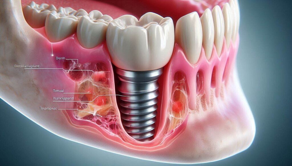
When the gum heals around an implant abutment or healing collar, an epithelium-lined sulcus forms, just as around a natural tooth. The junctional epithelium adheres to the implant surface via hemidesmosomes and a basal lamina. This epithelial adhesion is the first line of defense, and it is structurally similar to the natural attachment. The critical difference lies beneath this epithelium. The collagen fibers in the connective tissue zone do not insert into the implant. They cannot. The implant surface is impenetrable. Instead of inserting perpendicularly, the collagen fibers run parallel to the implant surface. They form a tight, encircling cuff, but it is a mechanical wrap, not a true anatomical insertion. This parallel orientation provides less resistance to bacterial penetration and mechanical disruption than the perpendicular insertion of natural tooth fibers. Furthermore, the blood supply to the peri-implant connective tissue is significantly less dense than around a natural tooth, because the implant lacks a periodontal ligament, which is a major source of vascularization. The implant-gum seal is a weaker, more vulnerable barrier. It is a functional scar, not a regenerative restoration of the original anatomy.
The Clinical Significance of the Weaker Implant-Gum Seal
This structural difference has direct, practical implications for the long-term health of your implant. The peri-implant soft tissue seal is more permeable to bacterial toxins. The bacteria in the biofilm that forms on the implant crown and abutment surface are physically closer to the underlying bone because the connective tissue cuff is less of an effective barrier. If daily oral hygiene is neglected, the inflammatory infiltrate can reach the bone more quickly than around a natural tooth. This is why peri-implant mucositis, the early, reversible inflammation of the gum, can progress to peri-implantitis, the destructive bone loss, more rapidly and aggressively than gingivitis progresses to periodontitis around a natural tooth.
The weaker soft tissue attachment also means that the implant-gum interface is more susceptible to mechanical trauma. Overzealous brushing with a hard-bristled toothbrush, aggressive flossing that snaps the floss against the gum, or probing by the dentist with a metal periodontal probe can inadvertently disrupt the delicate epithelial adhesion and cause recession. Once the gum recedes around an implant, the underlying titanium or zirconia abutment is exposed. This exposure is aesthetically compromising in the anterior zone, and it also exposes the smooth or textured implant surface, which is more plaque-retentive and difficult to clean. The lost gum tissue does not regenerate around an implant in the way it might around a natural tooth. The recession is often permanent.
The Role of the Abutment Surface and Biologic Width
The surface properties of the implant abutment, the component that connects the implant fixture to the crown, play a significant role in the quality of the soft tissue attachment. A smooth, highly polished abutment surface, typically machined titanium or polished zirconia, provides a less favorable substrate for bacterial adhesion and biofilm formation, and it supports a healthy, long junctional epithelium attachment. The biologic width, the dimension of soft tissue that forms around the implant, is similar in concept to the natural tooth but often forms a slightly longer zone, particularly if the implant is placed too deep or if multiple surgeries have been performed.
The restorative dentist must respect and manage this peri-implant biologic width. If the crown margin is placed too deep, violating the biologic width, the body will attempt to re-establish the necessary dimension by resorbing the bone away from the margin. This process, called bone remodeling or saucerization, leads to a stable but often aesthetically undesirable result, particularly if it occurs on the facial aspect of the implant. A skilled implant dentist designs the restoration and selects the abutment shape specifically to support the soft tissue seal. A custom-milled zirconia or titanium abutment is contoured to emerge from the gum with a profile that supports the gum tissue in a natural, scalloped shape and provides a smooth, cleansable surface for the junctional epithelium to adhere to. The attachment of the gum to the implant is not just a passive event; it is actively sculpted and planned by the clinician.
Conclusion
The gums do attach to a dental implant through a living biological seal consisting of an epithelium adhering via hemidesmosomes to the implant surface and an underlying connective tissue cuff, but this attachment is structurally weaker than a natural tooth’s because the collagen fibers run parallel, not perpendicular, to the implant, and the blood supply is less robust. This inherent vulnerability means the peri-implant seal is more susceptible to bacterial penetration and mechanical trauma, placing an absolute premium on your daily, meticulous, yet gentle biofilm disruption with specialized non-metallic tools. The quality of this vital soft tissue interface is a shared responsibility between the precision of your dentist’s abutment design and your lifelong commitment to protecting and cleaning the delicate gum collar that guards the foundation of your implant.
Frequently Asked Questions
Will my gum grow back if it recedes around my dental implant?
Unlike gum recession around a natural tooth, which can sometimes be reversed or grafted, the gum tissue around an implant rarely regenerates on its own once it has receded. Soft tissue grafting procedures can be performed to replace the lost tissue, but they are less predictable than around natural teeth due to the limited blood supply.
Why does my implant dentist use a plastic probe instead of a metal one?
A plastic or titanium periodontal probe is used to avoid scratching the smooth, polished surface of the implant abutment or the implant collar. A scratch from a metal probe would create a microscopic roughened area that is a haven for bacterial biofilm, potentially accelerating peri-implant disease.
Does the gum attach better to zirconia than to titanium?
Some histological studies suggest that the soft tissue attachment to zirconia, particularly the connective tissue fiber orientation, may be slightly more favorable than to titanium. However, both materials support a clinically successful soft tissue seal. The key factor is the smoothness and the contour of the abutment surface, not the bulk material.
Is it normal for the gums around my implant to bleed when I floss?
No. Bleeding is a cardinal sign of inflammation, specifically peri-implant mucositis. Healthy peri-implant tissue is pale pink and does not bleed when gently probed or cleaned. Bleeding indicates the presence of a biofilm-driven infection that must be addressed immediately with improved home care and a professional evaluation.
Can I have a gum graft around my dental implant to make it look more natural?
Yes. A connective tissue graft or a free gingival graft can be performed around dental implants to increase the thickness of the gum tissue, cover exposed surfaces, and improve the aesthetic appearance. These procedures are commonly performed, often at the second-stage uncovering surgery or before the final crown is made.
Additional Resource
The American Academy of Periodontology is the leading authority on the soft and hard tissues supporting teeth and implants. Their comprehensive resources detail the biology of periodontal and peri-implant tissues and the latest techniques for managing soft tissue complications. Visit: https://www.perio.org/