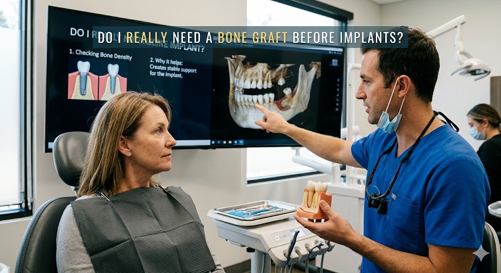
Do I Really Need a Bone Graft Before Implants?
Imagine walking into a dental office, feeling excited about finally replacing that missing tooth. You have researched the best implant specialists, saved up the money, and cleared your schedule for recovery. The finish line feels close. Then, the dentist looks at your X-ray, pauses, and says four words that change everything: “You need a bone graft.” Your heart sinks. Confusion sets in. Is this a legitimate requirement or an unnecessary upsell? You came for a single procedure, and suddenly you face months of additional healing and a higher bill. The question burning in your mind isn’t just about money or time. It is much simpler and more urgent: Do I really need a bone graft before implants?
The short answer is nuanced. For many people, yes, a bone graft is absolutely non-negotiable for long-term success. For others, it is entirely unnecessary. And for a growing third group, modern alternatives might bypass traditional grafting altogether. The key lies not in guesswork but in the cold, hard numbers of your own anatomy. Dental implants do not just sit in gum tissue like a removable denture. They fuse directly to living bone through a process called osseointegration. If that bone is too thin, too short, or too soft, the implant lacks a solid foundation. It is like trying to anchor a fence post in a shallow layer of sand instead of deep, compact earth. The post will wobble, fail, or eventually fall over.
Yet the landscape of implant dentistry has changed dramatically. A decade ago, patients with severe bone loss had only one path: major grafting surgery followed by a year of waiting. Today, innovations like zygomatic implants, ultra-short implants, and advanced imaging allow specialists to find or create stability in places previously deemed impossible. This does not mean bone grafts are obsolete. Far from it. Grafting remains the gold standard for creating predictable, long-lasting support. However, understanding why you need it—or if you truly do—empowers you to have a more intelligent conversation with your surgeon. It transforms you from a passive patient into an informed advocate for your own health.
The goal here is not to offer a simple yes or no. The goal is to pull back the curtain on the decision-making process. We will walk through the exact biological reasons bone loss happens, the red flags that make grafting unavoidable, the modern techniques that minimize discomfort, and the legitimate shortcuts that might apply to your specific case. By the end, you will not just have an answer to the question. You will have a comprehensive framework for understanding why that answer applies to you. No hype. No scare tactics. Just the unvarnished truth about what your jawbone actually needs to support a new smile for decades to come.
The Silent Collapse: What Happens to Your Jaw After Tooth Loss
To understand grafting, you must first understand loss. Most people think of tooth loss as a gap in their smile, a cosmetic inconvenience. The reality is far more mechanical and relentless. A natural tooth root does not just anchor the visible crown. It transmits chewing forces deep into the jawbone. Every time you bite, the periodontal ligament—a shock-absorbing tissue surrounding the root—sends tiny pressure signals to the bone cells. These signals tell osteoblasts, the body’s bone-building cells, that their work matters. The bone responds by staying dense and tall. It is a classic use-it-or-lose-it biological system.
When a tooth goes missing, that signaling stops abruptly. The brain no longer registers the need to maintain bone density in that specific spot. Osteoclasts, cells that resorb or break down bone tissue, begin to outpace osteoblasts. Within the first year of extraction alone, the alveolar ridge—the bony arch that holds teeth in place—can lose up to 25% of its width. A 2017 study published in the Journal of Oral and Maxillofacial Surgery confirmed that the most dramatic resorption occurs in the first three months post-extraction. The process then slows but never truly stops without intervention. Over a decade, patients can lose up to 60% of bone volume in the affected area.
The consequences go far beyond the implant site. Think of jawbone like an arch in architecture. Each tooth helps support its neighbors. Remove one block, and the adjacent teeth begin to drift, tilt, and super-erupt in unpredictable ways. The bite collapses slowly, leading to temporomandibular joint issues, facial asymmetry, and a sunken appearance that ages the face prematurely. For upper posterior teeth, the situation is even riskier. The maxillary sinus, an air-filled cavity above the upper molars, expands into the vacant space. This sinus pneumatization thins the bone floor to paper-thin dimensions, sometimes leaving less than one millimeter of bone separating the oral cavity from the sinus. Placing an implant there without addressing the bone volume would risk puncturing the sinus membrane entirely.
The Domino Effect on Facial Structure
Losing posterior support does more than complicate implant placement. The entire lower third of the face shrinks vertically. Patients who have worn dentures for decades often display a collapsed bite, deep wrinkle lines around the mouth, and a chin that protrudes forward unnaturally. This is not just aging. It is architectural collapse. The maxilla and mandible resorb inward and upward, reducing the distance between nose and chin. No amount of facial cream or filler can correct the underlying skeletal deficiency. Only restoring lost bone volume—or using advanced implant techniques that leverage remaining dense bone—can rebuild the framework.
Bone resorption explains why timing matters immensely. If you extract a tooth and plan to replace it with an implant, the ideal window is often immediate. A procedure called ridge preservation grafting involves placing bone graft material directly into the extraction socket at the time of removal. This simple step preserves the natural contours of the bony ridge and can eliminate the need for a far more invasive block graft later. Skipping this step to save a few hundred dollars often costs thousands and months of recovery down the line. The socket heals with a significant defect, and the surgeon must rebuild what nature already had perfectly shaped.
Critical Note for Readers
If your tooth extraction happened years ago, do not despair. The body adapts, but modern techniques can still rebuild what was lost. The key difference is that delayed grafting requires a separate surgical phase, more recovery time, and a higher degree of surgical skill. It does not mean implants are impossible. It simply means the road is longer. Your current bone dimensions, revealed through a cone-beam computed tomography (CBCT) scan, will dictate the most logical path forward.
| Time Since Extraction | Bone Loss Severity | Typical Intervention | Approximate Additional Healing Time |
|---|---|---|---|
| Immediate (0-2 weeks) | Minimal | Ridge preservation graft at time of extraction | 3-4 months (may heal concurrently with implant) |
| 3-6 months | Moderate (25-30% width loss) | Minor particulate graft or simultaneous implant with guided bone regeneration | 4-6 months |
| 1-3 years | Significant (up to 50% width loss) | Block graft or ridge split procedure | 6-9 months before implant placement |
| 5+ years | Severe (height and width loss, sinus expansion) | Major grafting (hip/ramus block, sinus lift) or zygomatic implants | 9-12+ months (or immediate loading for zygomatic) |
The Critical Millimeter: How Much Bone Do Implants Really Need?
A common refrain in dental consultations sounds deceptively simple: “You don’t have enough bone for implants.” But what does “enough” actually mean in measurable terms? Implant dentistry is a discipline of millimeters. Success or failure hinges on precise dimensions. A standard dental implant platform typically measures between 3.5 and 5 millimeters in diameter. The length can range from 6 millimeters for ultra-short designs up to 16 millimeters or more for standard implants in abundant bone. To fully contain and support an implant, surgeons generally follow the “rule of 1.5 millimeters.”
This guideline states that an implant should have at least 1.5 millimeters of healthy bone on the buccal (cheek-side) and lingual (tongue-side) aspects. So, a 4-millimeter-diameter implant requires a ridge at least 7 millimeters wide. For height, most clinicians aim for a minimum of 10 millimeters of bone above vital anatomical structures such as the inferior alveolar nerve in the lower jaw or the maxillary sinus in the upper jaw. These numbers are not arbitrary. They are the biological minimums necessary for a stable blood supply, long-term tissue health, and resistance to the micro-forces of chewing that gradually stress the bone-implant interface.
Anterior teeth in the aesthetic zone face even stricter requirements. The front of the mouth deals with thinner tissue, high smile lines, and relentless visual scrutiny. Here, bone thickness must reach at least 2 millimeters on the facial aspect to prevent the grayish hue of a titanium implant from showing through the gum tissue. Any less, and the result is not just functional failure but a cosmetic disaster that patients see every time they smile. This is why the “do I really need a bone graft” question often gets a firm yes in the aesthetic zone, even when the posterior regions might allow for compromises.
Beyond Width: The Intricacies of Bone Quality
Bone quantity, measured in millimeters, tells only half the story. Bone quality—its density and vascularity—profoundly influences implant success. Surgeons classify bone using the Lekholm and Zarb system, which ranges from Type 1 (dense, compact cortical bone) to Type 4 (soft, spongy cancellous bone with a thin cortical shell). The posterior maxilla notoriously exhibits Type 4 bone. It has limited blood supply and lower density, which compromises primary stability, the tight mechanical lock achieved at the time of placement. If a surgeon cannot achieve primary stability, the implant micro-moves during healing, and fibrous tissue encapsulates it instead of bone. This leads to early failure.
A graft does more than add volume. It transforms bone quality. Particulate grafting materials mixed with the patient’s own blood or bone marrow provide a scaffold for new bone formation. Over months, the graft incorporates and remodels into dense, well-vascularized bone capable of supporting an implant. This conversion is especially critical for patients with osteoporosis, a history of bisphosphonate use, or uncontrolled diabetes. Their native bone, even if dimensionally adequate, may lack the metabolic vitality to integrate an implant safely. Grafting offers a biological reboot, creating a healthier bed for osseointegration.
Technology now allows surgeons to measure bone density objectively before ever picking up a scalpel. CBCT scans generate Hounsfield unit readings that quantify density in specific regions. Values above 850 HU indicate excellent Type 1 or Type 2 bone. Values below 350 HU ring alarm bells. If your scan reveals ample width and height but dangerously low density, your surgeon may still recommend grafting or a longer healing period. This nuance explains why two patients with identical millimeter measurements might receive different treatment plans. One has dense bone and can proceed immediately. The other has soft, fatty marrow and requires augmentation to avoid a predictable failure.
A Note on Short Implants
The rise of ultra-short implants (4-6 millimeters in length) has shifted the conversation somewhat. These devices, with specially engineered surfaces that maximize bone contact, can sometimes bypass the need for vertical grafting in atrophic mandibles. However, they are not a universal solution. Short implants demand exceptional bone density and often require splinting multiple implants together to distribute forces. They work brilliantly for selected cases but fail catastrophically when pushed beyond their indications. A surgeon who casually dismisses grafting in favor of a short implant without rigorous analysis of your bite forces, bone quality, and opposing dentition is not doing you any favors. The right tool for the right scenario always wins.
| Implant Length | Typical Diameter | Minimum Bone Height Required | Best Application |
|---|---|---|---|
| Ultra-Short (4-6 mm) | 4.0-6.0 mm | 6-8 mm | Severely resorbed posterior mandible, dense bone |
| Short (7-8 mm) | 3.75-5.0 mm | 9-10 mm | Posterior areas with moderate resorption |
| Standard (10-13 mm) | 3.5-5.0 mm | 12-15 mm | Most single tooth and bridge cases |
| Long (14-16+ mm) | 3.5-5.0 mm | 16+ mm | Immediate extraction sites, pterygoid implants |
The Honest Signs You Almost Certainly Need a Bone Graft
Your body sends clear signals that bone volume has decreased beyond a safe threshold. These signs do not require a dental degree to recognize. They manifest in ways you can see, feel, or discover through specific diagnostic tests. Paying attention to them helps you walk into a consultation with realistic expectations. When a surgeon recommends grafting, and you have noticed several of these indicators, the advice is far more likely to be genuine clinical necessity rather than a profit-driven suggestion.
The first and most obvious sign is time. If your tooth has been missing for more than two years, the odds of needing augmentation skyrocket. The relentless process of resorption, described earlier, does not pause. It marches forward silently. A second powerful indicator is a visibly concave or sunken area along the jaw where the tooth used to be. Run your finger along the ridge. If you feel a sharp depression, almost like a ditch, the buccal plate—the outer wall of bone—has partially or fully collapsed. This external wall is critical for support and aesthetics, and its absence demands reconstruction.
1. Loose or Ill-Fitting Dentures
Patients who wear partial or full dentures often experience accelerated bone loss. A denture sits on top of the gum, transmitting compressive forces that the underlying bone interprets as detrimental pressure rather than stimulating tension. This pressure reduces blood flow and speeds resorption dramatically. If your denture once fit snugly but now rocks, clicks, requires adhesive to stay put, or falls out during speech, your supporting bone has flattened. You have lost not only teeth but the very foundation upon which a prosthesis rests. Implant-supported overdentures can halt this progression, but they first require a stable bone base. Grafting rebuilds the ridge to a height and contour that allows implants to bypass the soft, mobile tissue and anchor into solid bone.
2. A History of Periodontal Disease
Gum disease does not just bleed and cause bad breath. The bacterial infection triggers an inflammatory cascade that destroys both the soft tissue attachment and the underlying bone. Periodontal pockets measuring 6 millimeters or more indicate significant bone loss around the roots of teeth. If periodontal disease was the reason for your tooth loss, the infection likely compromised the bone well beyond the immediate socket walls. Even after extraction and healing, the ridge may be narrow, irregular, and lined with cortical bone of poor quality. Placing an implant into this compromised bed without grafting introduces the risk of peri-implantitis, a similar inflammatory destruction that causes late implant failure.
3. Sinus Proximity and Facial Structure Changes
For upper premolars and molars, the proximity of the maxillary sinus often dictates grafting necessity. You cannot feel the sinus floor without a scan, but you can recognize the consequences of its expansion. Patients missing upper back teeth who lie down and feel a sense of pressure or fullness in the cheek may be experiencing the thin bone separating the mouth from the sinus. More conclusively, a CBCT scan will measure the exact vertical bone available. Less than 5 millimeters of bone below the sinus floor leaves a surgeon with no safe option other than a sinus lift graft to place implants of adequate length. This procedure adds several months to the timeline but creates bone where none existed, making implant placement both possible and predictable.
4. Systemic Factors That Weaken Native Bone
Your medical history speaks volumes about your bone’s ability to heal around an implant. Smokers face a significantly higher risk of implant failure due to vasoconstriction and impaired healing. Uncontrolled diabetes interferes with bone metabolism and wound repair. Long-term corticosteroid use, autoimmune disorders, and certain medications like bisphosphonates all alter the biological landscape. In these scenarios, grafting serves a dual purpose. It adds volume and introduces growth factors and a fresh scaffold that can overcome the sluggish native healing response. A surgeon who recognizes these risk factors and recommends grafting is practicing preventive medicine, not overtreatment.
“The greatest misconception is that bone grafting is a punishment for waiting too long. It is not. It is a strategic rebuild that restores the anatomy to a state nature intended before disease and time took their toll.” — Dr. Michael Block, Oral and Maxillofacial Surgeon
When You Can Safely Skip the Graft: Real Exceptions
The previous section may sound like every patient with a missing tooth needs a bone graft. That is not accurate. There are specific, well-defined scenarios where grafting adds no value and only increases surgical time, cost, and morbidity. Understanding these exceptions protects you from unnecessary treatment. The key is rigorous diagnostic confirmation, not wishful thinking.
The most common scenario for graft-free implant placement is immediate implant placement into a fresh extraction socket. When a tooth requires removal due to fracture, trauma, or a non-restorable cavity, but the surrounding bone remains intact and infection-free, the surgeon can often place the implant at the same appointment. This technique, called immediate placement, relies on the socket walls to guide healing. Crucially, the socket must have four intact walls and a buccal plate at least 1 millimeter thick. If the surgeon can achieve primary stability by engaging the implant into native bone 3 to 5 millimeters beyond the root apex, the procedure often succeeds without any added graft material. Some clinicians still place a small amount of graft in the gap between the implant and the socket wall—a jump gap—but this is a minor augmentation, not a full reconstructive graft.
Ultra-Short and Narrow Implants in Selected Sites
When vertical bone height is limited but width is adequate, ultra-short implants offer a legitimate alternative to vertical ridge augmentation. The posterior mandible, above the inferior alveolar nerve, often presents this exact pattern. A traditional 10-millimeter implant would risk nerve damage, triggering a recommendation for a major block graft to add height. However, placing two or three ultra-short, wide-diameter implants and splinting them together with a bridge can distribute forces enough to compensate for reduced length. Studies with five-year follow-up show survival rates above 95% for properly selected cases. The patient avoids a second surgical site, potential nerve morbidity, and months of healing. The decision hinges on bone density. Soft, fatty bone cannot support a short implant. Dense, Type 1 bone often can.
Zygomatic and Pterygoid Implants: The True Graftless Solutions
For the completely edentulous upper jaw with severe atrophy, grafting once dominated the treatment plan. Patients faced bilateral sinus lifts, extensive bone grafts harvested from the hip, and up to two years of treatment before receiving a fixed set of teeth. Zygomatic implants changed that paradigm. These exceptionally long implants (30 to 52.5 millimeters) anchor not into the maxillary bone but into the zygomatic bone, the dense cheekbone that resists resorption even in advanced atrophy. Typically, a surgeon places two zygomatic implants in the posterior and two to four standard implants in the anterior maxilla. A fixed bridge attaches immediately or within a few days. No grafting. No sinus lift. No year of waiting.
Pterygoid implants similarly bypass the need for posterior maxillary grafting by engaging the pterygoid plate, a dense bony structure behind the maxilla. These techniques require advanced surgical training and carry their own risks, including potential orbital or nerve complications if performed incorrectly. They are not for every patient. But for those who qualify, they represent the ultimate answer to the question “Do I really need a bone graft before implants?” The answer, in these cases, is an emphatic no—as long as the zygomatic or pterygoid bone can provide the necessary anchorage.
- Intact fresh extraction sockets with four walls and apical bone beyond the root tip.
- Single-tooth gaps where adjacent teeth have preserved bone height and width.
- Posterior mandible with dense bone allowing ultra-short implant placement.
- Fully edentulous upper arch qualifying for zygomatic or pterygoid implant protocol.
- Areas where ridge expansion techniques can widen bone without grafting material.
| Scenario | Graft Needed? | Primary Reason |
|---|---|---|
| Immediate molar extraction, intact socket, no infection | Usually not | Native socket provides natural scaffold |
| Missing tooth >3 years, severe buccal concavity | Almost certainly | Buccal plate resorption beyond functional limits |
| Upper premolar with 3 mm of bone below sinus | Yes (sinus lift) | Insufficient bone for any standard implant |
| Fully edentulous maxilla with severe atrophy | Not necessarily | Zygomatic implants may bypass grafting entirely |
| Smoker with poorly controlled diabetes | Highly advisable | Compromised healing requires optimal bone bed |
The Grafting Experience: What Actually Happens
Fear of the unknown drives a huge portion of the anxiety around bone grafting. Patients imagine large, painful hip surgeries and months of incapacitation. The reality for the vast majority of modern grafting procedures is far more manageable. Understanding the workflow, the materials used, and the recovery timeline strips away the horror stories and replaces them with actionable expectations.
Grafting material comes from four broad sources. Autografts use the patient’s own bone, harvested from the chin, the back of the lower jaw (ramus), or the hip. They offer the gold standard of osteoconductivity, osteoinductivity, and osteogenicity—meaning they provide a scaffold, recruit the body’s own bone-forming cells, and contain live cells that can directly form bone. The downside is a second surgical site and the associated discomfort. Allografts use human bone from tissue banks, processed to remove all cellular material and sterilized. They are safe and avoid a second surgery site but lack the live cells of an autograft. Xenografts, typically bovine-derived, provide an excellent mineral scaffold that slowly resorbs as the patient’s own bone replaces it. Alloplastic grafts use synthetic materials like beta-tricalcium phosphate, offering unlimited supply without any human or animal origin. Most modern surgeons use a combination, often mixing autogenous bone scraped from the implant site with a xenograft or alloplast to maximize the biological response.
The Surgical Sequence and What You Feel
A typical grafting procedure for a single implant site begins with local anesthesia, the same as a dental filling. You feel pressure and vibration but no sharp pain. The surgeon makes an incision to expose the bone defect, then places the graft material precisely against the deficient area. A barrier membrane, often made of resorbable collagen, covers the graft to prevent soft tissue cells from invading the slower-growing bone cells—a critical step known as guided bone regeneration. The surgeon then sutures the gum tissue closed over the graft. The procedure itself often takes 45 to 90 minutes for a single site. Patients drive themselves home (if only local anesthesia is used) and manage the recovery with over-the-counter pain relievers and ice packs.
Post-operative swelling peaks at 48 hours and subsides significantly by day five. Bruising of the cheek or neck can occur, especially with larger grafts. Patients eat a soft diet for one to two weeks, avoiding chewing on the surgical side. Stitches typically dissolve on their own within two to three weeks. The graft then requires a consolidation period of three to nine months before it is ready for implant placement. A CBCT scan at the end of this waiting period confirms that the bone has matured and reached the necessary dimensions. Only then does the surgeon schedule the implant surgery. This staged approach, while requiring patience, yields the most predictable results for significant defects.
Simultaneous Approach: Graft and Implant Together
In cases of moderate bone deficiency, surgeons may combine grafting and implant placement into one surgery. This simultaneous approach works when the remaining native bone provides enough stability to anchor the implant while the grafted bone reinforces the defect area. The implant acts as a tent pole, supporting the membrane and maintaining space for the graft. This saves three to four months of healing time. The trade-off is a slightly higher risk of implant movement if the native bone fails to provide adequate initial stability. An experienced surgeon will make the call based on insertion torque values—a measure of how tightly the implant threads engage the bone. Values above 35 Ncm generally permit simultaneous grafting. Values below 20 Ncm demand a staged approach.
- Day 1-2: Intermittent ice, rest, soft cold foods, prescribed or OTC pain medication.
- Day 3-5: Swelling peaks then begins to fade, warm salt water rinses begin.
- Week 1-2: Soft diet strictly, avoid brushing directly over the surgical site.
- Week 3: Dissolving stitches disappear, gentle brushing resumes over the area.
- Month 3-6: Graft matures; follow-up CBCT scan to assess readiness for implant.
- Month 4-9: Implant placement surgery if staged; if simultaneous, implant already integrating.
Advanced Alternatives: When Innovation Bypasses Grafting
The field of implant dentistry does not stand still. A decade ago, a patient with a thin ridge had no choice but to accept a graft. Today, several techniques challenge the assumption that augmentation is the only path. These alternatives do not suit everyone, but for the right anatomical picture, they offer shorter timelines and less surgical complexity.
Ridge splitting, or bone expansion, represents a clever alternative for ridges that are wide enough in height but too thin horizontally. The surgeon uses specialized chisels or piezoelectric instruments to create a controlled split in the ridge, expanding the two cortical plates apart like opening a book. An implant is then placed into the widened space, and the gap fills with the patient’s own blood and bone debris, which remodels over time. No grafting material is necessary in many cases, though some surgeons add particulate graft for insurance. This technique works best in the upper jaw, where bone is more elastic, and demands a minimum ridge width of 3 millimeters to avoid fracturing the plates. Patients heal faster and avoid the cost of graft materials.
Tilted Implant Concepts (All-on-4 and All-on-6)
Full-arch rehabilitation for edentulous patients often encounters severe posterior bone loss, especially in the upper jaw. The All-on-4 concept, developed by Dr. Paulo Maló, uses tilted posterior implants to avoid the sinus and engage denser bone further back. By angling the posterior implants up to 45 degrees, the surgeon can place a longer implant that achieves bicortical anchorage—engaging both the crestal bone and the dense cortical bone near the nasal or sinus floor. This tilting distributes forces favorably and often eliminates the need for sinus grafting. The technique also allows for immediate loading, meaning the patient leaves the surgery with a fixed provisional bridge the same day. Long-term studies show cumulative survival rates above 98% at 10 years for the mandible and 96% for the maxilla. This is not a “shortcut” that compromises outcomes. It is a fundamentally different biomechanical approach that works with the available anatomy rather than trying to completely rebuild it.
Custom Subperiosteal Implants: A Niche Revival
A technology considered obsolete for decades has quietly re-emerged, powered by digital planning and 3D printing. Subperiosteal implants do not drill into the bone at all. Instead, a custom-designed metal framework sits on top of the bone, under the gum, with posts projecting through the tissue to support a bridge. The framework, printed from medical-grade titanium, exactly matches the patient’s jaw contour. This approach completely avoids bone grafting and offers an alternative for patients with severe atrophy who cannot tolerate or do not want zygomatic implants. The surgery is less invasive than harvesting large bone grafts, but the technology demands high precision. Poorly fitting subperiosteal frameworks can cause soft tissue breakdown and infection. Centers with extensive experience in digital workflow and additive manufacturing report promising mid-term results. For a specific subset of patients—especially those with knife-edge ridges that would require bilateral hip grafts—this option deserves consideration.
- Computer-guided ridge splitting: A digital surgical guide controls the split depth and angle, reducing fracture risk.
- Osseodensification burs: Special burs spin in reverse, compacting and densifying native bone during implant osteotomy rather than removing it, increasing primary stability in soft bone.
- Growth factor concentrates: Platelet-rich fibrin (PRF) spun from the patient’s own blood concentrates healing factors to accelerate graft maturation, allowing implant placement as early as 8-12 weeks.
- Dynamic navigation: Real-time tracking of implant drills on a CBCT allows placement of implants in extremely tight bone corridors without grazing nerves or sinuses.
The Financial Reality: What This All Costs
Money matters. Anyone who tells you that cost should not be a factor in medical decisions lives in a fantasy. The cost of a bone graft can double or triple the total investment for a single implant. Understanding the breakdown and the long-term value helps you make a decision you can live with, financially and emotionally.
A single tooth implant with no grafting, in an average U.S. market, costs between $3,000 and $4,500 for the entire process, including the abutment and crown. Adding a minor particulate graft and membrane typically adds $500 to $1,200. A more complex block graft, which requires harvesting bone from another site, adds $2,000 to $4,000. A bilateral sinus lift with extensive grafting can cost $5,000 to $8,000 before a single implant is placed. For full-arch cases, the grafting alone can run $15,000 to $25,000. These are significant numbers. They prompt the very reasonable question: Is there a cheaper way that still works?
The False Economy of Skipping a Necessary Graft
The short-term savings of refusing a recommended bone graft almost always evaporate catastrophically. An implant placed into inadequate bone has a drastically higher early failure rate. The implant either fails to integrate and simply falls out, or it develops peri-implantitis years later as the thin bone recedes around the neck. Removing a failed implant, cleaning out the infected site, rebuilding the lost bone, and replacing the implant costs two to three times the original fee for the grafted, properly planned case. You lose not just money but time—often an additional year or more of treatment. And you lose the original healthy bone that resorbed around the failing implant, making the salvage procedure far more complex. Paying for the right foundation the first time is not an upsell. It is the most cost-effective strategy per year of use.
Insurance and Financing Strategies
Dental insurance typically covers bone grafting under major restorative services, often at 50% of the contracted fee, up to an annual maximum that rarely exceeds $1,500 or $2,000. This leaves a significant out-of-pocket balance. Medical insurance may cover grafting when the cause of bone loss is a medical condition, trauma, or congenital defect. For large maxillofacial reconstructions, working with an oral surgeon who can bill medical insurance often shifts a portion of the cost away from your dental plan. Flexible spending accounts (FSAs) and health savings accounts (HSAs) allow pre-tax dollars to pay for grafting and implants. Third-party financing companies like CareCredit and Proceed Finance offer 0% interest promotional periods for 12 to 24 months, turning a large lump sum into manageable monthly payments. Some practices offer in-house membership plans that provide discounts on grafting and implant procedures. Exhaust all these avenues before assuming the procedure is financially out of reach.
| Procedure | Average Fee Range (USD) | Time Added to Treatment | Longevity Expectation |
|---|---|---|---|
| Immediate implant, no graft | $3,000 – $4,500 | 3-4 months to final crown | 25+ years with proper care |
| Implant + minor particulate graft | $4,000 – $6,000 | 4-6 months to final crown | 25+ years |
| Staged block graft + implant | $7,000 – $10,000 | 9-12 months before final crown | 20-25 years |
| Zygomatic full-arch (no graft) | $25,000 – $35,000 per arch | Immediate fixed bridge | 15-20+ years |
| Traditional full-arch with extensive grafts | $35,000 – $50,000 per arch | 12-18 months to final bridge | 25+ years |
“Patients sometimes view the graft as an added expense. I encourage them to reframe it as the most important investment in the project. The implant is the visible kitchen renovation. The graft is the foundation poured to bedrock. You do not skip the foundation to afford nicer countertops.” — Financial coordinator, specialty implant practice
Conclusion
Deciding whether you truly need a bone graft before dental implants comes down to the precise measurement of your remaining bone on a CBCT scan, the biological quality of that bone, and the specific location within your mouth. Skipping a necessary graft guarantees a far higher risk of implant failure, nerve damage, or sinus complications that cost far more to repair than the initial graft would have cost. Yet modern alternatives like zygomatic implants, tilted implant concepts, and ridge splitting now offer legitimate pathways that bypass extensive grafting for carefully selected patients. The safest path forward always involves selecting a surgeon who bases their recommendation on three-dimensional imaging and evidence-based millimeter requirements, not guesswork, and who can clearly explain exactly why your specific anatomy demands—or does not demand—the additional foundation work.
Frequently Asked Questions
1. How painful is a dental bone graft really?
Most patients compare the procedure to a tooth extraction. You receive local anesthesia, so you feel pressure and vibration but no sharp pain during the surgery. Post-operative discomfort typically peaks at 24-48 hours and is well-managed with over-the-counter anti-inflammatory medication and ice packs. A graft harvested from your own hip involves more significant soreness at the harvest site and is reserved for major reconstructions.
2. Can a bone graft fail, and what happens if it does?
Yes, grafts can fail, though modern techniques keep the failure rate below 5-10%. Smoking, infection, membrane exposure, and poor blood supply increase the risk. A failing graft typically presents as persistent drainage, pain, or mobility of the graft particles. If the graft fails, the surgeon debrides the area, allows it to heal for several weeks, and can regraft. A failed graft does not mean implants are impossible; it simply extends the timeline.
3. How long after a bone graft can I get my dental implant?
For minor socket grafts, the waiting period is usually 3-4 months. A larger ridge augmentation requires 4-6 months. Major block grafts or sinus lifts typically need 6-9 months of healing before the bone is mature enough to receive an implant and achieve reliable osseointegration. A follow-up CBCT scan confirms readiness.
4. Does dental insurance cover the cost of a bone graft?
Many dental insurance plans cover a percentage of bone grafting, typically 50% of the contracted fee, subject to your annual maximum. When grafting is required due to trauma or a medical condition, medical insurance may provide coverage. Always request a pre-treatment estimate and have your surgeon’s office check both medical and dental benefits before scheduling surgery.
5. Are there any long-term risks associated with not getting a recommended bone graft?
The primary risk is early or late implant failure. An implant placed in insufficient bone may fail to integrate, or it may develop peri-implantitis years later as the thin bone recedes. Additionally, an implant protruding through deficient bone can injure the inferior alveolar nerve (causing permanent lip numbness) or perforate the sinus membrane, leading to chronic sinus infections.
Additional Resource:
For an in-depth look at the science of bone regeneration and the specific grafting materials used, visit the American Academy of Periodontology’s patient resource page on regenerative procedures at https://www.perio.org/for-patients/periodontal-treatments-and-procedures/regenerative-procedures/
Disclaimer: This article is for informational purposes only and does not constitute medical or dental advice. Individual treatment needs vary based on a thorough clinical examination and three-dimensional imaging. Always consult a licensed dental professional to discuss your specific condition and treatment options.


