How Long Dental Implants Take to Osseointegrate?
The word “osseointegration” is the most important biological concept in implant dentistry. It is the direct, structural, and functional connection between living bone and the surface of a load-bearing dental implant. Without it, an implant is just a piece of metal or ceramic floating uselessly in a hole. The entire promise of a fixed, lifelong tooth replacement rests on this silent, microscopic process occurring beneath your gum. But how long does it really take? The answer is not a single, simple number. It is a range, a biological spectrum, that depends on the jawbone’s location, its density, the implant’s surface, and your own body’s healing capacity. This article is a deep dive into the timeline of osseointegration. We will dissect the cellular events week by week, compare healing in the upper and lower jaws, explore the factors that can accelerate or delay the process, and explain the critical distinction between “clinically ready” and “fully mature” bone fusion.
The Biological Definition of Osseointegration Time
The clinical definition of osseointegration time is the period from implant placement to the point where the bone-implant interface can safely withstand the controlled forces of a final prosthesis. This is not a moment when the bone suddenly “turns on.” It is a gradual process of increasing secondary stability, where the initial mechanical grip (primary stability) is replaced by a living, biological anchorage. The standard, evidence-based healing times are:
- Mandible (Lower Jaw): 3 to 4 months.
- Maxilla (Upper Jaw): 4 to 6 months.
These are the timelines published in the classic protocols by Brånemark and confirmed by decades of clinical studies. They represent the time for a standard root-form implant in healed, healthy bone with a conventional moderately-rough (SLA) surface. At the end of this period, a clinician can test the implant, confirm integration, and safely proceed with the final crown. This is the “unloaded healing” timeline, where the implant is not subjected to chewing forces.
The Cellular Timeline: A Week-by-Week Journey
What happens inside the bone during these months is a fascinating, carefully sequenced biological construction project.
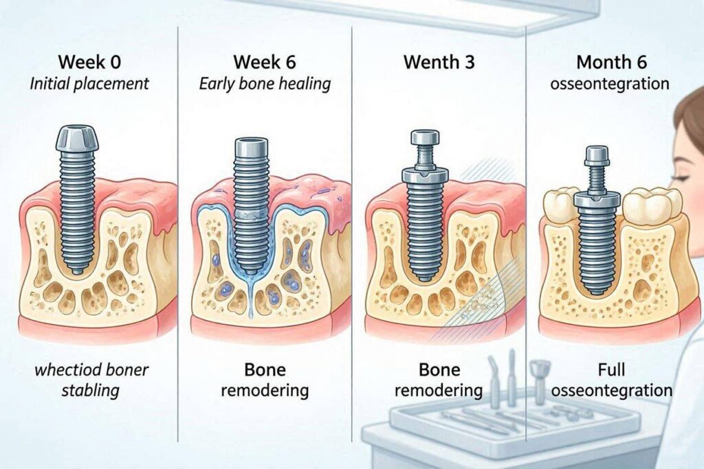
Week 1-2: The Hemostatic and Inflammatory Phase
The moment the implant is torqued into place, the space between its threads and the prepared bone fills with blood. A clot forms. Platelets degranulate, releasing platelet-derived growth factor (PDGF) and transforming growth factor-beta (TGF-β). These powerful cytokines call neutrophils and macrophages to the site to clean debris and prevent infection. Angiogenesis, the formation of new blood vessels, begins. This is a highly inflammatory, vascular phase. The implant has no biologic stability yet; it relies entirely on its mechanical grip.
Week 3-6: The Proliferative and Woven Bone Phase
Mesenchymal stem cells, attracted by the growth factors, differentiate into osteoblasts. These bone-forming cells migrate to the titanium dioxide surface. They begin secreting osteoid, a soft, unmineralized collagen matrix. This osteoid is then rapidly mineralized with calcium phosphate crystals. This first new bone is called woven bone. It forms quickly, within weeks, and provides the first biological grip, the beginning of secondary stability. However, woven bone is weak. Its collagen fibers are randomly oriented, like disorganized straw. The implant is entering the “stability dip,” where primary mechanical stability is decreasing due to bone remodeling, and the new biological stability is not yet strong enough. This is the most vulnerable period.
Month 2-4: The Remodeling and Lamellar Bone Phase
Osteoclasts, the bone-resorbing cells, begin to remove the weak woven bone. Osteoblasts follow, laying down new, highly organized lamellar bone in concentric layers. This is mature bone, with parallel collagen fibers, packed with strength. The interface transforms from a weak biological glue to a structural interlock of bone intimately fused with the microroughness of the implant surface. By month 3 in the dense mandible and month 4-6 in the maxilla, a sufficient volume of lamellar bone has formed to allow the implant to be loaded with a high degree of predictability. This is the clinical readiness point.
Month 6-12 and Beyond: The Maturation Phase
Even after the crown is placed and you are chewing, the bone continues to remodel and mature. The interface becomes increasingly dense and strong. The bone trabeculae align along the lines of stress transmitted by the implant (Wolff’s Law). This is a lifelong dynamic equilibrium. A one-year-old implant interface is typically stronger and more mature than a four-month-old one. When we say an implant “osseointegrates in 4 months,” we mean it reaches a clinically loadable threshold, not its ultimate, final strength.
Mandible vs. Maxilla: Why the Time Differs
The 3-4 month mandibular vs. 4-6 month maxillary split is not arbitrary. It reflects fundamental anatomical differences. The mandible, particularly the anterior region, is composed of dense, cortical type I or type II bone. It has a thick, compact outer layer and a dense core. This dense bone provides high primary stability, and its robust blood supply supports rapid intramembranous-like healing. The maxilla, particularly the posterior region, is composed of thinner cortical plates and a core of loose, spongy cancellous bone—type III or type IV. The blood supply is less robust, the primary stability is often lower, and the softer bone architecture needs more time for the osteoblasts to fill the space between the threads with strong lamellar bone. The stability dip is more pronounced in soft maxillary bone, so a longer, protected healing period is a wise biological safety margin.
Factors That Accelerate Osseointegration
The timeline can be shortened. This is the driving force behind modern implant surface technology. A hydrophilic, chemically active implant surface (such as Straumann’s SLActive or Dio’s HAS) dramatically changes the first hours of healing. A standard SLA surface is hydrophobic; it repels water. Blood proteins adhere slowly. A hydrophilic surface instantly attracts blood and its healing proteins. Osteoblast migration and woven bone formation are accelerated. Clinical studies on these surfaces have shown that implants can be safely loaded at 3-4 weeks in dense bone, instead of 3-4 months. This is a genuine biological acceleration. Good bone quality, a precise atraumatic surgical technique, and a young, healthy, non-smoking patient with excellent metabolic health are all biological accelerators. An implant placed with a flapless, guided technique that preserves the periosteal blood supply will often heal slightly faster than one placed with an extensive flap.
Factors That Delay Osseointegration
The timeline stretches when the healing environment is compromised. A simultaneous major bone grafting procedure resets the clock. The implant may be in contact with some native bone, but the majority of its surface is against graft particles. The graft must be revascularized and replaced by living bone, a process that takes 6-9 months. Loaded too early, the graft will fail. Compromised systemic health is a powerful decelerator. Uncontrolled diabetes, heavy smoking, vitamin D deficiency, and immunosuppression all slow the cellular machinery of bone repair. A patient on bisphosphonates has a fundamentally altered bone metabolism that makes osseointegration unpredictable and risky. Overheating the bone during surgery, using a dull drill, or causing excessive surgical trauma kills osteocytes and creates a zone of necrotic bone that must be resorbed and replaced before osseointegration can occur. This can add months to the healing or, if extensive, lead to a total failure.
Conclusion
The standard osseointegration time for a dental implant is 3 to 4 months in the dense lower jaw and 4 to 6 months in the softer upper jaw, a period in which the initial blood clot is transformed through woven bone into mature, load-bearing lamellar bone. This biological clock is influenced profoundly by the implant surface technology, with modern hydrophilic surfaces enabling early loading at weeks, while major bone grafting, smoking, and systemic disease can extend the healing period to many months or a year. The clinical decision to load an implant is always based on objective stability testing, confirming that this silent, miraculous process has reached a sufficient threshold for a lifetime of function.
FAQ
1. Can I make my implant osseointegrate faster with supplements?
If you are genuinely deficient in Vitamin D, calcium, or have a compromised diet, then correcting these systemic deficiencies under a physician’s supervision will support optimal bone healing. However, there is no “super-supplement” that will accelerate the genetically and biologically determined pace of osseointegration in a healthy individual.
2. Is an implant that took 6 months to integrate stronger than one that took 3 months?
Not necessarily. The final quality of the lamellar bone interface is similar. The shorter healing time in the mandible is a function of the dense, well-vascularized starting bone, not a weaker final result. Both achieve a robust, mature interface.
3. Can an implant fail to osseointegrate even after waiting 6 months?
Yes, late failure of osseointegration during the healing phase is possible if the initial stability was insufficient, if there was a subclinical infection, or if the implant was microscopically moving. This is confirmed by the diagnostic tests at the uncover appointment. A fibrous encapsulation will have formed instead of bone.
Additional Resource
To learn more about the biology of bone healing and implant integration, visit the Academy of Osseointegration: https://osseo.org/