How To Grow Bone For Dental Implant?
You have been told the words no dental patient wants to hear. “You don’t have enough bone for a dental implant.” You may have been missing a tooth for years, or you lost it to a traumatic extraction that took some of the surrounding bone with it. The jawbone, without the constant stimulation of a tooth root, has slowly and silently resorbed away. The implant you need, the titanium post that would replace the root, requires a certain volume of solid bone to anchor into. The question that follows this diagnosis is urgent and hopeful: can that lost bone be grown back? How do you grow bone for a dental implant?
The answer is one of the most remarkable chapters in modern surgical dentistry. Yes, lost jawbone can be rebuilt. It can be grown, regenerated, and augmented to create a sufficient foundation for a dental implant. This is not a pharmaceutical miracle or a distant future technology. It is a routine, predictable, and scientifically validated set of surgical procedures performed every day in periodontists’ and oral surgeons’ offices. This article is your guide to the biological principles and the surgical techniques that make bone regeneration possible.
The Biological Principle: Guided Bone Regeneration
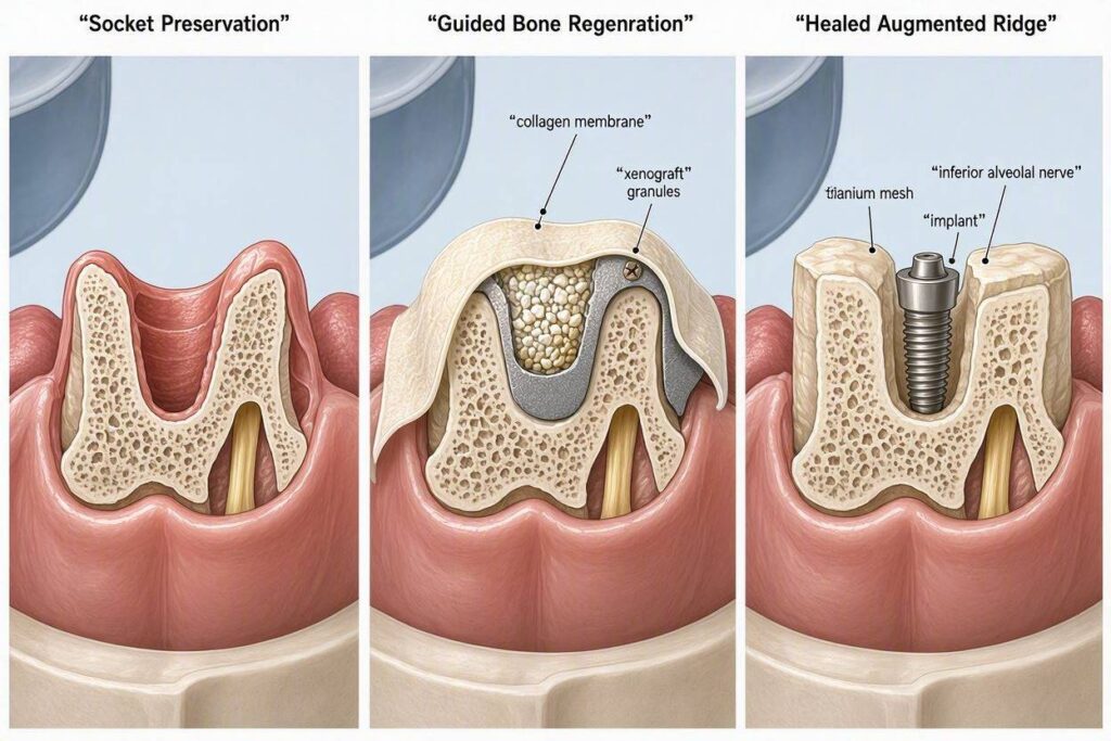
The foundation of all bone grafting for dental implants is a concept called Guided Bone Regeneration (GBR). It is based on a simple, elegant biological reality. The body has many different types of cells that can migrate into a healing wound. Bone cells (osteoblasts) are relatively slow-moving. Soft tissue cells, like fibroblasts from the gum and connective tissue, are fast-moving. If you simply fill a bone defect with a graft material and sew the gum closed, the fast soft tissue cells will invade the space and fill it with scar tissue before the slow bone cells have a chance to migrate in.
GBR uses a barrier membrane to protect the slow bone cells. The surgeon fills the bony defect with a particulate bone graft material. This graft acts as a scaffold, a three-dimensional framework. The surgeon then covers the graft with a specialized barrier membrane, which is tucked under the gum tissue. This membrane acts as a physical wall. It blocks the fast soft tissue cells from invading the graft space. It creates a protected, secluded chamber where only the slow, deliberate bone cells and their associated blood vessels can migrate in, populate the scaffold, and begin the process of turning the graft into living, vital bone. The membrane is the key. It guides the regeneration, ensuring bone, not scar tissue, fills the space.
Over a period of four to nine months, the particulate graft material is slowly resorbed and replaced by the patient’s own living bone. The membrane, if it is a resorbable type made of collagen, is slowly dissolved by the body’s enzymes. If it is a non-resorbable, titanium-reinforced membrane, it must be surgically removed in a second minor procedure. The result is a regenerated ridge of solid, vascular bone, ready to receive a dental implant.
The Raw Materials: Types of Bone Graft Materials
The scaffold that fills the defect can come from several sources. Each has its own profile of advantages, cost, and biological activity. The choice is made by the surgeon based on the size of the defect, the quality of the surrounding bone, and the patient’s preference.
- Autograft (Your Own Bone): This is the gold standard. The bone is harvested from the patient’s own body, typically from a nearby site in the jaw (like the chin or the back of the lower jaw) or from a distant site like the hip (iliac crest) for very large defects. Autograft is the only graft material that is osteogenic, meaning it contains living bone-forming cells. It is also osteoconductive (it acts as a scaffold) and osteoinductive (it releases growth factors that stimulate new bone formation). It is the complete package. The downside is the need for a second surgical site, with its own associated post-operative discomfort and morbidity.
- Allograft (Human Donor Bone): This is bone harvested from carefully screened and processed human donors, supplied by a regulated tissue bank. It is osteoconductive and mildly osteoinductive. It is completely safe and eliminates the need for a second surgical site in the patient. It is widely used and highly predictable. The bone is processed to remove all cells and DNA, leaving a mineral scaffold that the patient’s body slowly remodels into new bone.
- Xenograft (Animal-Derived Bone): The most common xenograft is deproteinized bovine bone mineral. It comes from cows, processed to remove all organic material, leaving a pure, natural mineral scaffold. Its structure is remarkably similar to human cancellous bone. It is highly osteoconductive and very slow to resorb, meaning it maintains the volume of the grafted space exceptionally well over time. It is one of the most widely used and researched bone graft materials in implant dentistry.
- Alloplast (Synthetic Bone): These are laboratory-created, synthetic bone substitutes. They are typically made of hydroxyapatite, beta-tricalcium phosphate, or bioactive glasses. They are osteoconductive, completely synthetic (eliminating any theoretical risk of disease transmission), and available in unlimited quantities. Some alloplasts are designed to be resorbed quickly, while others are more permanent.
The Cost of the Graft Material: The cost of the bone graft material is a direct line item on your surgical bill. An autograft costs nothing for the material itself, but the surgical time and morbidity are significant. An allograft or xenograft can add $300 to $800 to the procedure cost, depending on the volume required. A sophisticated synthetic alloplast with bioactive properties can be similarly priced. The surgeon’s fee for the grafting procedure is separate and reflects the complexity of the augmentation.
The Maxillary Sinus Lift: Growing Bone Upward
The most common and dramatic bone-growing procedure for dental implants is the maxillary sinus lift, also called a sinus augmentation. In the upper posterior jaw, the maxillary sinus is an air-filled cavity. After the upper molars and premolars are lost, the sinus floor can pneumatize, or drop down, leaving only a paper-thin shelf of bone between the mouth and the sinus. There is not enough vertical height to place an implant without penetrating the sinus.
A sinus lift adds bone to the floor of the sinus, effectively raising the sinus membrane and creating new vertical bone height. The surgeon makes a small window in the lateral wall of the sinus, carefully elevates the delicate sinus membrane without tearing it, and packs the created space with particulate bone graft material. The graft is not placed in a defect in the exposed jaw. It is placed inside the floor of the sinus, under the lifted membrane. Over six to nine months, this graft matures into solid bone, increasing the available vertical height from a millimeter or two to a robust 10-15 millimeters, plenty of room for a standard implant.
A sinus lift is a highly predictable, specialist-level procedure. The cost ranges from $1,500 to $3,000 per side and is a separate fee from the implant placement. Sometimes, if there is enough residual bone (at least 3-5 millimeters), the implant can be placed simultaneously with the sinus lift. If the bone is too thin, the sinus lift is performed first, and the implant is placed after a healing period.
Ridge Augmentation: Growing Bone Outward and Sideways
When a tooth is extracted, the buccal plate of bone, the thin wall on the cheek side, often resorbs rapidly. The result is a narrow, knife-edge ridge that is too thin to house an implant. This requires a lateral ridge augmentation.
The surgeon makes an incision, reflects the gum tissue, and exposes the narrow bony ridge. A particulate bone graft, mixed with the patient’s own blood or platelet-rich plasma, is packed against the deficient ridge, building it out to a width of 6-7 millimeters. The graft is covered with a barrier membrane, and the gum is sutured closed. Sometimes, for larger defects, a rigid block of bone (an autograft block from the patient’s chin or ramus, or a shaped allograft block) is screwed into place with tiny titanium fixation screws. This provides a rigid, immediate increase in bone volume.
Conclusion
Bone for a dental implant is grown through the biologically guided techniques of bone grafting. Using a scaffold of graft material and a protective barrier membrane, the surgeon creates a protected space where the body’s own slow-moving bone cells can regenerate lost volume. The maxillary sinus lift builds vertical height beneath the sinus floor. Ridge augmentation restores lost width and contour. These procedures are predictable, routine, and have transformed countless patients from non-candidates into successful implant recipients.
Frequently Asked Questions
How long does it take for a bone graft to heal before I can get an implant?
A typical socket preservation graft heals in three to four months. A more extensive ridge augmentation or a sinus lift usually requires six to nine months of healing before the new bone is mature and dense enough to receive an implant. A large block graft may require nine to twelve months.
Is bone grafting painful?
The post-operative discomfort from bone grafting is generally moderate and well-managed with prescribed pain medication and anti-inflammatories. The peak of swelling and discomfort is typically 48 to 72 hours after surgery. A sinus lift often has very little pain, though the patient must avoid blowing their nose or sneezing forcefully for several weeks to protect the graft. An autograft block from the chin or hip involves significantly more post-operative discomfort than a particulate graft.
Can I smoke during the bone graft healing period?
Absolutely not. Smoking is profoundly detrimental to bone graft healing. The nicotine-induced vasoconstriction and carbon monoxide-induced hypoxia starve the graft of oxygen and nutrients, dramatically increasing the risk of graft failure, wound dehiscence, and infection. A responsible surgeon will require complete smoking cessation before and during the entire graft healing period.
Additional Resource:
For a detailed, patient-focused explanation of bone grafting and sinus lift procedures, visit the American Academy of Periodontology: https://www.perio.org/consumer/bone-grafting