Peri-Implantitis: Symptoms, Causes, and Management
You notice a trace of blood when you brush around your implant crown. The gum looks slightly redder than the tissue surrounding your natural teeth. There is no pain, nothing that screams emergency, just a quiet unease that something is not quite right. You might dismiss these signs, attributing them to brushing too hard or eating something that irritated the gum. But these subtle signals represent the earliest warnings of a condition that has emerged as the leading cause of late implant failure. Peri-implantitis affects approximately 20 percent of implant patients at the implant level within ten years of placement, yet public awareness of this condition lags far behind understanding of gum disease around natural teeth. Recognizing the symptoms, understanding what causes the disease, and knowing the management options can mean the difference between saving an implant and losing it to progressive bone destruction.
Defining Peri-Implantitis
Peri-implantitis is not simply gum disease transferred to implants. It is a distinct pathological condition with its own characteristics, progression patterns, and treatment challenges. Understanding exactly what it is provides the foundation for effective prevention and management.
The Clinical Definition
Peri-implantitis is defined as an inflammatory condition affecting the tissues surrounding an osseointegrated dental implant, characterized by inflammation of the peri-implant mucosa and progressive loss of supporting bone beyond normal biological remodeling. This definition contains several elements that distinguish peri-implantitis from related conditions.
The inflammation must be present clinically, evidenced by bleeding on probing, redness, swelling, or suppuration. The bone loss must be progressive, meaning it increases over time when assessed through sequential radiographs. The bone loss must be beyond the normal crestal bone remodeling that occurs during the first year after implant placement, which is typically 1 to 1.5 millimeters.
This definition separates peri-implantitis from peri-implant mucositis, which involves inflammation of the soft tissues without bone loss, and from normal bone remodeling, which is a physiological adaptation rather than a pathological process. The distinction matters because mucositis is reversible with treatment, while peri-implantitis causes permanent architectural changes to the supporting bone.
How Peri-Implantitis Differs from Periodontitis
While peri-implantitis and periodontitis share similarities as plaque-induced inflammatory diseases affecting tooth-supporting structures, significant differences exist in their anatomy, progression, and treatment response.
The connective tissue around implants differs fundamentally from that around natural teeth. Natural teeth have periodontal ligament fibers that insert into cementum and bone, creating an organized, functional attachment. Implants lack these inserting fibers. The connective tissue around implants runs parallel to the implant surface rather than inserting perpendicularly, creating a less effective biological seal against bacterial invasion.
The vascular supply differs between teeth and implants. The periodontal ligament provides a rich vascular plexus around natural teeth, supporting immune surveillance and tissue repair. Implants lack this ligament blood supply, relying on vessels from the periosteum and the bone marrow spaces. This reduced vascularity may compromise the host response to bacterial challenge.
The inflammatory infiltrate in peri-implantitis extends closer to the bone surface than in periodontitis. The absence of a dense connective tissue barrier above the bone crest allows inflammatory mediators and bacteria to access the bone surface more readily. As a result, peri-implantitis lesions often progress faster and may be more destructive than periodontitis lesions of similar bacterial burden.
Prevalence and Risk Statistics
Peri-implantitis is common enough to warrant serious attention from both patients and clinicians. Systematic reviews report peri-implant mucositis prevalence of 40 to 65 percent at the implant level, meaning nearly half of implants develop reversible soft tissue inflammation at some point. Peri-implantitis prevalence is estimated at 18 to 22 percent at the implant level, with severe disease affecting approximately 10 percent of implants.
Patient-level prevalence, where at least one implant in the patient’s mouth is affected, is higher. Approximately 20 to 30 percent of implant patients develop peri-implantitis affecting at least one implant over a five to ten-year observation period. Patients with multiple implants and those with a history of periodontitis show significantly higher rates.
These statistics underscore the importance of ongoing maintenance. An implant that is successfully placed and restored is not guaranteed lifelong health. The biological environment that led to the original tooth loss, if not modified, will challenge the implant in the same way it challenged the natural dentition.
Recognizing the Symptoms
Peri-implantitis typically progresses silently in its early stages. The symptoms are often subtle, noticed by the clinician during a maintenance examination rather than by the patient at home. However, informed patients can recognize early warning signs and seek care before significant bone loss occurs.
Early Warning Signs
The earliest and most sensitive indicator of peri-implant inflammation is bleeding on gentle probing. Healthy peri-implant tissue should not bleed when probed with light pressure. Bleeding indicates ulceration of the sulcular epithelium, the thin tissue lining the crevice between the implant and gum. This is the same principle as bleeding gums around natural teeth, signaling inflammation that requires attention.
Patients may notice bleeding when brushing around the implant, when using interdental cleaning aids, or spontaneously in more advanced cases. The bleeding may be minimal, appearing as a pink tinge on the toothbrush or floss. Because dental implants are often perceived as invulnerable, patients may dismiss this bleeding as insignificant. It is not.
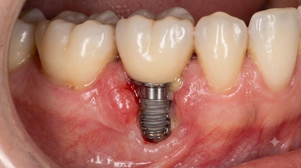
Redness and swelling of the peri-implant mucosa indicate active inflammation. The tissue loses its firm, pink appearance and becomes erythematous, edematous, and may appear shiny or puffy. The contour of the gum around the implant changes, with loss of the knife-edged margin and development of a rolled, swollen appearance.
An increase in probing depth around the implant, measured by the clinician during maintenance visits, indicates loss of attachment. While the patient cannot measure probing depths at home, they may notice that floss passes deeper around the implant than previously, or that food traps more readily in the area.
Advanced Symptoms
As peri-implantitis progresses, additional symptoms develop that are more noticeable to the patient. Suppuration, or pus, may exude from the peri-implant sulcus spontaneously or when pressure is applied to the surrounding tissue. The patient may notice a persistent bad taste, halitosis that does not resolve with oral hygiene, or visible discharge from around the implant.
Gum recession exposes structures that should be covered. The implant surface, with its roughened texture designed for osseointegration, may become visible above the gum line. This exposed surface cannot be effectively cleaned by the patient and accumulates bacterial biofilm, accelerating the disease process. The implant crown may appear longer than it did previously, a sign that the gum and bone have receded.
Pain is a late symptom and may not develop until the disease is advanced. When present, the pain is typically dull and aching, worse with biting or tapping on the implant. The pain originates from inflammation in the bone, not from the implant itself. The absence of pain in early and moderate peri-implantitis contributes to delayed diagnosis.
Implant mobility is a terminal symptom, indicating that bone loss has progressed to the point where osseointegration can no longer support the implant. By the time mobility develops, the implant has typically lost the majority of its bone support and cannot be saved. Mobility may be subtle initially, detected as a slight give when the implant is pressed, progressing to frank movement in all directions.
Radiographic Indicators
Radiographs provide the definitive assessment of bone support around implants. Periapical radiographs, taken with a standardized technique to allow comparison over time, reveal the bone level in relation to the implant threads.
Peri-implantitis appears radiographically as a radiolucent, or dark, area around the implant where bone should contact the implant surface. The bone loss may be horizontal, with the bone level reduced evenly around the implant, or vertical, creating a crater-like defect on one side. The pattern of bone loss provides information about the disease etiology and guides treatment planning.
The rate of bone loss is assessed through sequential radiographs taken at different time points. Stable bone levels over multiple years indicate a healthy implant, even if some bone loss occurred during the first year. Progressive bone loss over consecutive radiographs indicates active disease requiring intervention. The rate of progression varies among patients and among implants within the same patient.
Cone beam computed tomography, while not used routinely for implant maintenance due to radiation dose and cost considerations, provides three-dimensional visualization of bone defects when surgical treatment is being planned. The buccal and lingual bone plates, which cannot be assessed on two-dimensional periapical radiographs, are clearly visualized, revealing the full extent of bone loss.
The Causes of Peri-Implantitis
Peri-implantitis results from a complex interaction between bacterial biofilm, host response, and environmental and systemic factors. Understanding these causes informs both prevention and treatment strategies.
Bacterial Biofilm and Plaque Accumulation
The primary etiological agent of peri-implantitis is bacterial plaque accumulating on the implant surface. This biofilm, a complex community of microorganisms embedded in a protective extracellular matrix, triggers the host inflammatory response that drives tissue destruction.
The microbial composition of peri-implantitis lesions resembles that of chronic periodontitis, with a predominance of anaerobic gram-negative bacteria. Species commonly identified include Porphyromonas gingivalis, Tannerella forsythia, Treponema denticola, Prevotella intermedia, and Aggregatibacter actinomycetemcomitans. These periodontopathogens produce virulence factors that directly damage host tissues and stimulate destructive inflammatory responses.
The implant surface microtopography, intentionally roughened to promote osseointegration, unfortunately also promotes bacterial colonization once exposed to the oral environment. The roughened surface provides protected niches for bacterial attachment and makes mechanical debridement more difficult than on smooth natural tooth roots.
Plaque accumulation around implants occurs for several reasons. Inadequate oral hygiene allows biofilm to mature undisturbed. Restorations with overcontoured profiles or inadequate embrasure spaces prevent effective cleaning. Malpositioned implants create areas that are difficult for the patient to access with oral hygiene aids. The design of the implant-abutment connection creates a microgap that harbors bacteria.
Patient-Related Risk Factors
The host response to bacterial challenge determines whether plaque-induced inflammation progresses to tissue destruction. Some patients develop only mucositis, while others progress rapidly to peri-implantitis with significant bone loss. Several patient factors influence this susceptibility.
A history of periodontitis is the strongest predictor of peri-implantitis. The same host response characteristics that led to natural tooth loss predispose to implant bone loss. The oral microbiome of periodontitis patients contains higher proportions of pathogenic species. These patients require meticulous plaque control and frequent professional maintenance to prevent peri-implant disease.
Smoking dramatically increases peri-implantitis risk. The vasoconstrictive effects of nicotine reduce tissue perfusion and immune cell delivery. Carbon monoxide impairs oxygen transport. Tobacco chemicals directly impair fibroblast and osteoblast function. Smokers show higher rates of peri-implantitis, more rapid progression, and poorer response to treatment. Smoking cessation improves outcomes significantly.
Poorly controlled diabetes impairs host defense and wound healing. Hyperglycemia alters immune cell function, increases inflammatory mediator production, and impairs collagen synthesis. Diabetic patients with hemoglobin A1c above 7 percent are at increased risk for peri-implantitis. Good glycemic control reduces this risk to levels approaching non-diabetic patients.
Genetic factors influence peri-implantitis susceptibility. Polymorphisms in genes encoding inflammatory cytokines, particularly interleukin-1, affect the magnitude of the inflammatory response to bacterial challenge. Patients with specific interleukin-1 genotypes show increased peri-implantitis risk, though genetic testing is not routinely performed before implant placement.
Iatrogenic and Restorative Factors
Factors related to implant placement and restoration can create conditions that promote peri-implantitis independent of the patient’s oral hygiene efforts.
Excess cement left below the gum line during crown cementation is a well-documented cause of peri-implant bone loss. The cement acts as a foreign body, triggering inflammation that mimics or exacerbates plaque-induced disease. The rough cement surface harbors bacteria that are protected from mechanical debridement. Meticulous cement cleanup or the use of screw-retained restorations eliminates this risk.
Implant malposition contributes to peri-implant disease. An implant placed too close to an adjacent tooth or implant compromises the interimplant or interproximal bone. An implant that perforates the buccal bone plate lacks the bony support needed for tissue health. An implant positioned too far buccally or lingually creates restorative contours that impede hygiene.
Restorations with poor emergence profiles or overcontoured margins create plaque-retentive areas that the patient cannot clean. The crown should emerge from the implant with contours that allow effective oral hygiene access. Bulbous crowns, closed embrasures, and margins that extend subgingivally beyond necessity all contribute to plaque accumulation.
Non-passive fit of implant frameworks creates static stress on the implant components and the supporting bone. A multi-unit prosthesis that does not seat passively places continuous tension on the abutment screws and transfers stress to the bone-implant interface. This stress may contribute to bone resorption.
Biomechanical Factors
Excessive occlusal forces may contribute to peri-implant bone loss, either independently or in combination with bacterial inflammation. The relationship between occlusal overload and peri-implantitis remains a subject of research and debate.
Occlusal overload theory proposes that forces exceeding the bone’s adaptive capacity cause microfractures at the bone-implant interface, triggering bone resorption. Patients with bruxism or clenching, cantilevered prostheses, or premature occlusal contacts may subject implants to forces beyond what the supporting bone can withstand.
The combination of bacterial inflammation and occlusal overload may be particularly destructive. Inflammation from plaque reduces the bone’s capacity to withstand mechanical forces. Simultaneously, excessive forces may accelerate the spread of inflammation through the peri-implant tissues. Managing both biological and mechanical factors is essential for disease prevention.
Diagnosing Peri-Implantitis
Accurate diagnosis requires systematic clinical and radiographic evaluation. The diagnosis guides treatment decisions and establishes a baseline against which treatment outcomes can be measured.
Clinical Examination Protocol
The clinical examination begins with visual inspection of the peri-implant tissues. The clinician notes the color, contour, and texture of the mucosa surrounding the implant. Healthy tissue is pink, firm, and stippled, with a knife-edged margin at the implant. Inflamed tissue appears red or bluish, edematous, and may have a rolled or bulbous margin.
Probing is essential and must be performed gently, with light force of approximately 0.25 Newtons. Heavier probing force can penetrate the weaker connective tissue seal around implants, causing bleeding even in healthy tissue. The probe is walked circumferentially around the implant, recording probing depths at multiple sites. Increasing probing depths over time indicate attachment loss.
Bleeding on probing is recorded. Healthy peri-implant tissue should not bleed with gentle probing. Bleeding indicates inflammation of the sulcular epithelium and is the most sensitive indicator of peri-implant disease. The extent and severity of bleeding, recorded as bleeding on probing percentage, provides a measure of disease activity.
Suppuration is assessed by applying gentle pressure to the peri-implant tissues and observing for pus exuding from the sulcus. The presence of suppuration, even without significant probing depths or radiographic bone loss, indicates active infection requiring treatment.
Mobility testing determines whether the implant has lost osseointegration. Integrated implants should be absolutely immobile. Any perceptible movement indicates that the implant has failed and cannot be saved. Mobility is tested by applying controlled force with instrument handles, not by the patient’s report of perceived movement.
Radiographic Assessment
Standardized periapical radiographs using a paralleling technique allow accurate assessment of bone levels and comparison over time. The radiograph should show the entire implant and the surrounding bone with minimal distortion. The distance from a fixed reference point, typically the implant platform or the first thread, to the bone crest is measured.
Crestal bone loss of 1 to 1.5 millimeters during the first year after restoration is considered normal remodeling. Bone loss beyond this amount, or any progressive bone loss after the first year, indicates pathology. Serial radiographs taken at different time points are compared to determine whether bone loss is active or stable.
The pattern of bone loss is described. Horizontal bone loss reduces the bone level evenly around the implant. Vertical or angular bone loss creates defects on one or more implant surfaces. Circumferential bone loss creates a moat-like defect around the entire implant. The defect morphology influences treatment selection and prognosis.
Diagnostic Criteria Summary
A diagnosis of peri-implant health requires absence of clinical signs of inflammation, no bleeding on probing, no suppuration, and no bone loss beyond crestal bone remodeling. Probing depths should be stable over time, though baseline probing depths vary by tissue thickness.
Peri-implant mucositis is diagnosed when bleeding on probing is present, with or without increased probing depths, but no radiographic bone loss beyond crestal remodeling. The inflammation is confined to the soft tissue. This condition is reversible with treatment.
Peri-implantitis is diagnosed when inflammation, evidenced by bleeding and/or suppuration, is accompanied by progressive bone loss beyond crestal bone remodeling. Increased probing depths are typically present but may not be if bone loss is accompanied by gingival recession. The severity is classified by the extent of bone loss relative to implant length.
Non-Surgical Management
The initial treatment for peri-implantitis is non-surgical mechanical debridement, analogous to scaling and root planing for natural teeth. The goals are to reduce the bacterial load, eliminate inflammation, and arrest disease progression.
Mechanical Debridement Techniques
Removing biofilm and calculus from the implant surface presents challenges not encountered with natural teeth. The implant surface is intentionally roughened, providing protected areas for bacterial colonization. Metal instruments designed for natural teeth can scratch the implant surface, creating additional bacterial niches and potentially releasing titanium particles into the surrounding tissue.
Specialized instruments for implant debridement include plastic and graphite scalers, which are softer than titanium and do not scratch the implant surface. These instruments effectively remove soft deposits but are less effective against calculus. Titanium scalers are harder than plastic instruments but may still scratch the implant surface. Ultrasonic scalers with non-metal tips provide powered debridement without surface damage.
The debridement extends subgingivally to the base of the peri-implant pocket, removing biofilm from the implant surface and inflamed tissue from the pocket wall. Copious irrigation flushes debris and bacteria from the site. Local anesthesia is typically required for patient comfort during subgingival debridement.
The limitations of non-surgical debridement should be acknowledged. Complete removal of biofilm from the roughened implant surface in a deep pocket is technically challenging. The instrumentation may reduce bacterial load and inflammation without eliminating the infection entirely. Non-surgical treatment is most effective for peri-implant mucositis and early peri-implantitis with shallow to moderate probing depths.
Antimicrobial Adjuncts
Antimicrobial agents supplement mechanical debridement, particularly in sites that do not respond adequately to debridement alone. These agents are adjunctive, not primary, treatments.
Chlorhexidine mouthrinse, used for short periods after debridement, provides chemical plaque control while the patient’s mechanical hygiene improves. Chlorhexidine gel can be applied directly into the peri-implant pocket. The agent’s broad-spectrum antimicrobial activity reduces bacterial recolonization of the treated site.
Local delivery antibiotics, placed directly into the peri-implant pocket, provide high local drug concentrations without systemic exposure. Tetracycline fibers, minocycline microspheres, and doxycycline gel have been used. These agents release antibiotic over several days, suppressing the subgingival microbiota during the healing period.
Systemic antibiotics may be prescribed for cases with suppuration, multiple affected sites, or aggressive disease. Amoxicillin combined with metronidazole provides broad-spectrum coverage against the anaerobic gram-negative bacteria associated with peri-implantitis. The decision to use systemic antibiotics balances potential benefits against risks of adverse effects and bacterial resistance.
Patient-Performed Oral Hygiene
The success of professional treatment depends on the patient’s daily biofilm control. Without effective home care, professional debridement provides only temporary improvement before bacterial recolonization occurs.
Oral hygiene instruction specific to implant care is provided. Interdental brushes of appropriate diameter clean the mesial and distal surfaces of implant restorations. The brush should fit the embrasure space, cleaning the abutment surface without traumatizing the tissue. Water flossers with specialized implant tips deliver antimicrobial solutions into the peri-implant sulcus. Soft toothbrushes with modified technique clean the buccal and lingual surfaces.
Chlorhexidine mouthrinse may be prescribed for short-term use to supplement mechanical cleaning during the initial treatment phase. Long-term chlorhexidine use is limited by side effects including staining, taste alteration, and calculus formation. The goal is to transition the patient to effective mechanical cleaning without chemical dependence.
Surgical Management
When non-surgical treatment fails to resolve peri-implantitis, or when the disease presents with advanced bone loss, surgical intervention provides access for thorough debridement and the possibility of bone regeneration.
Access Flap Surgery
Access flap surgery opens the peri-implant tissues to allow direct visualization and debridement of the contaminated implant surface. This approach addresses the limitations of closed debridement, where the clinician works blindly within the pocket.
A full-thickness flap is elevated, exposing the implant and the surrounding bone defect. Granulation tissue is removed from the defect, exposing the bone walls and the implant surface. The implant surface is meticulously debrided under direct vision, using instruments appropriate for the specific surface characteristics.
Chemical detoxification of the implant surface may be employed. Citric acid, tetracycline solution, or hydrogen peroxide have been used to remove endotoxins and bacterial byproducts from the implant surface. The clinical benefit of chemical detoxification beyond mechanical debridement alone remains debated.
After debridement, the flap may be repositioned apically to reduce probing depths, eliminating the pocket where bacteria accumulated. This resective approach sacrifices some tissue height for improved cleansability. Alternatively, the flap may be replaced at the original level if bone regeneration is planned.
Regenerative Procedures
Bone defects around implants can, in selected cases, be grafted to regenerate lost support. This approach requires specific defect morphology and stringent patient selection.
Contained defects, where the bone walls surround the defect, are most amenable to regeneration. The walls contain the graft material and provide cells and blood supply for new bone formation. Non-contained defects, where one or more walls are missing, are less predictable and may require barrier membranes to create space for regeneration.
The implant surface within the defect must be thoroughly debrided before grafting. Any residual biofilm on the implant surface will compromise regeneration. Some clinicians advocate implantoplasty, where the exposed implant threads are removed with rotary instruments to create a smooth surface less hospitable to biofilm. Implantoplasty reduces implant diameter and potentially weakens the implant, so the decision requires careful consideration.
Bone graft material, typically a particulate allograft or xenograft, is placed into the defect. A barrier membrane may cover the graft to exclude soft tissue cells and allow bone cells to populate the defect. The flap is closed primarily, and the site heals for four to six months before re-evaluation.
Implantoplasty
Implantoplasty involves mechanical recontouring of the exposed implant surface to remove the roughened texture and threads. A smooth surface accumulates less biofilm and is more easily cleaned by the patient. The procedure is typically combined with access flap surgery.
Rotary instruments with diamond burs are used to carefully remove the implant threads and roughened surface in the supracrestal portion, the part of the implant above the bone level. Copious irrigation prevents overheating of the implant and surrounding bone. The surface is polished to a smooth finish.
Implantoplasty carries risks. The implant is weakened by the removal of material from its body. Titanium particles generated during the procedure may be deposited in the surrounding tissues, with unknown long-term consequences. The heat generated during recontouring can damage the bone if irrigation is inadequate.
Despite these concerns, implantoplasty combined with access flap surgery has shown favorable results in clinical studies, with reduced bleeding on probing and probing depths maintained over follow-up periods. The procedure is considered a valid option for managing exposed implant surfaces when regeneration is not feasible.
When Implant Removal Is Necessary
Not every implant affected by peri-implantitis can or should be saved. Recognizing when removal is the appropriate treatment prevents protracted attempts at salvage that ultimately fail.
Criteria for Implant Removal
Implant mobility indicates complete loss of osseointegration. No treatment can restore bone-to-implant contact once the implant is mobile. Removal is indicated, with the possibility of replacement after site healing and grafting.
Advanced bone loss exceeding 50 to 70 percent of the implant length, or bone loss extending to the apical third of the implant, offers a poor prognosis for disease resolution and long-term survival. The remaining bone support is insufficient to withstand functional forces, and the deep defect is unlikely to respond predictably to regenerative treatment.
Uncontrolled infection with suppuration that does not resolve with debridement and antimicrobial therapy may require implant removal to eliminate the infectious source. Persistent infection threatens adjacent implants and natural teeth and can cause systemic symptoms.
Implants placed in severely malpositioned sites, where the malposition contributes to the disease and cannot be corrected, may be better removed and replaced in a more favorable position. Attempting to maintain a poorly positioned implant often results in ongoing disease despite treatment.
The Removal Procedure and Site Management
Implant removal for peri-implantitis follows the same principles as removal for other causes of failure. The implant, which may retain some osseointegration in its apical portion, is removed with controlled force. The site is thoroughly debrided of granulation tissue, and the bone defect is evaluated.
Bone grafting of the removal site may be performed immediately or in a delayed procedure. Grafting preserves ridge dimensions for future implant placement. The site heals for three to six months, longer if grafting was performed, before reimplantation is considered.
The success rate for implants placed into previously failed sites after appropriate management is favorable, though somewhat lower than for primary implant placement. The factors that led to the initial peri-implantitis must be addressed to prevent recurrence in the replacement implant.
Prevention: The Most Effective Strategy
Preventing peri-implantitis is far more successful than treating established disease. Prevention begins before implant placement and continues throughout the implant’s lifetime.
Patient Selection and Risk Management
Identifying patients at elevated risk for peri-implantitis allows appropriate management before implant placement. Patients with active periodontitis should have their periodontal disease treated and stabilized before implant surgery. Smokers should be counseled on cessation and encouraged to quit before implant placement.
Medical conditions that affect healing and host defense should be optimized. Diabetic patients should achieve good glycemic control. Patients on medications that affect bone metabolism should be identified and managed appropriately. The implant treatment plan should account for any residual risk that cannot be eliminated.
Realistic expectations should be established. Patients with significant risk factors should understand that their implant maintenance requirements will be higher than those of low-risk patients. The commitment to meticulous oral hygiene and regular professional maintenance must be present before implant treatment begins.
Surgical Factors in Prevention
Proper implant positioning within the bony envelope prevents thin or missing bone plates that predispose to recession and bone loss. The implant should be surrounded by at least 2 millimeters of bone on all sides. The platform should be positioned at the appropriate depth relative to the planned gingival margin.
Adequate keratinized tissue around the implant facilitates oral hygiene and provides a stable soft tissue seal. If keratinized tissue is inadequate, soft tissue grafting at the time of implant placement or during the restorative phase can augment the tissue zone.
The implant-abutment connection should be designed to minimize microgap and bacterial leakage. Platform-switched connections, where the abutment is narrower than the implant platform, may reduce crestal bone loss by shifting the inflammatory infiltrate away from the bone crest.
Restorative Factors in Prevention
Screw-retained restorations eliminate the risk of cement-induced peri-implantitis. When cement retention is used, the cement margin should be positioned no deeper than 2 millimeters subgingivally, and meticulous cement removal technique is essential.
Restoration contours should facilitate, not impede, oral hygiene. Embrasure spaces should allow passage of interdental cleaning aids. Crown margins should be smooth and well-adapted to the abutment. Overcontoured restorations create plaque traps that the patient cannot clean.
Occlusion should be carefully managed to prevent overload. The implant crown should share occlusal forces with the natural dentition, with no premature contacts or heavy excursive contacts. Patients with bruxism should be provided with protective night guards.
Maintenance Protocols
Professional maintenance visits at intervals of three to six months, individualized by risk, allow early detection of peri-implant inflammation before bone loss occurs. The maintenance visit includes assessment of oral hygiene effectiveness, professional cleaning of implant surfaces with non-metal instruments, probing and bleeding assessment, and periodic radiographic evaluation of bone levels.
Reinforcement of oral hygiene instruction at each maintenance visit addresses any deficiencies that have developed. The patient’s technique may drift over time. New oral hygiene aids may become available. The maintenance visit provides an opportunity to optimize the patient’s home care.
Early intervention when mucositis is detected prevents progression to peri-implantitis. Professional cleaning combined with improved home care resolves mucositis in the vast majority of cases. The maintenance protocol treats mucositis as a preventable precursor that must be addressed promptly.
The Patient’s Role in Long-Term Success
The patient’s daily efforts and commitment to maintenance ultimately determine the long-term health of their implants. Professional treatment provides periodic intervention, but the implant resides in the patient’s mouth between visits.
Daily Oral Hygiene for Implants
Effective daily biofilm removal requires appropriate tools and technique. Interdental brushes of the correct size clean the mesial and distal implant surfaces. The brush should pass through the embrasure space with slight resistance, cleaning the abutment and the underside of the crown. Water flossers provide additional cleaning, particularly for patients with multiple implants or complex prostheses.
Soft toothbrushes with a systematic brushing technique clean all accessible implant surfaces. Electric toothbrushes with pressure sensors prevent excessive force that could traumatize the peri-implant tissues. Two minutes of brushing twice daily, with attention to the implant restoration margins, is the minimum standard.
Disclosing tablets or solutions, which stain plaque for visualization, can be used periodically to assess cleaning effectiveness. Areas of retained plaque indicate where technique requires modification. This objective feedback improves oral hygiene performance.
Lifestyle Modifications
Smoking cessation is the single most impactful behavioral change for implant health. The relationship between smoking and peri-implantitis is dose-dependent, with heavy smokers at highest risk. Even reduction in smoking frequency improves outcomes, though complete cessation is the goal.
Dietary choices affect plaque formation and inflammation. Frequent sugar consumption promotes plaque accumulation and acid production. A diet rich in refined carbohydrates supports the bacteria that cause peri-implant disease. Nutritional adequacy, including adequate vitamin C and zinc, supports tissue health and immune function.
Stress management may influence peri-implant health through effects on immune function and parafunctional habits. Patients under stress may increase clenching or grinding behavior. Stress-related immunosuppression could affect the host response to bacterial challenge.
Conclusion
Peri-implantitis is a destructive inflammatory condition characterized by bleeding, suppuration, increased probing depths, and progressive bone loss around dental implants, affecting approximately one in five implants within ten years of placement. The primary cause is bacterial plaque accumulation on the implant surface, with risk amplified by a history of periodontitis, smoking, poorly controlled diabetes, and restorative factors including excess cement and poor prosthetic design. Management follows a stepped approach beginning with non-surgical mechanical debridement and improved oral hygiene, progressing to surgical access for thorough debridement with or without bone regeneration when non-surgical treatment fails, and ultimately requiring implant removal when mobility develops or bone loss becomes advanced. Prevention through careful patient selection, optimal implant positioning, hygienic restorative design, consistent home care, and regular professional maintenance remains the most effective strategy, as established peri-implantitis with significant bone loss is challenging to treat and may not resolve completely despite appropriate therapy.
Frequently Asked Questions
Can peri-implantitis be cured?
Peri-implant mucositis, the reversible precursor, can be cured with professional cleaning and improved oral hygiene. Established peri-implantitis with bone loss can often be arrested, preventing further destruction, but the lost bone does not regenerate spontaneously. Regenerative procedures can restore some bone in favorable defects. The disease is managed long-term rather than definitively cured.
How do I know if I have peri-implantitis?
The earliest sign is bleeding when brushing or flossing around the implant. Other signs include redness, swelling, gum recession, bad taste, and in advanced cases, pus and implant mobility. Professional evaluation with probing and radiographs is necessary for definitive diagnosis. Many cases cause no pain until advanced.
How often should I have my implants checked?
Professional maintenance every three to six months is recommended, with the interval individualized based on risk factors. Patients with a history of periodontitis, smokers, and those with previous peri-implantitis require more frequent visits. Annual radiographs monitor bone levels.
Is peri-implantitis contagious?
The bacteria that cause peri-implantitis can be transmitted through saliva, similar to periodontitis bacteria. Couples and family members often share similar oral bacteria. However, the development of disease depends on host susceptibility and local factors, not simply the presence of the bacteria.
Can antibiotics treat peri-implantitis?
Antibiotics alone cannot resolve peri-implantitis because they do not remove the bacterial biofilm from the implant surface. Antibiotics may be used as adjuncts to mechanical debridement. Systemic or local delivery antibiotics suppress the bacterial load during the healing phase after debridement.
Does peri-implantitis hurt?
Early and moderate peri-implantitis is typically painless. The absence of pain contributes to delayed diagnosis. Pain develops when inflammation involves the bone significantly or when infection becomes acute with abscess formation. By the time pain occurs, the disease is often advanced.
Can I get peri-implantitis years after implant placement?
Yes. Peri-implantitis can develop at any time after implant placement, from months to decades. The risk continues throughout the implant’s lifetime. Late development is common when oral hygiene deteriorates, medical conditions develop, or risk factors such as smoking increase.
Will removing the implant fix the problem?
Removing the affected implant eliminates the local infection and allows the site to heal. If the implant has failed due to advanced bone loss, removal is the appropriate treatment. The bone defect heals over several months, and a replacement implant can be placed once the site is healthy and any contributing factors have been addressed.
Additional Resource
European Federation of Periodontology: Peri-Implant Diseases Guidelines
https://www.efp.org/for-patients/peri-implant-disease/