What Causes Tooth Implant Infection
A dental implant is a marvel of modern medicine, but it exists in a hostile environment. Your mouth is a warm, wet ecosystem teeming with billions of bacteria. The battle against infection does not end when the implant fuses to your bone. It begins. A tooth implant infection, known in the clinical world as peri-implant disease, is the most common cause of late implant failure. This comprehensive guide explains exactly what causes these infections, how they progress from reversible gum inflammation to destructive bone loss, and the specific risk factors that make some patients more vulnerable. Understanding the enemy is the first step in defeating it before it ever takes hold.
The Biofilm: The Root Cause of Implant Infection
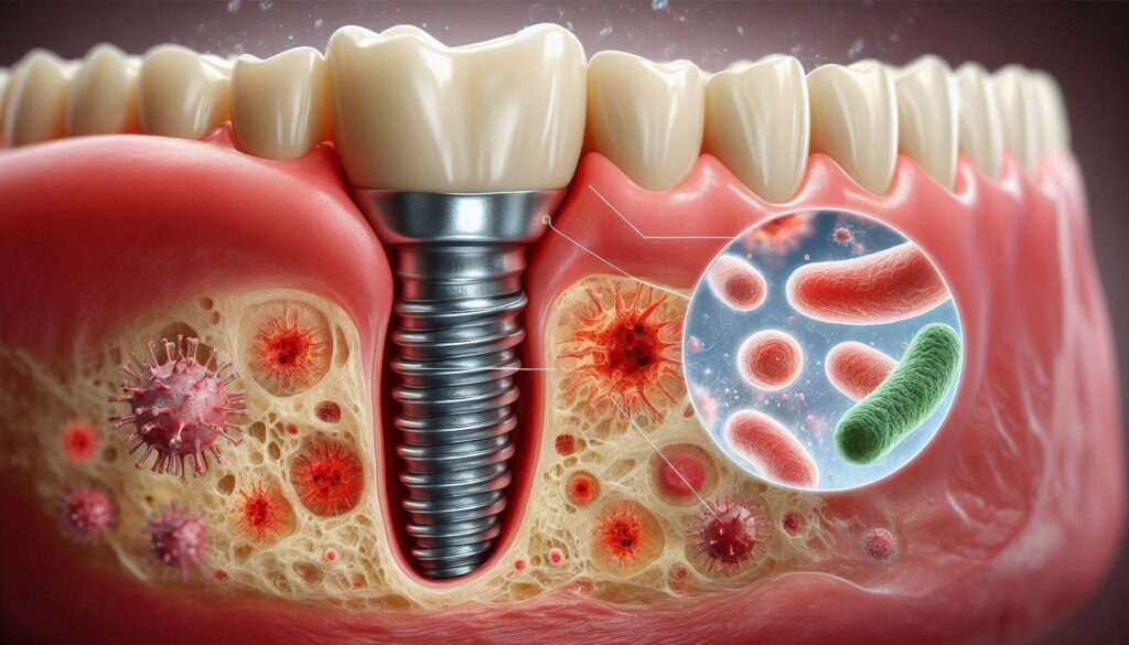
The single cause of a tooth implant infection is bacterial biofilm. This is a sticky, colorless, highly organized community of bacteria that adheres to surfaces in the mouth. It forms on natural teeth, and it forms with equal enthusiasm on the smooth titanium or zirconia surfaces of dental implants. The critical difference is how the surrounding tissues respond to this biofilm challenge. Around a natural tooth, the periodontal ligament and the dense connective tissue fibers that insert into the root cementum create a robust, resilient biological seal. This seal is a formidable barrier against the invasion of bacteria and their toxic byproducts.
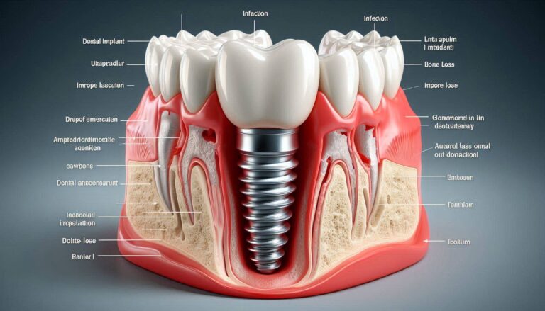
Around an implant, this seal is structurally and biologically weaker. The collagen fibers in the peri-implant mucosa run parallel to the implant surface rather than inserting perpendicularly into it. The attachment is a hemidesmosomal epithelial adhesion, a delicate interface. The blood supply to the peri-implant tissues is also less robust than around a natural tooth. This makes the soft tissue cuff around an implant more vulnerable to penetration by bacterial toxins. When biofilm is allowed to accumulate undisturbed along the gumline and in the sulcus around the implant, the bacteria provoke an inflammatory response. This initial stage, confined to the soft gum tissue, is called peri-implant mucositis. The gums become red, swollen, and bleed easily when probed or brushed. The implant is still solidly integrated in bone. This is the critical, curable stage.
The Progression from Mucositis to Peri-Implantitis
If the biofilm is not mechanically disrupted and removed, the inflammatory infiltrate extends deeper. The delicate epithelial seal breaks down. The inflammatory process reaches the underlying bone. This is the transition point from reversible mucositis to destructive peri-implantitis. The body’s own immune response, intended to fight the bacteria, becomes the agent of destruction. Pro-inflammatory cytokines, such as interleukin-1 and tumor necrosis factor-alpha, are released by immune cells in the infected tissues. These cytokines stimulate the activity of osteoclasts, the cells that resorb bone.
The bone begins to melt away from the implant surface. This bone loss is painless and often asymptomatic in its early stages. The patient may notice bleeding when brushing, a persistent bad taste, or, in advanced cases, gum recession that exposes the dark metal threads of the implant. The definitive diagnosis is made by the dentist, who uses a thin plastic or titanium probe to measure the depth of the pocket around the implant and takes a radiograph to compare the current bone level to the baseline image taken when the crown was placed. A crater-shaped defect of radiolucency around the implant is the hallmark image of peri-implantitis. The cause of this destruction is the body’s uncontrolled inflammatory response to a chronic, untreated biofilm infection. The implant itself is an innocent scaffold. The bacteria and the host response are the disease.
The Major Risk Factors That Accelerate Implant Infection
While biofilm is the necessary cause, a constellation of risk factors dramatically increases a patient’s susceptibility to implant infection and accelerates the rate of bone loss. The single most powerful predictor of future peri-implantitis is a history of chronic periodontitis. The pathogenic bacteria that caused the patient’s gum disease, including Porphyromonas gingivalis, Tannerella forsythia, and Treponema denticola, are anaerobic, gram-negative, highly virulent organisms. They do not disappear when the diseased teeth are extracted. They persist in the oral microbiome, colonizing the tongue, the tonsils, and the remaining teeth.
When an implant is placed in a mouth with a history of periodontitis, these same pathogens rapidly colonize the new implant surface. The patient’s immune system, genetically programmed to mount a hyper-inflammatory response to these specific bacteria, reacts with the same bone-destroying cascade that led to the original tooth loss. This is why placing an implant in a patient with untreated, active periodontal disease is a predictable failure. The patient must be periodontally stable before implant surgery. But “stable” does not mean “cured.” Periodontal disease is a chronic condition managed through meticulous lifelong maintenance. A patient with a periodontal history who lapses from their three-month professional recall schedule has a dramatically elevated risk of developing peri-implantitis.
Smoking, Diabetes, and Compromised Host Defenses
Smoking is the other towering risk factor. The chemicals in tobacco smoke, particularly nicotine and carbon monoxide, create a profoundly immunocompromised environment in the oral cavity. Nicotine is a potent vasoconstrictor. It reduces blood flow to the gums, depriving the tissues of oxygen and the white blood cells needed to fight infection. The visible inflammation, the redness and bleeding that is an early warning sign of disease, may be masked in a smoker because the blood vessels are constricted. The disease can progress silently, with significant bone loss occurring before any bleeding or swelling is noticed. Smokers have a significantly higher prevalence of peri-implantitis and a poorer response to treatment.
Uncontrolled diabetes mellitus is a systemic disease that impairs the body’s defense mechanisms on multiple fronts. Hyperglycemia impairs neutrophil function, the first-responder immune cells that fight bacterial invasion. It damages the microvasculature, reducing healing capacity. It increases the levels of inflammatory mediators in the gingival crevicular fluid. A diabetic patient with a poorly controlled HbA1c is a breeding ground for peri-implant infection. The risk is modifiable. A well-controlled diabetic patient with an HbA1c below 7 percent has a risk profile much closer to a non-diabetic patient. The key is strict glycemic control, maintained consistently over the life of the implant.
Iatrogenic Causes: When the Restoration Contributes to Infection
Not all implant infections are purely a consequence of patient behavior or systemic disease. Some are iatrogenic, meaning they are inadvertently caused by the dental treatment itself. A poorly designed or poorly fitted implant restoration can create an environment that is impossible for the patient to clean. An over-contoured crown, with a bulky emergence profile that traps food and plaque, becomes a chronic source of inflammation. A crown margin that does not fit precisely against the abutment leaves a microscopic gap, a dead space where bacteria can proliferate, protected from the patient’s oral hygiene efforts.
Perhaps the most significant iatrogenic cause is retained cement. In a cement-retained implant crown, the crown is glued onto the abutment with a dental cement. If excess cement is not meticulously and completely removed from the subgingival space around the implant, it becomes a foreign body nidus for bacterial colonization. The rough surface of the cement is a perfect plaque trap. It sits in the sulcus, constantly irritating the tissue and harboring bacteria. The bone loss from retained cement can be rapid and severe. This is a completely preventable cause of implant infection. Many dentists now prefer screw-retained implant crowns, which eliminate the risk of retained cement entirely, because the crown is secured with a small titanium screw rather than glue. If your implant crown is cement-retained, you should ask your dentist specifically about their protocol for ensuring complete cement removal.
The Importance of the Prosthetic Margin and Oral Hygiene Access
The junction where the implant abutment meets the implant fixture, and the junction where the crown meets the abutment, are potential weak points. A precision, machined fit between these components creates a seal that is resistant to bacterial penetration. A poorly machined connection, with a microscopic gap, can become a bacterial reservoir, leaking inflammatory byproducts into the surrounding bone and causing a persistent, low-grade infection that is resistant to treatment.
The design of the prosthesis must also allow you, the patient, to clean it effectively. If a full-arch implant bridge is designed with an overly bulky contour or if the access for a water flosser and interdental brushes is blocked, the patient is set up for failure. The dentist has a profound responsibility to design a prosthesis that is not just aesthetic and functional but also cleansable. The best implant restoration is one that the patient can easily maintain for a lifetime. Infection is often a failure of design, not just a failure of patient effort.
Recognizing the Early Signs of a Developing Infection
Because peri-implant infections are often painless in their early stages, you cannot rely on discomfort as your warning system. You must become a vigilant observer of the subtle signs. The earliest and most consistent symptom of peri-implant mucositis is bleeding. If your gums bleed when you brush, water floss, or insert an interdental brush around your implant, the tissue is inflamed. Healthy peri-implant tissue does not bleed. This is the equivalent of a dashboard warning light. Do not ignore it and assume it is normal. It is a sign of an active, albeit reversible, infection.
Other early warning signs include redness and swelling of the gum around the implant, a persistent bad breath or metallic taste that does not resolve with brushing and tongue cleaning, and, in more advanced cases, the appearance of pus or exudate when pressure is applied to the gum. A sign of established bone loss is gum recession, where the gum pulls away and exposes the darker metal of the implant post. The most ominous sign is mobility. A healthy, integrated implant has absolutely no movement. If you ever feel your implant shift or move, it is a dental emergency. This indicates a catastrophic loss of osseointegration and requires immediate professional evaluation. The window of opportunity for simple, non-surgical treatment closes as the disease progresses from mucositis to established peri-implantitis. Early detection, at the bleeding and probing depth stage, is the key to saving the implant.
Conclusion
A tooth implant infection is fundamentally caused by a bacterial biofilm that flourishes on the implant surface, initiating a reversible soft-tissue inflammation called peri-implant mucositis, which, if not disrupted by daily hygiene, progresses into the destructive bone loss of peri-implantitis driven by the body’s own hyper-inflammatory immune response. The risk of this infection is massively amplified by a history of periodontitis, smoking, and uncontrolled diabetes, and it can be directly caused by iatrogenic factors like retained dental cement or a poorly contoured restoration that defies cleaning. Your daily, meticulous mechanical disruption of the biofilm, combined with regular professional maintenance, is the only defense against this silent, progressive, and potentially implant-ending disease.
Frequently Asked Questions
Can an infected dental implant be saved?
Yes. Peri-implant mucositis is completely reversible with improved home care and a professional cleaning. Peri-implantitis with bone loss can often be arrested through surgical access, decontamination of the implant surface, and bone grafting, but the lost bone is rarely fully regained. Early detection is everything.
What does an infected implant look like?
In early stages, the gum around the implant looks red and swollen and bleeds easily. In advanced stages, you may see gum recession exposing the metal implant threads, pus, or a visible fistula. On an X-ray, a saucer-shaped dark shadow around the implant indicates bone loss.
Can I put hydrogen peroxide on my infected implant?
You should not self-treat with undiluted hydrogen peroxide or any other home remedy. Peri-implant diseases require professional mechanical debridement with specialized non-metal instruments to disrupt the biofilm without scratching the implant surface. Contact your implant dentist immediately.
Is implant infection more likely in the upper jaw?
There is no definitive evidence that infection risk is inherently higher in the maxilla, though the softer bone quality may allow for slightly more rapid bone loss once peri-implantitis is established. The risk is driven by hygiene, periodontal history, and smoking, not jaw location.
What is the best mouthwash to prevent implant infection?
No mouthwash can substitute for the mechanical disruption of biofilm with a water flosser and interdental brushes. An alcohol-free, chlorhexidine-based mouthwash may be prescribed for short-term use after surgery or during an acute infection flare-up, but the daily foundation of prevention is physical cleaning, not chemical rinsing.
Additional Resource
The American Academy of Periodontology is the leading professional organization for the prevention, diagnosis, and treatment of periodontal and peri-implant diseases. Their patient resources include detailed information on peri-implantitis and its management. Visit: https://www.perio.org/