What Is the Healing Process of a Dental Implant?
You leave the surgical office with a new titanium post anchored in your jawbone and a head full of questions about what happens next. The procedure itself is behind you, but the journey is far from complete. Beneath your gum tissue, hidden from view, a remarkable biological process is unfolding. Your body is making a decision about this new addition: accept it as part of you or isolate it as foreign material. That decision, made at the cellular level over weeks and months, will determine whether your implant succeeds or fails. Understanding the healing process transforms the waiting period from an anxious mystery into a fascinating and predictable biological sequence. This guide walks you through every phase of dental implant healing, from the moment you leave the surgical chair to the day your new crown is ready for function.
The Biological Foundation of Implant Healing
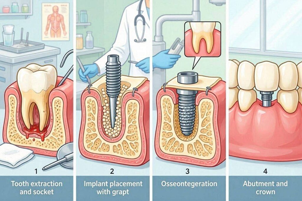
Dental implant healing is not a passive process where the body simply tolerates the implant. It is an active, energy-intensive biological program that creates a living connection between inert metal and dynamic bone. Understanding the fundamental biology sets the stage for understanding everything that follows.
What Osseointegration Actually Means
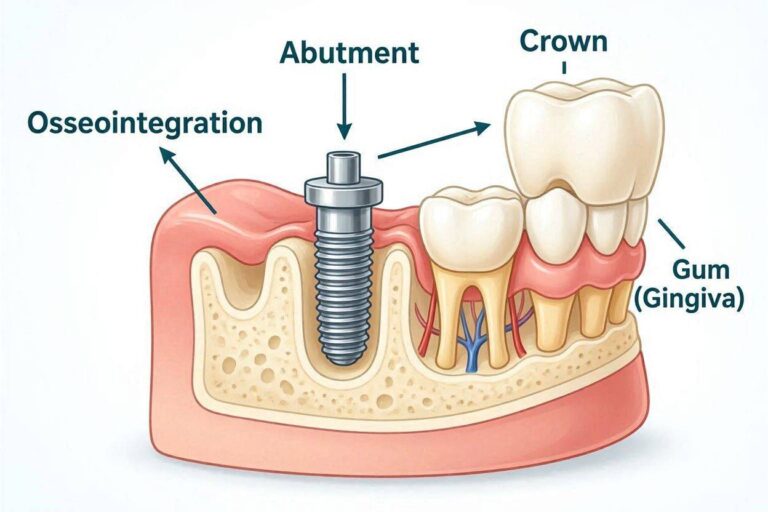
Osseointegration describes the direct structural and functional connection between ordered living bone and the surface of a load-bearing implant. This definition, established by Professor Per-Ingvar Brånemark who discovered the phenomenon, emphasizes that the connection is both anatomical and functional. The bone is not simply touching the implant. It is attached to it in a way that transmits forces from the implant to the skeleton.
At the microscopic level, osseointegration means that bone cells have deposited mineralized bone matrix directly onto the titanium oxide surface of the implant. There is no intervening layer of fibrous tissue, cartilage, or other material. The bone-to-implant contact occurs at the nanometer scale, with bone matrix proteins intertwined with the oxide layer.
This direct bone contact is what gives integrated implants their clinical rigidity. When the surgeon tests an integrated implant, it feels like it is part of the bone because biologically, it is. The implant cannot be removed without cutting it from the bone, just as a broken tooth root cannot be removed without surgical intervention.
Why Titanium Integrates With Bone
Titanium’s ability to osseointegrate is not shared by most materials. Stainless steel, for example, develops a fibrous capsule when placed in bone, isolating it from direct bone contact. Understanding why titanium behaves differently reveals the sophistication of the biological response.
The titanium dioxide layer that forms spontaneously on the implant surface is the key. This oxide layer is highly biocompatible, meaning the body does not recognize it as foreign in the way it recognizes other materials. Proteins in the blood and tissue fluids adsorb onto the oxide surface in a conformation that does not trigger the inflammatory foreign body response.
The oxide layer’s surface charge and wettability promote the adsorption of specific proteins that facilitate cell attachment. Fibronectin, vitronectin, and other cell adhesion proteins bind to the titanium dioxide surface in orientations that expose their cell-binding domains. Osteoblasts, the bone-forming cells, recognize these domains through integrin receptors and attach to the implant surface.
The microtopography of modern implant surfaces amplifies this response. Roughened surfaces created by sandblasting, acid etching, or anodization increase the surface area available for protein adsorption and cell attachment. The microscopic peaks and valleys provide mechanical interlocking for the forming bone and may directly stimulate osteoblast differentiation through mechanotransduction.
The Cellular Players in Implant Healing
Implant healing involves a coordinated sequence of cellular events, each phase preparing the way for the next. Understanding the major cell types and their roles illuminates the healing timeline.
Platelets are the first responders, arriving at the implant surface within seconds of placement. They adhere to the adsorbed protein layer and to each other, forming a provisional matrix on the implant surface. Activated platelets release growth factors including platelet-derived growth factor, transforming growth factor-beta, and vascular endothelial growth factor. These signaling molecules initiate the healing cascade.
Neutrophils and macrophages arrive within hours, part of the innate immune response. These cells clean the wound, removing bacteria, debris, and damaged tissue. Macrophages play a particularly important role, transitioning from a pro-inflammatory to a pro-healing phenotype as the wound environment changes. They release additional growth factors that recruit the cells responsible for new tissue formation.
Mesenchymal stem cells, the precursors to bone-forming osteoblasts, migrate to the implant surface from the surrounding bone marrow and periosteum. These cells are attracted by growth factor gradients established by platelets and macrophages. Upon reaching the implant surface, they differentiate into osteoblasts and begin the work of bone formation.
Endothelial cells form new blood vessels through angiogenesis, sprouting from existing vessels in the surrounding bone. Adequate blood supply is essential for delivering oxygen, nutrients, and additional cells to the healing site. The new vessels grow into the fibrin clot and the developing granulation tissue.
Osteoblasts are the bone-forming cells that create osseointegration. They synthesize and secrete the organic matrix of bone, primarily type I collagen, and then mineralize this matrix with calcium phosphate crystals. Osteoblasts on the implant surface lay down bone directly onto the titanium dioxide layer.
Phase One: Hemostasis and Initial Clot Formation
The healing process begins the moment the implant is seated in its prepared site and continues through the first hours after surgery.
The First Minutes After Placement
When the surgeon places the implant into the prepared osteotomy site, the space between the implant threads and the surrounding bone fills with blood. This blood comes from vessels cut during the drilling process and from the marrow spaces in the surrounding cancellous bone. The implant is bathed in blood containing all the cellular and molecular components necessary for healing.
The blood contacting the titanium surface immediately begins to change. Plasma proteins adsorb onto the oxide layer within milliseconds to seconds. The specific proteins that adsorb, and their conformation on the surface, influence everything that follows. Fibrinogen, fibronectin, and other adhesion proteins create the provisional matrix onto which cells will later attach.
The coagulation cascade activates through both the intrinsic and extrinsic pathways. Thrombin converts fibrinogen to fibrin, creating a three-dimensional mesh that stabilizes the clot. Platelets become entrapped in this fibrin mesh and begin releasing their granule contents, flooding the local environment with growth factors.
The Protective Role of the Blood Clot
The blood clot that forms around the implant serves multiple essential functions beyond simply filling space. Understanding these functions explains why clot disruption is so detrimental to implant healing.
The clot provides the provisional matrix for cell migration. The fibrin mesh, with its adsorbed proteins and embedded growth factors, creates the scaffold onto which cells crawl as they move toward the implant surface. Without this matrix, cell migration is haphazard and bone formation is compromised.
The clot releases a sustained supply of growth factors as platelets degranulate and then as macrophages process the clot during later phases. These growth factors establish the chemotactic gradients that attract mesenchymal stem cells and other healing cells to the implant surface.
The clot protects the implant surface from oral fluids and bacteria during the initial healing period. In two-stage implant procedures where the implant is buried under closed gum tissue, this protection is supplemented by the soft tissue closure. In one-stage procedures with a healing abutment, the clot is protected by the overlying tissue and the patient’s careful avoidance of the surgical site.
Why Clot Stability Matters
Micromovement of the implant during the clot phase disrupts the delicate fibrin scaffold and can cause the healing process to divert toward fibrous encapsulation rather than osseointegration. The critical threshold for micromovement appears to be approximately 100 to 150 microns. Movement below this threshold may not disrupt healing. Movement above it directs mesenchymal stem cells toward fibroblast rather than osteoblast differentiation.
This is why achieving primary stability at implant placement is so critical. The implant must be firmly anchored in the bone at the time of surgery so that functional forces during the healing period do not cause excessive micromovement. The surgeon tests stability at placement and may modify the treatment plan if adequate stability is not achieved.
Patient behavior during the initial healing period also affects clot stability. Chewing on the implant site, manipulating the area with the tongue, and aggressive rinsing or spitting can all disrupt the forming clot. The post-operative instructions to maintain a soft diet and avoid the surgical site exist to protect this vulnerable phase of healing.
Phase Two: Inflammatory Phase
The inflammatory phase overlaps with hemostasis and extends through the first three to five days after implant placement. This phase is essential for healing, not a complication to be suppressed.
The Purpose of Post-Surgical Inflammation
Inflammation carries a negative connotation for many patients, who associate it with infection and complications. However, the acute inflammatory response to implant placement is a normal and necessary part of the healing process. Without it, the subsequent phases of healing cannot proceed.
The inflammatory response serves to clean the wound. Neutrophils and macrophages phagocytose bacteria, debris, and damaged tissue fragments from the surgical site. This cleaning function reduces the risk of infection and prepares the site for new tissue formation.
Inflammatory cells, particularly macrophages, release the growth factors and cytokines that orchestrate the next phases of healing. These signaling molecules recruit mesenchymal stem cells, stimulate angiogenesis, and activate the cells that will form new bone. The inflammatory phase establishes the molecular environment for repair.
The inflammatory phase also increases vascular permeability, allowing cells and plasma proteins to exit the bloodstream and enter the healing site. This increased permeability produces the swelling that patients experience after surgery. The swelling is a visible manifestation of the healing process at work.
What Patients Experience During This Phase
The inflammatory phase corresponds to the period when patients experience the most noticeable post-operative symptoms. Understanding these symptoms as normal healing phenomena rather than complications reduces anxiety.
Swelling peaks during this phase, typically reaching its maximum 48 to 72 hours after surgery. The swelling is a direct result of increased vascular permeability and fluid accumulation in the tissues. It resolves as the inflammatory phase transitions to the proliferative phase and the lymphatic system clears the excess fluid.
Pain and discomfort are most pronounced during the inflammatory phase, corresponding to the release of inflammatory mediators that sensitize nerve endings. The pain should be manageable with prescribed or over-the-counter analgesics. Pain that is not controlled by medication or that intensifies after day three may indicate infection rather than normal inflammation.
Redness and warmth at the surgical site reflect increased blood flow, another component of the inflammatory response. These signs should be confined to the immediate surgical area and should begin to diminish as the inflammatory phase resolves.
The Transition from Inflammation to Proliferation
The inflammatory phase does not end abruptly but transitions gradually into the proliferative phase as the cellular population in the wound changes. Macrophages shift from a pro-inflammatory to a pro-healing phenotype. Neutrophil numbers decline. Fibroblasts and endothelial cells become the dominant cell types.
This transition is orchestrated by changes in the growth factor environment. The initial burst of pro-inflammatory cytokines gives way to growth factors that promote tissue formation. The fibrin clot is gradually replaced by granulation tissue, a highly vascularized provisional tissue that will serve as the substrate for new bone formation.
The clinical correlate of this transition is the improvement patients experience after the first three to five days. Swelling begins to subside. Pain decreases. The surgical site feels less tender. These improvements signal that the healing process is progressing normally from its inflammatory to its proliferative phase.
Phase Three: Proliferative and Granulation Tissue Phase
The proliferative phase spans approximately day four through day fourteen after implant placement, though the timing varies by individual and by the specific surgical procedure.
Angiogenesis and New Blood Vessel Formation
Adequate blood supply is essential for all subsequent healing. The proliferative phase establishes this supply through angiogenesis, the formation of new blood vessels from existing ones. Without new vessels, the forming bone would be starved of oxygen and nutrients.
Endothelial cells in the walls of existing blood vessels are activated by growth factors released during the inflammatory phase, particularly vascular endothelial growth factor. These cells degrade the basement membrane of the parent vessel, proliferate, and migrate into the surrounding tissue as solid cords of cells.
The cords of endothelial cells develop a lumen, forming new capillaries. These capillaries connect to create a vascular network throughout the granulation tissue. The new vessels are initially leaky, which contributes to the continuing but diminishing edema during this phase. Over time, the vessels mature and develop tighter junctions.
The vascular network that develops during this phase will supply the osteoblasts that form new bone. The density and organization of this vascular network influence the rate and quality of bone formation. Areas with poor vascularity heal more slowly and may form less bone.
Granulation Tissue Formation
Granulation tissue is the provisional tissue that fills the healing wound. Its name derives from the granular appearance it presents on wound surfaces, caused by the numerous small blood vessels protruding through the tissue surface.
The granulation tissue matrix is produced by fibroblasts that have migrated into the wound. These fibroblasts synthesize collagen and other extracellular matrix components that provide structural support for the developing tissue. The matrix is initially disorganized but becomes more structured over time.
Granulation tissue serves as the scaffold for the next phase of healing. The matrix components bind growth factors and provide attachment sites for migrating cells. The vascular network delivers oxygen and nutrients. The cellular population includes the mesenchymal stem cells that will differentiate into osteoblasts when bone formation begins.
Around dental implants, granulation tissue fills the space between the implant surface and the surrounding bone. The tissue is in direct contact with the implant surface, and the cells within it will determine whether bone or fibrous tissue forms at this interface.
Wound Contraction and Early Tissue Maturation
During the later portion of the proliferative phase, the wound begins to contract. Myofibroblasts, specialized fibroblasts with contractile properties, pull the wound edges together, reducing the wound volume. This contraction is more relevant for soft tissue healing than for the bone-implant interface, but it contributes to the overall closure and stabilization of the surgical site.
The granulation tissue begins to mature, with collagen fibers becoming more organized and the vascular density decreasing. This maturation prepares the tissue for the next phase, in which the provisional matrix will be replaced by specialized tissues, bone in the case of the implant interface.
For implants placed with a one-stage protocol, the healing abutment protruding through the gum is surrounded by maturing granulation tissue that will form the peri-implant soft tissue collar. The tissue around the abutment gradually adapts, creating the seal that will protect the underlying bone-implant interface.
Phase Four: Bone Formation and Osseointegration
The bone formation phase is when osseointegration actually occurs. This phase begins within the first week after implant placement and continues for months, overlapping with the proliferative phase and extending well beyond it.
Osteoblast Migration and Differentiation
Mesenchymal stem cells that migrated into the granulation tissue during the proliferative phase now differentiate into osteoblasts under the influence of bone morphogenetic proteins and other osteoinductive growth factors. These signals direct the stem cells toward the bone-forming lineage rather than the fibroblast lineage.
The differentiation process involves the sequential activation of genes characteristic of the osteoblast phenotype. The cells begin expressing alkaline phosphatase, an enzyme essential for mineralization. They produce type I collagen, the primary organic component of bone matrix. They express osteocalcin and other bone-specific proteins.
Osteoblasts migrate to the implant surface, attracted by the chemotactic gradients established during earlier phases. The cells that reach the implant surface attach to the adsorbed protein layer through integrin receptors. This attachment is a critical step. Cells that successfully attach to the surface will proceed to form bone directly on it. Cells that do not reach the surface will form bone that approaches but does not contact the implant.
Woven Bone Formation
The first bone formed at the implant surface is woven bone, a relatively disorganized type of bone that forms rapidly. Woven bone is characterized by randomly oriented collagen fibers and a high proportion of osteocytes, the cells that osteoblasts become once they are surrounded by mineralized matrix.
Woven bone formation begins within the first week after implant placement and is well underway by two to four weeks. The bone forms directly on the implant surface in areas where osteoblasts have attached. In the spaces between the implant threads and the prepared bone wall, woven bone forms from both the implant surface outward and the bone wall inward, eventually meeting to fill the space.
Woven bone provides the initial mechanical stability that supplements the primary stability achieved at implant placement. As the woven bone matures and remodels, the implant stability typically dips slightly during the first few weeks after placement, then progressively increases as bone formation and maturation continue.
This dip in stability, sometimes called the stability dip, corresponds to the period when the bone damaged during drilling is being resorbed and replaced by new bone. The primary mechanical stability from the initial implant-bone contact decreases before the secondary biological stability from new bone formation increases. The implant is most vulnerable during this transition.
Lamellar Bone Formation and Maturation
Woven bone is a temporary tissue. Over weeks to months, it is replaced by lamellar bone, the mature, organized bone that provides long-term implant support. This replacement occurs through the coupled processes of bone resorption by osteoclasts and bone formation by osteoblasts, collectively termed bone remodeling.
Lamellar bone is characterized by highly organized collagen fibers arranged in parallel sheets, or lamellae. This organization gives lamellar bone its superior mechanical properties compared to woven bone. The mineralization density of lamellar bone is higher, providing greater strength and stiffness.
The remodeling process that converts woven to lamellar bone also adapts the bone architecture to the mechanical forces transmitted through the implant. Bone is deposited where forces are highest and resorbed where forces are lowest, optimizing the bone structure for its load-bearing function. This is Wolff’s law applied to implant support.
The maturation of lamellar bone continues for many months. While osseointegration sufficient to support a restoration is typically achieved by three to four months in the mandible and four to six months in the maxilla, the bone around the implant continues to remodel and mature for a year or longer. Annual remodeling continues throughout the implant’s lifetime as part of normal bone maintenance.
Phase Five: Soft Tissue Healing Around Implants
While bone healing is occurring at the implant interface, the overlying soft tissue is undergoing its own healing process. The quality of this soft tissue healing is critical for the long-term health and esthetics of the implant restoration.
Epithelial and Connective Tissue Attachment
The soft tissue surrounding an implant differs fundamentally from the gingiva around natural teeth. Understanding these differences illuminates both the strengths and vulnerabilities of the peri-implant soft tissue seal.
Around natural teeth, connective tissue fibers insert perpendicularly into the cementum, creating a tight seal that resists bacterial penetration. The junctional epithelium attaches to the tooth surface through hemidesmosomes and a basal lamina, forming an epithelial barrier.
Around implants, the connective tissue fibers run parallel to the implant surface rather than inserting into it. This parallel orientation creates a weaker mechanical seal than the perpendicular fiber insertion around teeth. The junctional epithelium attaches to the implant surface in a manner similar to its attachment to teeth, forming an epithelial barrier.
The biological width around implants, the dimension of soft tissue from the bone crest to the gingival margin, is typically 3 to 4 millimeters. This includes approximately 2 millimeters of connective tissue and 1 to 2 millimeters of junctional epithelium. Maintaining this biological width is important for stable soft tissue levels around the implant.
The Formation of the Peri-Implant Soft Tissue Seal
The soft tissue around healing abutments or implant-supported temporary restorations matures over several weeks. The tissue adapts to the contour of the abutment or restoration, forming a collar of keratinized tissue that provides the first line of defense against bacterial invasion.
The maturation of the soft tissue seal involves the development of a sulcus, a shallow crevice between the tissue and the abutment surface. The sulcus depth around healthy implants is typically 2 to 4 millimeters, similar to natural teeth. Deeper probing depths may indicate peri-implant disease or excessive tissue thickness.
The quality of the soft tissue, including its thickness and the presence of keratinized tissue, influences its resistance to recession and inflammation. Thicker tissue with an adequate zone of keratinized gingiva is more resistant to recession and provides better long-term esthetic stability.
Soft Tissue Maturation Timeline
Immediately after implant placement or abutment connection, the soft tissue is edematous and inflamed from the surgical procedure. Over the first two weeks, the acute inflammation resolves and the tissue begins to adapt to the abutment contours.
By four to six weeks, the soft tissue has matured sufficiently for final impression procedures. The tissue collar is stable, with a defined sulcus and consistent contour. Impressions taken before this maturation may capture tissue that is still changing, resulting in a restoration that does not fit optimally as the tissue continues to remodel.
Complete soft tissue maturation continues for six to twelve months after restoration delivery. During this period, the tissue remodels in response to the restoration contours, oral hygiene, and functional forces. Minor changes in tissue contour and position are normal during this maturation period.
The Complete Healing Timeline
Understanding the chronological sequence of implant healing helps patients set realistic expectations and recognize when healing may be deviating from the normal course.
Week by Week Breakdown
| Time Period | Bone Healing Events | Soft Tissue Events | Patient Experience |
|---|---|---|---|
| Day 1-3 | Clot formation, inflammatory cell infiltration | Swelling, initial wound closure | Swelling peaks, discomfort managed with medication |
| Day 4-7 | Early granulation tissue, beginning of woven bone formation | Swelling resolves, tissue begins adaptation | Decreasing discomfort, return to normal activities |
| Week 2 | Active woven bone formation | Epithelial closure, early tissue maturation | Minimal discomfort, sutures dissolve or removed |
| Week 3-4 | Continued woven bone formation, early remodeling | Soft tissue collar forming around abutment | No discomfort, adjusting to healing abutment |
| Month 2 | Transition from woven to lamellar bone | Soft tissue maturation continues | Normal sensation, awaiting restoration |
| Month 3-4 | Lamellar bone maturation, sufficient for loading (mandible) | Soft tissue mature enough for impressions | Impressions for final restoration |
| Month 4-6 | Continued maturation (maxilla) | Soft tissue stable | Final crown delivery |
| Month 6-12 | Ongoing remodeling, full bone maturation | Final soft tissue adaptation | Implant in full function |
This timeline represents typical healing in healthy patients with good bone quality. Individual healing rates vary, and the surgeon evaluates readiness to proceed based on clinical and radiographic assessment rather than rigid adherence to the calendar.
Factors That Accelerate or Delay Healing
Several factors influence the rate and quality of implant healing. Younger patients tend to heal faster than older patients, though chronological age is less important than biological age and overall health. Good bone quality heals faster than poor bone quality, which is why mandibular implants typically heal faster than maxillary implants.
Smoking delays healing through vasoconstriction, reduced oxygen delivery, and direct toxic effects on healing cells. Smokers should be counseled that their healing timeline may be extended and their risk of complications is higher than non-smokers.
Systemic conditions including diabetes, autoimmune diseases, and immunocompromised states affect healing. Well-controlled diabetes with hemoglobin A1c below 7 percent allows healing approaching that of non-diabetic patients. Poorly controlled diabetes significantly impairs bone formation and increases infection risk.
Medications that affect bone metabolism, including bisphosphonates and certain biologics, can interfere with normal bone healing around implants. Patients taking these medications require careful evaluation and may not be candidates for implant placement.
How Surgeons Determine Healing Is Complete
The decision to proceed with the restorative phase is based on multiple assessments, not a single test or timeline. Clinical evaluation includes assessing the soft tissue health, the absence of inflammation, and the stability of the implant when tested manually or with instruments.
Radiographic evaluation confirms that bone appears to be in contact with the implant surface without radiolucent lines. Comparison with immediate post-placement radiographs shows that the bone-implant interface has matured as expected. Bone levels should be stable at the implant platform.
Resonance frequency analysis, when available, provides an objective measure of implant stability. The implant stability quotient should be stable or increasing compared to placement values. Values above 60 to 65 generally indicate sufficient stability for loading.
Insertion torque measured during abutment connection provides additional information. If the implant was placed with a two-stage protocol, the surgeon assesses stability when the cover screw is removed and the healing abutment placed. An implant that has osseointegrated will feel solidly fixed.
Healing Complications and Their Recognition
Most implant healing proceeds without complication, but patients should be aware of the signs that healing may be deviating from the normal course.
Infection During Healing
Post-operative infection presents differently than normal inflammatory healing. While some swelling and discomfort are expected, infection produces worsening symptoms after the initial improvement expected during the first week.
Signs of infection include increasing pain after day three or four, swelling that increases rather than decreases after day three, redness that expands beyond the immediate surgical site, purulent discharge, and systemic symptoms including fever and malaise.
Infection during the healing period threatens osseointegration. The inflammatory environment favors fibrous tissue formation over bone formation. Prompt treatment with antibiotics and possibly surgical drainage may salvage the implant if intervention occurs early.
Failed Osseointegration
Failed osseointegration means the implant has not achieved bone-to-implant contact and is instead surrounded by fibrous tissue. This failure may be discovered when the implant is uncovered for the restorative phase or may present with symptoms during the healing period.
Signs of failed osseointegration include persistent discomfort, mobility when the implant is tested, a radiolucent line around the implant on radiographs, and a dull sound when the implant is percussed rather than the sharp ring of an integrated implant.
Failed osseointegration cannot be reversed. The implant must be removed, the site allowed to heal, and a new implant placed after adequate bone healing and any necessary grafting. The replacement implant typically requires the same healing timeline as the original.
Early Bone Loss
Some crestal bone loss around implants during the first year is considered normal remodeling related to the establishment of biological width. However, excessive early bone loss indicates a problem with healing or with the implant position or restoration.
Bone loss greater than 1.5 millimeters during the first year, or progressive bone loss that continues after the first year, is considered pathological. Causes include infection, excessive occlusal forces, implant malposition, or a host response that is incompatible with implant maintenance.
Early detection of bone loss through radiographic monitoring allows intervention before the implant is compromised. Improved oral hygiene, professional debridement, and occlusal adjustment may arrest early bone loss and prevent progression to implant failure.
Optimizing Healing Through Patient Behavior
The patient’s actions during the healing period directly influence healing outcomes. Understanding what helps and what harms empowers patients to protect their investment.
Nutrition for Optimal Bone Healing
Bone formation requires adequate nutritional substrates. Protein provides the amino acids for collagen synthesis. Calcium and phosphate are the minerals that form bone crystal. Vitamin D regulates calcium absorption and bone mineralization. Vitamin C is essential for collagen cross-linking.
A balanced diet with adequate protein, dairy products or calcium-fortified alternatives, and fruits and vegetables provides the necessary nutrients for healing. Patients with restricted diets may benefit from supplementation after consultation with their surgeon.
Hydration is important for all healing processes. Adequate fluid intake maintains blood volume, supports nutrient delivery to the healing site, and facilitates waste removal. Water is the preferred fluid, with alcohol avoided during the healing period due to its negative effects on healing and potential interactions with medications.
Activities That Promote Healing
Gentle physical activity promotes circulation and overall health without stressing the surgical site. Walking and normal daily activities are encouraged. Strenuous exercise should be avoided for the period specified by the surgeon, typically three to seven days.
Stress management supports healing through its effects on immune function and inflammation. Chronic stress increases cortisol levels, which can impair wound healing. Relaxation techniques, adequate sleep, and maintaining normal routines as much as possible support the healing process.
Compliance with post-operative instructions, including medication schedules, oral hygiene modifications, and dietary restrictions, directly affects healing outcomes. These instructions are based on decades of clinical experience and research into what promotes successful osseointegration.
Behaviors That Impair Healing
Smoking is the single most damaging behavior for implant healing. The vasoconstrictive effect of nicotine reduces blood flow and oxygen delivery to the healing site. Carbon monoxide from tobacco smoke binds to hemoglobin, further reducing oxygen availability. The chemicals in tobacco directly impair fibroblast and osteoblast function. Smokers should quit before implant placement and remain abstinent throughout the healing period.
Alcohol consumption impairs wound healing through multiple mechanisms including nutritional effects, immune suppression, and direct toxicity to healing cells. Alcohol should be avoided during the initial healing period and limited thereafter.
Poor oral hygiene allows bacterial accumulation that can infect the surgical site or cause peri-implant inflammation. While the surgical site itself must be protected from direct brushing during the initial healing period, the rest of the mouth should be kept clean to reduce the overall bacterial load.
Conclusion
The healing process of a dental implant proceeds through distinct biological phases beginning with hemostasis and clot formation in the first hours, progressing through an inflammatory phase that peaks at 48 to 72 hours, transitioning to a proliferative phase with granulation tissue formation and angiogenesis over the first two weeks, and culminating in bone formation and remodeling that continues for months. Osseointegration, the direct structural connection between living bone and the implant surface, begins with woven bone formation within the first week and matures through remodeling into lamellar bone over three to six months, at which point the implant can support functional loading. Soft tissue healing parallels bone healing, with the peri-implant mucosa forming a protective seal around the implant or abutment that matures over four to six weeks. The patient’s role in protecting the healing site through diet modification, oral hygiene, avoidance of smoking and alcohol, and compliance with post-operative instructions directly influences the success of this complex biological process and the long-term survival of the implant.
Frequently Asked Questions
How long does it take for a dental implant to fully heal?
Complete healing including bone maturation continues for six to twelve months after implant placement. However, the implant can typically support a restoration after three to four months in the lower jaw and four to six months in the upper jaw. The surgeon determines when healing is sufficient based on clinical and radiographic evaluation.
What does normal healing feel like?
Normal healing involves swelling that peaks at two to three days and resolves over one to two weeks, discomfort that is manageable with medication and decreases after the first few days, and a gradual return to normal sensation. After the first two weeks, the implant site should be essentially comfortable, though some awareness of the healing abutment is normal.
How do I know if my implant is healing properly?
Proper healing is characterized by decreasing swelling and discomfort, pink and firm gum tissue around the implant site, absence of pus or persistent bleeding, and radiographic evidence of bone contact with the implant surface. Your surgeon evaluates healing at follow-up appointments and confirms readiness for restoration.
Can I feel the implant healing?
Osseointegration itself is not perceptible. The bone formation and remodeling occur at the microscopic level without sensation. After the initial post-operative period, a healing implant should be essentially symptom-free. Pain, swelling, or other symptoms after the first two weeks may indicate a problem.
Why does healing take longer in the upper jaw?
The maxilla typically has less dense bone than the mandible, with a thinner cortical layer and more porous cancellous bone. This lower density bone provides less initial stability and requires more time for the new bone formation and maturation that create osseointegration. The posterior maxilla, where bone density is lowest, requires the longest healing times.
What happens if I accidentally chew on the implant side?
Occasional light contact is unlikely to cause problems if the implant has good primary stability. However, repeated loading or heavy chewing on the implant site during healing can cause micromovement that disrupts osseointegration. Contact your surgeon if you have concerns about accidental loading.
Can healing be accelerated?
There are no proven methods to significantly accelerate implant osseointegration beyond the body’s natural healing rate. Good nutrition, adequate sleep, stress management, and avoidance of smoking and alcohol support optimal healing but do not dramatically shorten the timeline. Attempts to rush the process by loading the implant early increase failure risk.
Is it normal to see the implant through the gum?
In one-stage procedures, the healing abutment is intentionally visible through the gum. The implant body itself should not be visible. If you see what appears to be the implant body threads through the gum, this indicates tissue recession or bone loss that requires professional evaluation.
Additional Resource
Academy of Osseointegration: Patient Information
https://osseo.org/patient-resources/