Can MRI Be Done with Dental Crowns What You Should Know
Preparing for a medical scan often brings up many questions about your health history. If you have restorative work in your mouth, you might wonder: can MRI be done with dental crowns safely? It is a common concern for many patients who want to ensure their upcoming diagnostic procedure goes smoothly.

can mri be done with dental crowns
Modern imaging technology is highly advanced, yet understanding the materials inside your mouth remains essential for peace of mind. Most modern restorations are non-magnetic, meaning they typically do not interfere with the powerful magnets used during your scan. By learning about the composition of your specific hardware, you gain the confidence needed to communicate effectively with your medical team.
Key Takeaways
- Most modern restorative materials are safe for magnetic resonance imaging.
- Always inform your technician about any metal implants or restorations.
- Non-magnetic materials prevent image distortion during the scanning process.
- Open communication with your dentist helps clarify the composition of your hardware.
- Understanding your health history reduces unnecessary anxiety before your appointment.
Understanding MRI Technology and Dental Materials
Many patients wonder how their dental restorations behave when exposed to the powerful forces of an MRI machine. Prioritizing MRI safety for dental work requires a basic understanding of how these sophisticated diagnostic tools function in a clinical setting.
How Magnetic Resonance Imaging Works
Magnetic Resonance Imaging, or MRI, uses a powerful magnetic field and radio waves to generate detailed images of your internal structures. Unlike X-rays, this technology does not rely on ionizing radiation to capture data.
The machine aligns the hydrogen atoms in your body using a strong, constant magnetic field. Once aligned, radio frequency pulses are applied to disrupt this alignment, causing the atoms to emit signals that a computer translates into high-resolution images.
The Interaction Between Magnetic Fields and Metal
The primary concern regarding MRI safety for dental work involves how specific metals react to the intense magnetic pull. Some materials are ferromagnetic, meaning they are strongly attracted to magnets, which can cause movement or heating.
“The safety of a patient with metallic implants depends largely on the magnetic properties of the specific alloy used in their dental restoration.”
— Radiology Imaging Standards
Most modern dental crowns are crafted from materials that are non-ferromagnetic, making them generally safe for the imaging environment. The following table outlines how different materials typically behave during a scan:
| Material Type | Magnetic Property | MRI Compatibility |
| Pure Gold | Non-ferromagnetic | Safe |
| Ceramic/Porcelain | Non-metallic | Safe |
| Stainless Steel | Ferromagnetic | Requires Caution |
| Titanium Alloys | Paramagnetic | Generally Safe |
Understanding these material properties is a vital step in ensuring MRI safety for dental work. Always consult with your healthcare provider to confirm that your specific dental hardware is compatible with the imaging equipment being used.
Can MRI Be Done with Dental Crowns
If you are wondering whether your dental crown will interfere with an MRI, you are not alone. Many patients feel anxious about how their dental hardware might react to the powerful magnetic fields used in imaging technology. Fortunately, the vast majority of people can undergo these scans without any complications.
General Safety Guidelines for Dental Restorations
When you arrive for your appointment, the medical team will prioritize MRI safety for dental work by asking you to complete a screening form. This document helps the staff identify the specific materials present in your mouth. Radiologists follow strict protocols to ensure that any metallic components are compatible with the high-strength magnets in the scanner.
Transparency is key during this process. By providing an accurate history of your dental procedures, you allow the technician to make an informed decision about your safety. Most modern facilities are well-equipped to handle patients with various types of dental restorations, ensuring that your diagnostic needs are met safely.
Why Most Crowns Are MRI-Safe
The primary reason that can mri be done with dental crowns is that most modern restorations are crafted from non-ferromagnetic materials. Unlike older hardware that might contain iron or nickel, today’s porcelain, ceramic, and high-noble metal crowns do not react to the magnetic pull of the scanner. This makes dental restoration safety a standard expectation for most patients.
Because these materials are inert, they do not pose a risk of movement or heating during the imaging process. You can feel confident and relaxed knowing that your crown is unlikely to interfere with the quality of your images or your physical well-being. If you have specific concerns about the age or composition of your crown, a quick conversation with your dentist can provide extra peace of mind.
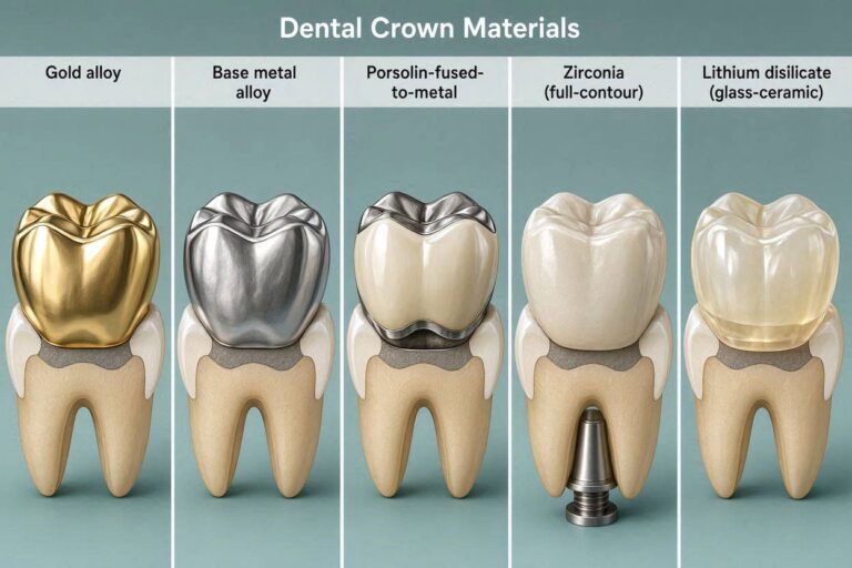
Common Materials Used in Dental Crowns
Understanding the composition of your dental work is a vital step in ensuring your dental restoration safety during medical imaging. Dental crowns are crafted from various dental crown materials, each reacting differently to the magnetic fields found in an MRI machine.
Knowing exactly what your crown is made of allows your radiologist to assess potential risks before your scan begins. This preparation helps ensure a smooth experience and high-quality diagnostic results.
Porcelain and Ceramic Crowns
These restorations are highly popular because they mimic the appearance of natural teeth. Because they contain no metal, they are generally considered the gold standard for porcelain crown MRI safety.
- They are non-conductive and non-ferromagnetic.
- They do not cause image distortion or artifacts.
- They are safe for all types of MRI scans.
Gold and Precious Metal Alloys
Gold has been used in dentistry for decades due to its durability and biocompatibility. While these crowns contain metal, they are typically made from noble alloys that are not ferromagnetic.
This means they do not move or heat up significantly in the presence of a magnetic field. However, they may still cause minor signal loss in the immediate vicinity of the tooth.
Porcelain-Fused-to-Metal (PFM) Crowns
PFM crowns combine the strength of a metal base with the aesthetic appeal of porcelain. The presence of metal in dental crowns can sometimes lead to small artifacts on an MRI scan.
The extent of this interference depends on the specific alloy used in the substructure. Most modern PFM crowns are safe, but it is important to inform your technician about their presence.
| Material Type | Ferromagnetic | MRI Impact |
| All-Ceramic | No | None |
| Gold Alloy | No | Minimal |
| PFM | Varies | Low to Moderate |
How Metal in the Mouth Affects MRI Image Quality
When you have metal in dental crowns, it is natural to wonder how this might influence your medical imaging results. While these restorations are typically safe, they can occasionally create visual interference during your scan.

Edit
Full screen
Delete
MRI image artifacts
Understanding Artifacts in Imaging
In the world of radiology, these visual disturbances are known as MRI image artifacts. These appear as dark spots or streaks that can obscure the surrounding anatomy on your scan.
Think of these artifacts as a form of “visual noise” that happens when the scanner’s signal is interrupted. While they can be distracting, they do not necessarily mean your scan is invalid or dangerous.
The Impact of Ferromagnetic Materials
The primary cause of these distortions is the presence of ferromagnetic materials. These substances react strongly to the powerful magnetic fields generated by the MRI machine.
When these materials are present, they can pull on the magnetic field lines, causing a localized MRI image distortion. This effect is most common with older, non-precious metal alloys that contain iron, nickel, or cobalt.
Techniques Radiologists Use to Minimize Distortion
Fortunately, modern medical technology offers several ways to manage these effects. Radiologists are highly skilled at adjusting scan parameters to ensure you receive a clear and accurate diagnostic result.
They may use specific software sequences designed to reduce the impact of metal on the final image. Additionally, technicians might adjust your head position to move the dental work further away from the area of interest, which helps significantly improve the overall clarity of the scan.
Potential Risks and Safety Considerations
Although rare, some patients may experience minor sensations due to their dental hardware during an imaging scan. Being aware of these possibilities helps you stay calm and prepared for your appointment. Proactive communication with your medical team remains the best way to ensure a smooth experience.
Heating and Discomfort During the Scan
One of the primary concerns for patients with metal restorations involves MRI heating risks. In specific cases, the radiofrequency energy used by the scanner can cause a slight increase in temperature within metallic components. While this is usually negligible, some individuals might notice a mild, localized warmth in their mouth.
This sensation is typically temporary and fades quickly once the scan concludes. If you feel any unusual heat, it is important to remain still and use the provided call button. Your comfort is a priority, and technicians are trained to adjust settings to minimize these effects.
Movement of Dental Hardware
Concerns regarding the physical movement of dental work often stem from the presence of ferromagnetic dental materials. These specific metals can react to the powerful magnetic field of the scanner. While modern dental crowns are rarely made of highly magnetic alloys, the potential for a slight pulling sensation exists in older or non-standard restorations.
It is important to note that most dental crowns are securely bonded to the tooth structure. This strong attachment prevents any actual displacement of the hardware. Feeling a slight vibration or pressure is a common report, but it does not indicate that your crown is coming loose.
When to Alert the Medical Staff
Clear communication is essential if you notice anything unexpected during your procedure. You should immediately notify the technician if you experience sharp pain, significant discomfort, or a feeling of shifting in your dental work. These symptoms can also help the team identify potential MRI image distortion, which might affect the clarity of your results.
The following table outlines common sensations and the recommended actions for patients:
| Sensation | Likelihood | Recommended Action |
| Mild warmth | Low | Continue scan; report after |
| Slight vibration | Very Low | Stay still; notify technician |
| Sharp pain | Extremely Rare | Press call button immediately |
| Shifting sensation | Extremely Rare | Stop scan; consult dentist |
By staying alert and keeping your technician informed, you ensure that your safety is maintained throughout the entire imaging process. Your peace of mind is just as important as the quality of the diagnostic images produced.
Communicating with Your Radiologist and Dentist
Preparing for your scan involves more than just arriving on time; it requires a proactive approach to sharing your dental history for MRI. Clear communication between you, your dentist, and the radiology team is the cornerstone of a successful imaging experience. By taking these steps early, you ensure that everyone is on the same page regarding your safety.
Providing Your Dental History
Before your appointment, gather specific details about your dental work to share with the facility. You should know the types of materials used in your crowns, bridges, or implants. If you are unsure, a quick call to your dentist can provide the necessary clarity.
Keep a simple list of your dental restorations ready. This proactive step helps the radiologist determine if any specific adjustments are needed for your scan. Having this information handy makes your MRI scan preparation much smoother and less stressful.
Questions to Ask Before Your Appointment
Do not hesitate to ask questions when you speak with the imaging center staff. Being informed helps you feel more confident about the procedure. Consider asking the following:
- Are there specific protocols for patients with metal dental crowns?
- Will my dental work cause significant image distortion?
- Is there anything I should do to prepare my mouth before the scan?
“Effective communication is the bridge between confusion and clarity.”
— Anonymous
The Importance of Pre-Scan Screening Forms
The pre-scan screening form is a vital tool for your safety. It is designed to identify any potential risks, including metallic objects that might react to the magnetic field. Always fill out these forms with complete honesty and attention to detail.
Accurate radiologist communication starts with these documents. If you have any doubts about a specific item in your mouth, mention it to the technician immediately. They are trained to handle these situations and will appreciate your diligence in maintaining a safe environment.
What to Expect During Your MRI Appointment
Your journey toward a successful MRI scan begins long before you step into the imaging room. Proper MRI scan preparation is essential to ensure that your experience is both safe and efficient. By following the instructions provided by your clinic, you can focus on staying relaxed throughout the procedure.
Preparing for the Imaging Session
When you arrive, you will likely be asked to complete a safety questionnaire. This is the perfect time to provide your dental history for MRI records to the staff. Being thorough with this information helps the team understand exactly what materials are present in your mouth.
- Remove all jewelry, watches, and hairpins.
- Wear comfortable, metal-free clothing if possible.
- Inform the staff about any permanent dental hardware you cannot remove.
The Role of the MRI Technician
The MRI technician is your primary point of contact during the scan. They are trained to guide you through every step, ensuring you feel completely supported while inside the machine. Effective radiologist communication is a two-way street, so do not hesitate to ask questions if you feel unsure about the process.
Your technician will monitor you from a separate control room. They can see and hear you at all times, providing a sense of security. If you feel any discomfort, you can communicate with them immediately using the provided call button or intercom system.
Managing Anxiety About Dental Hardware
It is natural to feel a bit of anxiety regarding your dental work when entering a magnetic environment. Many patients worry about their crowns or bridges, but remember that most modern dental materials are perfectly safe. Focusing on your breathing can help you remain still, which is the most important factor for high-quality images.
“The key to a successful scan is staying as still as possible, which is much easier when you feel informed and prepared.”
— Medical Imaging Specialist
If you feel overwhelmed, try to visualize a calm setting or listen to music if your facility offers it. Trusting your medical team to handle the technical details allows you to concentrate on your own comfort. You are in capable hands, and the staff is there to make the process as smooth as possible.
Distinguishing Between Crowns, Implants, and Fillings
Not all dental restorations are created equal when it comes to medical imaging. It is essential to understand the nuances of dental implants vs crowns to ensure your safety during a scan. Each type of hardware serves a different purpose and is crafted from distinct materials that react uniquely to magnetic fields.

Edit
Full screen
Delete
porcelain crown MRI safety
Why Implants Require Different Precautions
Dental implants are more complex than standard crowns because they involve a metal post anchored directly into the jawbone. These posts are typically made of titanium, which is generally non-ferromagnetic and safe for MRI machines. However, the internal components or the abutment may vary in composition.
Because of this, radiologists often require specific screening protocols for patients with implants. Always inform your technician if you have an implant so they can adjust the imaging parameters accordingly. This extra step helps prevent potential image distortion near the jaw area.
The Difference Between Amalgam Fillings and Crowns
Many people confuse simple fillings with more extensive dental work. Amalgam fillings are composed of a mixture of metals, including silver, tin, and mercury. While these are usually stable, they can sometimes cause minor artifacts on an image compared to modern dental crown materials.
Crowns, on the other hand, are designed to cover the entire tooth structure. When considering porcelain crown MRI safety, it is helpful to know that ceramic or all-porcelain options are completely inert. They do not interact with the magnetic field at all, making them the ideal choice for clear imaging results.
Assessing Your Specific Dental Work
To prepare for your appointment, take a moment to review your dental records. Knowing whether your restoration is made of gold, porcelain, or a metal alloy allows your medical team to plan effectively. If you are unsure about the materials used in your mouth, your dentist can provide a detailed breakdown.
| Dental Work Type | Common Material | MRI Safety Profile |
| Porcelain Crown | Ceramic/Glass | Excellent (Non-magnetic) |
| Dental Implant | Titanium | Safe (Low interference) |
| Amalgam Filling | Metal Alloy | Safe (Minor artifacts) |
| PFM Crown | Metal + Porcelain | Variable (Check with dentist) |
When to Be Concerned About Dental Hardware
While modern dentistry prioritizes safety, some older hardware requires a closer look before your scan. Most patients undergo imaging without issue, but non-standard dental restorations can occasionally complicate the process.
Identifying Older or Non-Standard Restorations
Older dental work often utilized different metal alloys than those found in today’s offices. If you have restorations that are several decades old, they may contain materials that react differently to magnetic fields.
It is helpful to review your dental records if you are unsure about the composition of your crowns or bridges. Identifying these materials early allows your medical team to adjust their protocols accordingly.
Signs That You Should Consult Your Dentist First
You should reach out to your dental provider if you have experienced recent sensitivity or if you know you have complex hardware. If you have been told in the past that your dental work is “magnetic,” this is a clear sign to seek professional clarification.
“Clear communication between your dental and medical teams is the cornerstone of a safe and effective diagnostic experience.”
Consulting your dentist is also wise if you have large, multi-unit bridges or custom-made appliances. They can provide documentation regarding the specific alloys used, which helps radiologists predict potential MRI image artifacts.
Navigating Complex Dental Situations
Managing your expectations is key when dealing with complex oral health histories. It is important to understand the distinction between dental implants vs crowns, as implants often involve different structural components that require specific screening.
While MRI heating risks are generally low for standard crowns, larger metallic structures can sometimes cause localized warmth. To ensure your comfort, consider the following steps:
- Request a copy of your dental material specifications.
- Inform the MRI technician about the location of your most extensive work.
- Ask if the imaging center uses specific software to reduce signal distortion.
By taking these proactive measures, you can navigate your appointment with confidence. Being informed is the best way to ensure your health remains the top priority during any imaging procedure.
Conclusion
Most patients with dental crowns undergo magnetic resonance imaging without any complications. Modern dental materials are designed to be compatible with the powerful magnets used in these diagnostic tools. You can feel confident about your upcoming appointment by staying informed about your specific dental history.
Proactive communication remains the best way to manage your health. Share details about your dental work with your radiologist before the scan begins. This simple step helps the medical team adjust settings to produce clear images while keeping you comfortable.
Some individuals may possess non-standard dental restorations that require extra attention. If you have older hardware or unique alloys, reach out to your dentist for clarification. Understanding the composition of your dental work provides peace of mind during the screening process.
Your health journey involves many steps, and clear information makes the path easier. Fill out all pre-scan forms with care to ensure your medical team has the full picture. Taking these precautions helps you focus on your recovery and overall wellness.
Do you have questions about your specific dental situation? Reach out to your primary care provider or dental specialist today. Getting the right answers now leads to a smoother experience at the imaging center.
FAQ
Is it safe to undergo an MRI if I have a dental crown?
Yes, in the vast majority of cases, it is perfectly safe! Most modern dental crowns are made from non-ferromagnetic materials like porcelain, ceramic, or zirconia. Brands like Ivoclar Vivadent and their popular IPS e.max crowns are specifically designed to be biocompatible and do not react to the powerful magnetic fields used in Magnetic Resonance Imaging.
Will a gold crown be pulled out of my mouth by the MRI magnet?
Not at all. Gold is a non-magnetic metal. Even if your crown is a high-noble alloy from a manufacturer like Argen, it will not be attracted to the magnet. The MRI machine only exerts a pull on ferromagnetic metals, such as iron, nickel, or cobalt. Your precious metal restorations will remain securely in place.
What are “artifacts,” and will my crowns cause them?
Artifacts are essentially distortions or “shadows” that appear on the MRI image caused by metal. While materials like titanium or zirconia are safe, they can occasionally interfere with the clarity of the scan, especially if the imaging is focusing on your head or neck. Radiologists at facilities like RadNet are trained to use specific software sequences to minimize these distortions and ensure a clear diagnosis.
Do I need to tell the MRI technician about my Porcelain-Fused-to-Metal (PFM) crowns?
Yes, it is always best to be transparent. PFM crowns contain a metal substructure beneath the porcelain. While these are typically safe, knowing the exact composition helps the MRI technician prepare the equipment. If your PFM was fabricated using Dentsply Sirona materials, for example, the technician can better predict how it might affect the image quality.
Is there a risk of my dental work heating up during the scan?
While extremely rare, some metallic dental components can experience localized heating due to the radiofrequency energy of the MRI. This is more common with older, non-standard restorations or long bridges. If you feel any warmth or tingling near your 3M ESPE dental work during the session, you should immediately use the squeeze-ball or intercom to alert the staff.
How do dental implants differ from standard crowns during an MRI?
Unlike a crown that sits on top of a tooth, a dental implant—such as those produced by Nobel Biocare or Straumann—is surgically anchored into the jawbone. These are almost always made of Grade 4 or 5 Titanium, which is MRI-compatible. However, because they are integrated into the bone, the screening process is slightly more rigorous to ensure the implant is fully “osseointegrated” and stable before the scan.
Should I contact my dentist before my MRI appointment?
If you have older dental work or are unsure what materials were used, a quick call to your dentist is a smart move. You can ask for your dental records to see if your restorations are ceramic, gold, or a base metal alloy. Having this specific information ready for your pre-scan screening form will help the medical team at the imaging center provide the safest and most accurate results.
Can I keep my Invisalign aligners in during the MRI?
It is generally recommended to remove any removable hardware, including Invisalign clear aligners or partial dentures, before entering the MRI suite. While the plastic itself isn’t magnetic, some aligners use small metal “attachments” or buttons that could cause image artifacts. Always follow the instructions provided by the radiology staff during your preparation.