
The Silent Threat: Recognizing When a Tooth Infection Spreads to the Brain
It often begins as a dull ache, a slight sensitivity to hot or cold coffee, or a tender spot on the gum you might ignore. You might pop a painkiller and go about your busy day. But beneath that seemingly minor dental annoyance, a dangerous cascade of events can be brewing. A severe tooth abscess is not just a local problem; it is a full-body threat. In extremely rare but critical cases, bacteria from an infected tooth can travel through your bloodstream or tissue planes and reach the brain.
This reality is terrifying, yet knowledge remains your most powerful defense. This guide is not meant to scare you into panic but to arm you with the awareness necessary to act before it is too late. The journey from a nagging toothache to a life-threatening brain infection involves distinct stages and symptoms that demand immediate recognition. We will walk through exactly how this spread happens, the critical warning signs you must never dismiss, and the precise moments when grabbing your car keys to head to the emergency room becomes an act of saving your own life.
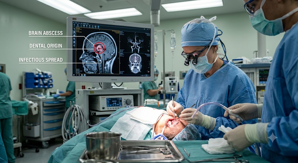
The Anatomy of a Dental Emergency: How Infection Travels
To understand the gravity of the situation, you must first understand the landscape. Your mouth is a bustling ecosystem of bacteria. Most of these microorganisms are harmless or even helpful, but when a cavity, crack, or gum disease allows bacteria to penetrate the hard outer layers of a tooth, trouble begins. Inside the tooth lies the pulp, a soft chamber filled with nerves, blood vessels, and connective tissue. When the pulp dies or becomes necrotic, it turns into a breeding ground for bacteria.
The Birth of an Abscess
A tooth abscess is a pocket of pus that forms as your immune system wages war against this bacterial invasion. White blood cells rush to the area, sacrificing themselves to engulf the invaders, and the resulting accumulation of dead cells and bacteria forms pus. The abscess exerts relentless pressure on the surrounding bone and tissues. You feel this as a throbbing, persistent, sharp pain that can radiate to your jawbone, neck, or ear. The pressure builds because the infection has nowhere to drain. If it does not find a path out through the gum or the tooth itself, it will take the path of least resistance through the deeper planes of your face.
The Dangerous Terrain of the Face
Your face and neck are not solid blocks of tissue. They are divided into fascial spaces, which are potential gaps between layers of muscle and connective tissue. Think of them as virtual highways running from your jaw to your brain. The bacteria from a lower molar can easily slip into the submandibular space, the sublingual space, or the deeper lateral pharyngeal space. From there, the infection can climb upward.
Ludwig’s angina represents one of the most feared complications, a rapidly spreading cellulitis of the floor of the mouth and neck. While this itself is life-threatening by closing off the airway, it also demonstrates how quickly an infection ignores anatomical boundaries. The infection can track further up through the skull base via foramina, which are small holes that allow nerves and blood vessels to pass through. This direct route transforms a dental problem into a neurological nightmare. The proximity of the upper teeth to the sinuses also creates a shortcut. An abscess at the root of an upper molar or premolar can perforate the thin sinus floor, causing a sinus infection that then erodes the bony wall separating the sinus from the brain cavity.
The Critical Timeline: From Toothache to Brain Abscess
The progression is not instantaneous, which makes patient awareness crucial. The transition from a localized dental issue to a cerebrovascular or intracranial emergency typically follows a roadmap. Recognizing the stop signs along this path can prevent a tragedy.
Stage One: The Localized Dental Infection
This stage focuses entirely on the tooth. Pain is the hallmark here. It is a deep, gnawing ache that does not go away. It intensifies when you lie down because the change in head position increases blood pressure in the infected area. Hot foods make it excruciating; cold water might provide fleeting relief. You might see a pimple-like bump on the gum called a fistula. This little bump is a drainage point, a pressure-release valve. If the abscess drains through a fistula, the pain might decrease significantly. Do not mistake this relief for healing. The infection is still active and silently destroying jawbone.
Stage Two: Spread to Surrounding Facial Tissues
When the infection escapes the bony confines of the jaw, swelling begins. The face distorts. The cheek puffs up, the area under the jaw becomes hard and tender, and the skin turns red and hot to the touch. This is cellulitis. A critical warning sign here is trismus, the medical term for lockjaw. If your jaw muscles become inflamed, you will struggle to open your mouth. This signals that the infection has invaded the masticator space, a deep compartment near the base of the skull. Trismus is not just a mechanical inconvenience; it is a red flag that the infection has traveled beyond simple extraction reach.
Stage Three: Vascular Invasion and Septicemia
Bacteria enter the bloodstream directly through eroded blood vessels. The cavernous sinus, a large collection of veins at the base of the brain, drains blood from the face. The valveless nature of facial veins allows infection to travel backward into this sinus. This is cavernous sinus thrombosis. Alternatively, bacteria floating freely in the blood can reach the heart and settle on the valves, or they can be pumped into the brain, lodging in tiny capillaries and starting a new colony. You will now experience systemic signs: high spiking fevers, chills, rapid heart rate, and confusion. This marks the shift from a surgical dental issue to a critical medical emergency. The body begins to spiral into sepsis, a dysregulated immune response that attacks vital organs.
Stage Four: The Intracranial Infection
Once inside the skull, the infection manifests as one of several entities. A brain abscess is a localized pus collection within the brain tissue itself. Subdural empyema is a collection of pus between the outer protective layer of the brain and the brain itself, spreading like a sheet over the brain’s surface. Meningitis is the inflammation of the meninges, the protective membranes covering the brain and spinal cord. The brain has no space to spare. The rigid skull box constrains it. Any extra volume from pus or swelling rapidly increases intracranial pressure. This pressure squeezes the brain stem, eventually causing respiratory arrest. This final stage can unfold with terrifying speed, sometimes within a day of the first neurological symptom.
Recognizing the Danger: Tooth Infection Spread to Brain Symptoms
This is the core of the guide, the detailed signposts that separate a bad dental day from a catastrophic health crisis. When a tooth infection spreads to the brain, the body sends out a sequence of unmistakable distress signals. You must treat these symptoms as a medical emergency equivalent to a stroke or heart attack.
Severe Neurological Headaches
A toothache is a localized, identifiable pain. A brain-related headache is an entirely different beast. The pain is not throbbing like a tooth; it feels like a deep, unrelenting pressure from the inside of the skull. Patients frequently describe it as the worst headache of their life. It refuses to yield to standard over-the-counter pain relievers. The pain intensifies when you lean forward, cough, or strain, because these actions momentarily increase intracranial pressure. The headache is often diffuse, meaning you cannot pinpoint it with one finger, unlike the discrete ache of a bad tooth. If your dental abscess pain suddenly transforms into a blinding, explosive headache, the infection may have crossed the blood-brain barrier.
High and Uncontrollable Fever Patterns
A low-grade fever might accompany a large dental abscess, but a brain-bound infection kicks the immune system into absolute overdrive. Fevers spike rapidly above 103 degrees Fahrenheit (39.4 degrees Celsius). The chills are violent, shaking the entire body, known as rigors. The pattern often becomes cyclical; a fever spike leads to profuse sweating as the temperature breaks, only to rise again hours later. This spiking and plunging pattern reflects the body’s losing battle against bacterial toxins flooding the bloodstream. Anti-fever medication like acetaminophen or ibuprofen might reduce the temperature marginally, but it will not normalize it. This persistent, high-grade fever combined with a known dental infection forms a dire warning.
Sudden and Dramatic Mental Status Changes
One of the most terrifying aspects for family members is the change in mental clarity. The lucid person you know becomes confused, disoriented, and drowsy. They might not know the date, the location, or recognize familiar faces. The lethargy progresses rapidly. It becomes difficult to keep the person awake. This altered consciousness arises from the brain swelling and the toxic effect of inflammation on neural circuits. Do not dismiss this as fatigue or a reaction to pain medication. A drowsy patient with an infection is an alarm bell for cerebral involvement. Confusion often heralds the onset of intracranial pressure problems and can escalate to a stupor, where the patient only responds to painful stimuli, and eventually coma.
Ocular Symptoms and Vision Disturbances
The eyes share a close anatomical and neurological relationship with the cavernous sinus and the brain. When the cavernous sinus becomes clotted with infected material, blood backs up. This leads to a specific set of eye changes. The eyelids swell and droop, a condition called ptosis. The eye itself protrudes forward because of the venous congestion behind it, which is known as proptosis. The movement of the eyeball becomes restricted, causing severe double vision. Cranial nerves III, IV, and VI, which control eye movement and pupil constriction, run right through the cavernous sinus and become paralyzed. A fixed and dilated pupil on one side is an ominous sign of uncal herniation, where the brain is herniating through the tentorium due to extreme pressure. Any new double vision, protrusion of the eye, or an inability to move the eye in a specific direction demands immediate neurosurgical evaluation.
Focal Neurological Deficits and Seizures
As a brain abscess grows or as empyema spreads, it compresses specific brain regions. This produces focal signs. A person might experience weakness in one arm and leg on the opposite side of the body, mimicking a stroke. Speech may become garbled (dysarthria) or the patient might be unable to understand or produce language (aphasia). Sensory disturbances such as numbness or tingling on one side occur. Seizures are a common presenting feature of a brain abscess. The seizure can be generalized tonic-clonic, where the whole body shakes and the person loses consciousness, or it can be focal, where just one limb twitches uncontrollably while the person remains aware. A first-time seizure in an adult with a recent dental infection history is a brain abscess until proven otherwise. These episodes represent electrical storms triggered by the irritation of the cortical surface.
The Telltale Nuchal Rigidity
Meningeal irritation causes a hallmark physical exam finding: neck stiffness. The medical term is nuchal rigidity. The patient finds it intensely painful or impossible to touch their chin to their chest. This passive stretching inflames the infected meninges. It differs from the sore neck associated with jaw swelling. In that scenario, the lateral neck movement or swallowing hurts. With meningeal signs, the stiffness is in the nape of the neck and the major restriction is in forward flexion. Brudzinski’s sign, where lifting the head causes the knees to bend, and Kernig’s sign, where extending the bent knee while the hip is flexed causes pain, are clinical signs of this irritation. Neck rigidity coupled with severe headache and fever spells bacterial meningitis until cultures come back negative.
Recognizing Cavernous Sinus Thrombosis
This specific entity deserves a dedicated spotlight because of its association with dental infections, particularly those involving the upper canines and premolars. The veins draining these teeth feed directly into the angular vein, which empties into the superior ophthalmic vein and then the cavernous sinus. The symptoms present dramatically. High fever, headache, and periorbital swelling that starts with one eye and rapidly becomes bilateral. The classic finding is a chemotic conjunctiva, where the clear membrane over the eye swells up like a blister. The patient appears toxic and very unwell. This condition carries a 30% mortality rate even with aggressive therapy, and about half of survivors have permanent neurological deficits. The window for intervention is alarmingly narrow.
A Comparative Look: Dental Infection vs. Brain Infection Symptoms
To clarify the transition from a local to a systemic and neurological crisis, the following table contrasts the symptom profiles. This comparison can help you objectively analyze a confusing situation.
| Body System | Localized Dental Abscess | Infection Spread to Brain |
|---|---|---|
| Pain Profile | Throbbing, localized to a specific tooth; worsens with biting or hot/cold stimuli. | Diffuse, severe, bursting headache; “worst pain of my life”; worsens with coughing or lying flat. |
| Fever Response | Low-grade or absent; may feel slightly warm. | High, spiking fevers ( >103°F) with shaking chills (rigors). |
| Facial Appearance | Localized gum pimple, cheek swelling on one side. | Periorbital edema, protruding eyes, swollen forehead, possible bilateral facial puffiness. |
| Cognitive State | Alert, anxious about pain, normal conversation. | Confusion, extreme drowsiness, disorientation, progressing to stupor or coma. |
| Eye Function | Normal vision, no double vision. | Double vision, fixed/dilated pupil, inability to move eye, eyelid drooping. |
| Neck Mobility | Pain in jaw/neck lymph nodes; can still touch chin to chest without sharp pain. | Severe stiffness; impossible to touch chin to chest without excruciating pain. |
| Neurological Signs | None. | Seizures, weakness on one side, garbled speech, facial droop. |
The Urgency of the Red Line: When to Call 911
Many people hesitate. The instinct is to call a dentist in the morning, to avoid a costly emergency room bill, to hope that a fresh round of antibiotics will kick in. With the constellation of symptoms described above, waiting is a gamble you simply cannot afford. The mortality rate for brain abscesses has improved from near 100% to under 10% thanks to modern scanning and surgery, but this statistic relies heavily on the timing of intervention. Every hour of delay allows the bacterial colony to expand and the intracranial pressure to surge.
You must bypass the dentist’s voicemail and dial emergency services immediately if you see the “Red Line” signs. The red line is a visual concept. It represents the crossing of the infection from a local facial space into the central nervous system. If the person with a known tooth abscess becomes confused, cannot stay awake, or has a seizure, you call an ambulance. If they have a high fever, a stiff neck, and are suddenly seeing double, you call an ambulance. If they complain of an explosive headache that painkillers do not touch, you call an ambulance. Do not drive yourself if you are the one experiencing these symptoms; a seizure or loss of consciousness behind the wheel transforms a medical crisis into a double-fatality accident scene.
When you speak to the emergency dispatcher, be concise. Say, “This person has a severe tooth infection and is now showing signs of confusion, a fever of 104, and a stiff neck.” This information allows the dispatcher to pre-alert the receiving hospital to suspect an intracranial infection, potentially activating neurology, neurosurgery, and radiology teams before you even arrive.
Inside the Hospital: The Path to a Diagnosis
Upon arrival at the emergency department, a triage nurse trained to recognize sepsis and neurological emergencies will prioritize the patient based on vital signs. The high-speed diagnostic sequence begins.
The Immediate Physical Examination
The emergency physician will perform a rapid but thorough neurological assessment. They will test the cranial nerves by shining a light into the eyes to check pupillary response and tracking a finger to check eye movements. They will ask the patient to smile, raise eyebrows, and stick out the tongue. Strength will be tested by having the patient push and pull against the doctor’s hands. The fundoscopic exam, where the doctor uses a handheld ophthalmoscope to look at the back of the eye, is crucial. The presence of papilledema—swelling of the optic disc—indicates a dangerous rise in intracranial pressure. The neck will be gently flexed to test for meningeal signs. The doctor will auscultate the heart and lungs to check for murmurs that suggest endocarditis, a common co-condition where bacteria settle on heart valves.
The Gold Standard: Contrast-Enhanced Imaging
Laboratory blood work will show a massively elevated white blood cell count and elevated inflammatory markers like C-reactive protein and procalcitonin. Blood cultures will be drawn to identify the specific bacterial culprit. However, imaging is the definitive step. A CT scan without contrast is fast and can rule out hemorrhage or large masses, but it can miss a small early abscess or subtle meningitis. The critical scan is the MRI with gadolinium contrast. Gadolinium leaks across the disrupted blood-brain barrier and rings around an abscess brightly, creating a classic ring-enhancing lesion. This image tells the neurosurgical team exactly where the pus is, its size, and the degree of shift of the brain’s midline structures. For suspected cavernous sinus thrombosis, a CT venogram or MR venogram maps the venous flow blockage.
The Role of Lumbar Puncture
If the imaging suggests meningitis or increased pressure, a lumbar puncture or spinal tap may be performed, but with extreme caution. If there is a large mass effect from an abscess, tapping the spine can create a pressure gradient that sucks the brain downward through the foramen magnum, causing instantaneous death. Therefore, CT imaging almost always precedes a lumbar puncture. The cerebrospinal fluid extracted will be clear and sparkling in a normal person. In bacterial meningitis, it is cloudy and under high pressure. The lab analyzes cell counts, glucose, and protein, and a Gram stain provides an immediate clue about the bacteria type.
The Bacterial Rogues’ Gallery: What’s Lurking in the Mouth
The mouth is a complex polymicrobial environment. Dental abscesses are rarely caused by a single pathogen. They are usually a mixed bag of aerobic and anaerobic bacteria that synergize to cause tissue destruction. Understanding the microbiology explains the aggressive antibiotic cocktail required.
The Streptococcus Family
Viridans group streptococci are the most common culprits. Streptococcus anginosus, also known as the Streptococcus milleri group, deserves special attention. These bacteria have a notorious ability to cause abscesses in distant organs, particularly the brain and liver. They are normal mouth flora but become deadly invaders once they breach the mucosal barrier. They tend to form thick-walled pus collections.
The Anaerobic Invaders
Anaerobic bacteria thrive in oxygen-poor environments like a necrotic tooth root or a brain abscess core. Peptostreptococcus, Prevotella, and Fusobacterium species dominate. Fusobacterium necrophorum carries a specific syndrome, Lemierre’s syndrome, where an oropharyngeal or dental infection leads to septic thrombophlebitis of the internal jugular vein and septic emboli to the lungs and brain. This bacteria is aggressive and causes a severe systemic illness in previously healthy young adults. The abscesses often contain foul-smelling pus due to the production of short-chain fatty acids.
The Aggressive Staphylococcus
Staphylococcus aureus, including methicillin-resistant strains, can contaminate a dental extraction socket or an existing abscess from skin flora. S. aureus abscesses are particularly destructive because they produce coagulase enzymes that wall off the infection and toxins that destroy tissue. They cause a robust inflammatory response and high fevers.
The Aggressive Treatment Protocol: A Two-Pronged Attack
A brain abscess of dental origin requires a simultaneous and coordinated assault from infectious disease specialists and neurosurgeons. Medical management alone is rarely sufficient for large or multiple abscesses.
The Antibiotic Regimen
Given the mixed flora, empiric therapy starts before culture results return. A triple-therapy approach is common. The patient receives a beta-lactam antibiotic like ceftriaxone or cefepime to cover strep and staph. They add metronidazole, which has excellent penetration into the central nervous system and abscess cavities, to kill the anaerobes. Often, vancomycin is the third agent to ensure coverage of MRSA until sensitivities prove otherwise. Antibiotics are intravenous, high-dose, and prolonged. A typical course lasts six to eight weeks, often transitioning to oral rifampicin combinations later. Weekly monitoring of kidney and liver function is necessary because these drugs are toxic.
The Neurosurgical Decision: Drainage vs. Excision
The decision to operate depends on the “trapping” effect and mass effect. A cerebellar abscess causing brainstem compression requires emergent evacuation. Abscesses greater than 2.5 centimeters in diameter, regardless of location, are usually aspirated. The procedure is a stereotactic aspiration, where a thin needle is navigated to the exact pus center using a three-dimensional coordinate system attached to the head. The pus is sucked out and sent for culture. This often results in immediate clinical improvement as pressure is relieved. In some cases, where the abscess is superficial and well-encapsulated, a full craniotomy and excision of the entire capsule can be performed. This eliminates the source but carries higher risks of inducing seizures.
Definitive Dental Source Control
Treating the brain abscess without removing the infected tooth is like emptying a boat without plugging the hole. Once the patient is neurologically stable and cleared for general anesthesia, the offending tooth must be extracted. Sometimes this is done during the same hospitalization. The extraction is performed carefully, with infection present, to prevent bacteremia from spreading. The socket is curetted and irrigated heavily. Antibiotics continue to bathe the surgical site.
Realistic Recovery and Permanent Scars
Surviving a brain infection of dental origin is a life-altering event. It is a victory, but the battle leaves marks. Rehabilitation is a long road. Physical therapy addresses hemiparesis, the one-sided weakness that persists. Speech and language pathologists help rewire the brain for word-finding and swallowing. Occupational therapy restores fine motor skills for daily living tasks.
The cognitive and emotional sequelae can be the most painful. Short-term memory dysfunction is common; the hippocampus is sensitive to hypoxia and inflammatory insult. Patients may struggle to recall conversations or learn new tasks. Executive function—planning, organizing, multitasking—can be significantly impaired. Frontal lobe abscesses can cause personality changes, ranging from impulsivity and irritability to apathy and flattened affect. Seizure disorders develop in about one-third of brain abscess survivors, necessitating lifelong anti-epileptic medication. The risk of recurrence is low if the source has been completely eradicated, but any new neurological symptom thereafter warrants immediate re-imaging. The psychological trauma of realizing a tooth nearly killed you requires compassionate counseling, not just stitches and drugs.
The Prevention Protocol: A Daily Defense Strategy
The power to prevent this nightmare lies almost entirely in your daily routine. The mouth-body connection is not a marketing slogan; it is a physiological reality. Preventing a tooth infection spread to the brain is a straightforward, affordable, and non-negotiable set of actions.
The Non-Negotiable Oral Hygiene Ritual
Mechanical disruption of the biofilm, the sticky plaque, is the goal. Brushing twice daily with a fluoride toothpaste is the foundation, but technique trumps speed. Angle the bristles at 45 degrees toward the gumline and make small, gentle, circular vibrations. Do not scrub horizontally across the tooth necks; this causes erosion and gum recession. Electric toothbrushes with pressure sensors and timers have been shown to remove significantly more plaque than manual brushes for the average user.
Flossing or using interdental brushes must happen once every 24 hours to disrupt the plaque colonies between teeth where a brush cannot reach. The bacteria in these tight spaces cause more abscesses than those on the cheek side of the tooth surface. Swishing vigorously with an antimicrobial mouth rinse that contains essential oils or chlorhexidine provides an additional chemical reduction in bacterial load. However, chlorhexidine can stain teeth with long-term use, so it is a prescription tool for active disease, not a daily cosmetic rinse.
The Dietary Terrain Modification
Your diet determines the acidity and bacterial growth in your mouth. Sucrose and high-fructose corn syrup are the metabolic fuel for Streptococcus mutans. It is not just the amount of sugar; it is the frequency. Sipping a soda over an hour bathes the teeth continuously in acid and sugar, providing a constant feast for bacteria. Water remains the only tooth-safe beverage between meals. Chewing xylitol-sweetened gum after meals stimulates salivary flow, and xylitol actively inhibits bacterial adhesion. Crunchy fibrous vegetables and fruits like apples, celery, and carrots act as natural toothbrushes, scrubbing the surfaces and stimulating gums.
The Professional Surveillance Schedule
The six-month dental recall is not an arbitrary appointment designed by insurance companies. It is the surveillance window that catches demineralization before it becomes a cavitation, and a small cavity before it reaches the nerve. For patients with a history of abscesses or dry mouth, a three to four-month recall is protective. Dental radiographs, bitewing X-rays, reveal the silent cavities growing between teeth that are invisible to the naked eye. A panoramic X-ray every few years monitors the bone health and the position of impacted teeth. If you have a dental crown, bridge, or root canal treatment, these restorations have lifespans. Cracks can develop, leaking bacteria beneath them. Regular scrutiny catches these failures early. The cost of a crown replacement is astronomically lower than a neurosurgical ICU stay, both financially and in terms of human suffering.
Immediate First Aid: Bridging the Gap Before the Dentist
You have a swollen face at 2 AM, and the dental office opens at 9 AM. You are in pain and scared, but you do not yet have confusion or a stiff neck. The goal of interim management is not to cure the infection but to prevent its downward spiral into the fascial spaces while you wait for definitive care.
Thermal Therapy
Apply a cold compress or an ice pack wrapped in a thin towel to the outside of your cheek over the swollen area. Hold it for 15 minutes on, 15 minutes off. The cold constricts blood vessels, reducing the flow of inflammatory mediators into the tissue. This decreases swelling and provides a mild numbing effect. Do not apply heat. Heat causes vasodilation, which increases swelling and can encourage the pus to spread through the path of least resistance into deeper spaces. This old wives’ tale of a hot water bottle can convert a localized abscess into a spreading cellulitis.
Head Elevation and Posture
Gravity is your ally. Sleep propped upright on pillows or in a recliner. Keeping your head above your heart reduces the hydrostatic pressure in the infected vessels. This is why the throbbing pain intensifies when you lie flat. Elevation helps minimize the pulsatile pressure that drives the bacteria into the venous system. It also helps with breathing if the swelling is tracking toward the airway.
Pain Control and What to Avoid
Non-steroidal anti-inflammatories like ibuprofen are preferable to narcotics at this stage because they target the prostaglandin-mediated inflammatory swelling. However, you must take these with food to protect the stomach. Critically, never take leftover antibiotics from a previous illness or a friend. A single incomplete dose can suppress the infection without killing it, buying time for the deep-track spread without causing the dramatic external swelling that would drive you to seek care. This “masking” can be fatal. Warm salt water rinses can help keep the mouth clean and may encourage a superficial gum fistula to drain, releasing pressure. But never push, poke, or squeeze the swelling. External pressure on an abscess can force bacteria into the bloodstream, a dangerous bacteremia event.
Understanding the Special Risk of Upper Teeth Infections
The location of the offending tooth is a significant factor in the trajectory of the spread. The old dental axiom, “the upper canine is the eye tooth,” holds anatomical truth. An abscess at the root tip of an upper canine or lateral incisor can perforate the thin buccal plate of bone and point just below the eye. If it drains backward, it tracks into the infraorbital region and links directly to the angular vein. This vein has no valves, a critical design flaw in the context of infection. Blood flowing through it can reverse direction, carrying bacteria into the superior ophthalmic vein and then into the cavernous sinus. Therefore, an abscess over an upper front tooth must be treated with extreme urgency. You should never wait for facial swelling below the eye to resolve on its own. The risk of intracranial extension is higher with these anterior maxillary teeth than with most others.
Lower molars carry a different risk. The roots of the second and third lower molars extend deep into the mandible, often below the attachment of the mylohyoid muscle. When these abscesses rupture through the lingual plate of the mandible, they enter the submandibular space directly. This is the classic beginning of Ludwig’s angina. The floor of the mouth rises, the tongue is pushed up and back against the soft palate, and the airway becomes a narrowing slit. The patient speaks with a hot-potato voice, leans forward to drool because they cannot swallow their own saliva, and becomes stridorous. This airway emergency requires intubation before the anatomy becomes obliterated by swelling.
The Pediatric Concern: A Different Diagnostic Challenge
Children are not miniature adults when it comes to dental abscesses. The communication barrier makes early detection difficult. A child with a toothache may simply refuse food or cry and point to an ear. The referred pain patterns in children are broad. An infected lower molar can cause an earache, leading to a misdiagnosis of otitis media if the dentist is not consulted. The thinner bones and developing dentition allow rapid spread. A facial swelling in a child can double in size within hours.
In toddlers, a traumatic fall that chips a front tooth can cause the pulp to die without changing the tooth color significantly for months. This silent necrosis smolders and eventually erupts as a gum boil above the tooth. Parents should be vigilant for a small bump on the gum above a discolored or previously bumped front tooth. This is a dental fistula and indicates a chronic, low-grade abscess that may have already damaged the developing permanent tooth bud underneath. While intracranial spread remains extremely rare in healthy children, the risk of septicemia and the systemic impact on a small body mass is elevated. A febrile child with facial swelling requires immediate pediatric dental and medical consultation.
A Note on Antibiotic Resistance and the Need for Culture
The era of casually prescribing penicillin for every toothache is fading under the shadow of antimicrobial resistance. Brain abscesses of dental origin often yield a polymicrobial broth that resists simple penicillin. The frequent use of clindamycin for penicillin-allergic patients has also driven up resistance rates among common anaerobes. This is why the neurosurgical aspiration of a brain abscess serves a dual purpose: it decompresses the brain and provides a specimen for microbial identification.
The microbiology lab performs a culture and sensitivity that grows the bacteria and tests it against a panel of antibiotics. The results, usually available in 48 to 72 hours, allow the infectious disease team to narrow the antibiotic spectrum, reducing toxicity and the collateral damage to the gut microbiome. This targeted therapy is the standard of modern care. The days of blind, lifelong-suppressive antibiotics without source removal are over. The physical removal of the necrotic tooth and the drainage of pus remain the cornerstone of cure, as antibiotics cannot penetrate the acidic, oxygen-depleted core of a large abscess without surgical reduction of the bacterial load.
Comparative Analysis: Abscess Progression Speed in Different Patients
The timeline of infection spread is not uniform across all individuals. A healthy, immunocompetent young adult often mounts a violent and rapid inflammatory response, walling off the infection with fibrin and producing copious pus. This causes pain and swelling fast, driving the patient to seek care early. This is actually a protective mechanism; the symptoms align with the severity.
In contrast, an elderly patient or one with uncontrolled diabetes may experience a silent, smoldering progression. The high blood sugar levels in diabetics impair the migration of neutrophils and their ability to phagocytose bacteria. The immune response is blunted. Pain may be minimal, swelling gradual, and fever absent. The infection can spread extensively through the deep planes before being recognized, and at that point, the patient presents with a brain abscess and the original dental source is a forgotten, small, non-painful dead tooth. This population requires proactive dental screening to remove septic foci before elective surgeries or chemotherapy.
Pregnant women experience a relative immunosuppression and hormonal changes that increase gum inflammation and the risk of pregnancy epulis, a benign but vascular gum swelling. A dental abscess in pregnancy requires aggressive treatment. The second trimester is the safest window for necessary extractions. Letting an abscess smolder during pregnancy risks septic preterm labor and exposes the mother to a cascade of inflammatory mediators that can affect the fetus. The belief that dental treatment is dangerous during pregnancy is a harmful myth; a raging dental infection is far more dangerous.
The Unmistakable Signs of a Cavernous Sinus Thrombosis
Because of its unique and terrifying presentation, this condition must be highlighted again with specific and unmistakable detail. If you see these features in the context of a recent facial or dental infection, do not call a primary care office; call an ambulance.
The patient complains of a severe headache, often behind the eye. A high fever spikes abruptly. The eye on the side of the infection begins to swell, and the conjunctiva turns a bright, angry red and swells out, looking like a glob of jelly. This is chemosis. Within hours, the other eye begins to show the same signs because the cavernous sinuses communicate across the midline. The eyes bulge. The nerves running through the sinus fail, causing the eyes to freeze in a forward gaze with dilated pupils. The forehead, eyelids, and bridge of the nose become puffy and edematous. The skin may take on a dusky, purplish hue from venous stasis. Lethargy and nuchal rigidity follow quickly. The mortality rate hovers around 30%, and survivors often have residual double vision, ptosis, or even unilateral blindness from central retinal vein occlusion. An upper canine tooth abscess can cause this. That single fact should recalibrate how we perceive dental urgency.
The Critical Airway Emergency: Ludwig’s Angina
Before the infection reaches the brain, it may kill the patient by closing the throat. Ludwig’s angina is a rapidly progressive cellulitis of the submandibular, sublingual, and submental spaces. It is a non-pus-forming, putty-like swelling that hardens the floor of the mouth like wood. The tongue is forced superiorly and posteriorly, like a cork in a bottle, sealing the airway.
The patient assumes a desperate posture: sitting bolt upright, hands on knees, leaning forward, drooling. The voice sounds thick and muffled. An inspiratory high-pitched sound, stridor, signals that the airway diameter is shrinking to a critical, straw-like opening. This is a cannot-intubate, cannot-ventilate nightmare scenario for anesthesiologists. Definitive management is an awake fiberoptic nasal intubation or a surgical airway, a cricothyrotomy, to bypass the obstruction before the patient asphyxiates. If the brain is the concern, the airway is the priority. You must secure the airway first, then worry about the intracranial complications.
Long-Term Neurological Outcomes and Quality of Life
Modern neurosurgery and intensive care have shifted the metric from simple survival to functional survival. A person surviving a dental-origin brain abscess faces a long trajectory. Early, aggressive inpatient rehabilitation is the standard. The neuroplasticity of the brain allows other regions to take over lost functions, but this rewiring takes months to years.
Seizure disorders often emerge late, even months after the abscess has been treated and the scans are clean. The scarring gliosis left behind acts as an epileptogenic focus. Patients are placed on prophylactic anti-seizure medications like levetiracetam or phenytoin, but these drugs cause their own side effects: fatigue, dizziness, and mood changes. Cognitive fatigue, the inability to sustain mental effort, remains a persistent complaint. Simple tasks like calculating a tip at a restaurant or reading a dense paragraph become exhausting. Support groups and cognitive behavioral therapy become essential tools for managing the new normal. The psychological trauma—understanding that a tooth, a mundane body part, almost ended a life—requires processing. Many survivors become intense advocates for dental health, haunted by the randomness of the infection.
A Detailed Guide to Clinical Examination at Home
While you are not a clinician, a basic observational checklist can help you articulate the severity of the situation to a medical professional over the phone. First, look at the face in good light. Is the swelling confined to the jaw, or has it crept up to the eye or down to the neck? Asymmetry around the eye is a red flag. Ask the person to smile and show their teeth. A new asymmetry or droop is a stroke signal that overlaps with brain infection signs.
Ask the person to look up and down and side to side with their eyes. Complaints of double vision or visibly jerky, uncoordinated eye movements are crucial findings. Take their temperature with a reliable thermometer; a reading over 101.5 degrees Fahrenheit combined with a swollen face elevates the urgency. Ask them to touch their chin to their chest. If they grimace and cannot, document it. Ask them to perform simple serial sevens—subtracting 7 from 100 sequentially—to screen for confusion. If they cannot do this, or if they cannot recall the current month, the brain is not functioning correctly. These simple tests provide objective data that bypasses the patient’s tendency to minimize their own symptoms.
The Final Barrier: Overcoming Dental Anxiety and Financial Fear
A significant driver of these catastrophic outcomes is avoidance. The reasons are almost never trivial to the person experiencing them. Severe dental phobia, a recognized anxiety disorder, paralyzes individuals. The sounds, smells, and sensations of a dental drill trigger a fight-or-flight response that makes booking an appointment feel impossible. Financial constraints are equally crippling. The choice between a root canal and the monthly rent payment is a false choice that many face directly.
However, the cost of inaction is an exponential curve. A $150 extraction becomes a $1,500 root canal and crown. That untreated tooth becomes a $15,000 hospital admission for intravenous antibiotics and incision and drainage. That becomes a $150,000 neurosurgical ICU stay, assuming survival, plus months of lost wages and permanent disability. Hospitals have emergency Medicaid provisions and charity care programs, but the dental clinic might not. Yet, federally qualified health centers and dental schools offer sliding-scale fees based on income. The only irreversible choice is waiting until the infection makes the choice for you. Sedation dentistry, from nitrous oxide to oral conscious sedation and IV sedation, exists precisely to bridge the gap for phobic patients. A compassionate dentist can prescribe an oral anxiolytic to be taken the night before and the morning of the appointment, allowing the patient to navigate the door.
Reconstructing the Damage: The Aftermath in the Jaw
After the brain crisis has passed and the offending tooth has been extracted, the site in the jaw requires attention. A significant abscess destroys the cancellous bone, leaving a radiolucent void. Over time, this defect fills in with new bone through a process called guided bone regeneration if the clot is protected. Sometimes, the surgeon places a bone graft, allograft or xenograft, into the socket to preserve the ridge width and height for a future implant or bridge.
If the extracted tooth was a visible one, the patient emerges from this near-death experience with a missing tooth and a sunken facial profile. Dental rehabilitation with osseointegrated implants or precision partial dentures restores the masticatory function and the smile, providing a psychological closure to the trauma. This prosthetic phase is not merely cosmetic; it prevents the drifting of adjacent teeth and the super-eruption of opposing teeth, which would create a lifetime of occlusal instability and temporomandibular joint issues. The full circle of care, from neurosurgery back to restorative dentistry, highlights the interdependence of medical and dental disciplines.
The Mouth as a Sentinel: Systemic Disease Detection
While discussing the spread of infection, one must acknowledge that the mouth often reflects systemic disease. Uncontrolled diabetes leads to periodontal abscesses. Leukemia can present with gingival swelling and necrosis that mimics a dental abscess. Bisphosphonate-related osteonecrosis of the jaw, from medications like alendronate for osteoporosis or zoledronic acid for cancer, can cause non-healing extraction sockets that become secondarily infected and may spread. This reinforces the need for a thorough medical history by the treating dentist. A dental abscess that does not behave typically, that fails to drain or respond to standard antibiotics, may be a sign of a deeper immune dyscrasia. The dentist becomes the diagnostician, the sentinel who spots the oral manifestations of a systemic crisis.
A Global Perspective on the Crisis
In low-resource settings without access to advanced CT scans or neurosurgery, a tooth abscess spreading to the brain remains a uniformly fatal event. The work of organizations providing mobile dental camps and training local healthcare workers in basic extraction techniques saves lives directly. The extraction of a grossly decayed, purulent molar in a rural clinic prevents a slow, agonizing death from sepsis or cerebral herniation. This stark reality underlines the privilege of accessible emergency care and the importance of public health water fluoridation and school-based sealant programs. These measures are not just about cavity prevention; they are about preventing fatal brain infections.
Conclusion
A tooth infection spreading to the brain transforms a preventable dental issue into a catastrophic neurological emergency that can kill or permanently disable within days. The cardinal warning signs that demand an immediate emergency response include an explosive headache, high fever with rigors, confusion, eye protrusion, and a stiff neck that makes touching the chin to the chest impossible. Survival hinges on a rapid, coordinated attack combining intravenous antibiotics, emergency neurosurgical drainage of the abscess, and definitive removal of the septic tooth, all while preserving the airway. The path to avoiding this terrifying progression is anchored in consistent daily oral hygiene, early dental treatment of cavities, and recognizing that the cost of preventive care is infinitely smaller than the price of a life-altering brain infection.
Additional Resources
For authoritative and continuously updated information on sepsis, brain abscesses, and oral health, consult the Sepsis Alliance at sepsis.org. This organization provides educational resources for recognizing the signs of sepsis, a complication that often overlaps with the systemic spread of dental infections, and offers support networks for survivors navigating the long-term aftermath.
Frequently Asked Questions
Can a tooth infection really kill me?
Yes. While rare, a severe tooth abscess can spread to the brain, heart, or airway, causing cavernous sinus thrombosis, endocarditis, brain abscess, or airway obstruction from Ludwig’s angina. Each of these conditions carries a significant risk of death if not treated urgently.
How long does it take for a tooth infection to reach the brain?
There is no fixed timeline. It can take weeks of slow, smoldering spread or escalate over a single weekend. Progression speed depends on the virulence of the bacteria, the anatomical location of the tooth, and the strength of the individual’s immune response. The swelling typically spreads stepwise from the jaw to the deep facial planes, then to the skull base.
Will antibiotics alone cure a brain abscess from a tooth?
No. A large, established brain abscess almost always requires surgical aspiration or drainage. Antibiotics cannot penetrate the thick capsule and necrotic core of a mature abscess effectively without the pus being physically removed. Prolonged, high-dose intravenous antibiotics serve as adjunctive therapy.
What is the “death tooth” and how does it relate to the brain?
The term “death tooth” is a colloquialism for a tooth that has a necrotic, non-vital pulp. This dead tissue inside the tooth is a sterile inflammatory focus until bacteria enter through a crack or cavity. Once colonized, it becomes a sealed bacterial incubator that can flush bacteria into the surrounding jawbone and eventually into the bloodstream or deep facial spaces leading to the brain.
Is an upper tooth or a lower tooth more dangerous for spreading to the brain?
Upper teeth, particularly the canines and premolars, pose a higher direct risk for cavernous sinus thrombosis because their draining veins connect directly to the valveless facial vein network that feeds the brain’s venous sinuses. Lower molars carry a higher risk of spreading to the neck and causing airway compromise, which is also life-threatening but blocks breathing before reaching the brain in many cases.
Disclaimer: This article is for informational purposes only and does not constitute medical advice. If you suspect you have a dental or medical emergency, call emergency services or seek immediate evaluation at a hospital emergency department.


