Why Bone Grafting For Dental Implant?
Bone grafting is frequently mentioned alongside dental implant treatment, often to the surprise of patients who assumed they could simply replace their missing tooth with an implant. The recommendation for bone grafting can feel like an unexpected obstacle adding cost and treatment time. Understanding why bone grafting is necessary and what it accomplishes helps patients appreciate its role in successful implant treatment.
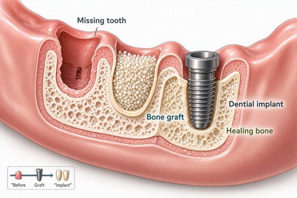
Bone grafting is required for dental implants when the jawbone lacks sufficient volume, density, or quality to support implant placement. After tooth extraction, the surrounding bone begins to resorb, losing up to 25% of its width in the first year and continuing to shrink over time. Bone grafting rebuilds this deficient foundation, creating adequate bone for implant stability and long-term success. Without sufficient bone, implants cannot achieve the primary stability and osseointegration essential for function.
The Biology of Bone Loss After Tooth Extraction
Understanding why bone disappears after tooth loss explains why grafting becomes necessary.
How Teeth Maintain Bone
The jawbone exists to support teeth. The presence of tooth roots stimulates bone maintenance through the forces transmitted during chewing. When you bite and chew, forces travel through the tooth into the periodontal ligament and surrounding bone. This mechanical stimulation signals bone cells to maintain bone density and volume.
This process, called Wolff’s Law, governs bone biology throughout the body: bone that is loaded maintains itself; bone that is not loaded resorbs. The jawbone is no exception. Tooth roots provide the necessary loading to preserve jawbone volume.
What Happens When a Tooth Is Lost
When a tooth is extracted, the mechanical stimulation from that root ceases. The body interprets the absence of loading as a signal that the bone is no longer needed. Osteoclasts (bone-resorbing cells) begin removing bone tissue. Osteoblasts (bone-building cells) reduce their activity.
The result is progressive bone resorption that follows a predictable pattern:
First Year After Extraction:
The alveolar ridge (the bone that held the tooth) loses approximately 25% of its width. Height loss of 2-4 millimeters is common. The outer (facial) plate of bone, which is thinner than the inner plate, resorbs more rapidly.
Subsequent Years:
Bone loss continues at a slower but steady rate. Over many years, significant vertical and horizontal bone loss occurs. In long-term denture wearers, the jawbone may resorb to the point where only a thin ridge of bone remains above the mandibular nerve or sinus floor.
Factors That Accelerate Bone Loss
Periodontal Disease:
Teeth lost to gum disease often have already experienced bone loss around their roots before extraction. The inflammatory process that destroys bone in periodontitis continues to affect the ridge after tooth removal.
Traumatic Extractions:
Rough extraction technique that fractures the bony socket walls causes more bone loss than atraumatic extractions that preserve the socket architecture.
Denture Wear:
Conventional dentures rest on the gum tissue and transmit forces to the underlying bone. Unlike natural teeth that stimulate bone maintenance, dentures apply compressive forces that may accelerate bone resorption. Long-term denture wearers often develop severe ridge atrophy.
Systemic Conditions:
Osteoporosis, diabetes, and other conditions affecting bone metabolism may accelerate post-extraction bone loss.
Time Without Tooth Replacement:
The longer a tooth has been missing, the more bone has been lost. Patients who delay implant treatment for years after extraction are more likely to need bone grafting.
| Time After Extraction | Typical Bone Loss | Implant Feasibility |
|---|---|---|
| Immediate (same day) | None | Excellent (immediate placement possible) |
| 3-6 months | 25% width loss, 2-3mm height loss | Usually adequate; minor grafting sometimes needed |
| 1-3 years | 40-60% width loss, 3-5mm height loss | Grafting often required |
| 5-10+ years | Severe atrophy | Major grafting or alternative implant protocols required |
When Bone Grafting Is Necessary
Not every implant case requires bone grafting. Specific clinical scenarios predict the need.
Insufficient Bone Width
The implant must be surrounded by at least 1-1.5 millimeters of bone on all sides. If the ridge is too narrow to accommodate the planned implant diameter, bone grafting widens the ridge.
A ridge that measures 4 millimeters wide cannot accept a 4-millimeter diameter implant with adequate surrounding bone. Ridge augmentation grafting adds width to the bony ridge, creating sufficient dimensions for implant placement.
Insufficient Bone Height
The implant must have adequate length for stability, typically 8-10 millimeters minimum. The available bone height is limited by anatomical structures: the mandibular nerve in the lower jaw and the maxillary sinuses in the upper jaw.
When insufficient bone exists between the ridge crest and these structures, grafting adds vertical height. Sinus lift procedures augment bone in the upper back jaw. Onlay or interpositional grafts add height in other areas.
Extraction Socket Deficiencies
Even immediately after extraction, the socket may have defects that compromise implant placement. The buccal (cheek-side) plate of bone is often thin or missing entirely. Periodontal disease or infection may have destroyed bone before extraction.
Socket preservation grafting at the time of extraction places bone graft material into the socket to maintain ridge dimensions during healing. This proactive approach often eliminates the need for more extensive grafting later.
Previous Implant Failure
When an implant fails and is removed, the resulting defect requires grafting to rebuild bone for a replacement implant. Peri-implantitis often destroys significant bone that must be regenerated before a new implant can be placed.
“Bone grafting is not an obstacle to implant treatment; it is the solution that makes implant treatment possible when nature has not provided an adequate foundation. The graft creates what time and tooth loss have taken away, restoring the patient’s ability to receive the benefits of implant-supported teeth.” — American Academy of Periodontology
Types of Bone Grafting Procedures
Different clinical situations require different grafting approaches.
Socket Preservation Grafting
This is the most common and least invasive grafting procedure. Immediately after tooth extraction, bone graft material is packed into the empty socket. A membrane may cover the graft to protect it during healing. The gum tissue is sutured closed or left to heal around a membrane.
Purpose: Maintain ridge dimensions for future implant placement.
Healing Time: 3-6 months before implant placement.
Cost: $300-$700 per socket.
Socket preservation is far simpler and less expensive than rebuilding a ridge that has already resorbed. Whenever implant placement is planned, socket preservation at the time of extraction is strongly recommended.
Ridge Augmentation (Horizontal or Vertical)
When the ridge has already resorbed, bone graft material is placed along the deficient ridge to rebuild width or height. The graft may be particulate (granules) or block form (a solid piece of bone). A membrane often covers the graft. Titanium mesh or tenting screws may support the membrane and create space for bone formation. The gum tissue is closed over the graft for submerged healing.
Purpose: Rebuild ridge dimensions sufficient for implant placement.
Healing Time: 4-9 months before implant placement.
Cost: $800-$3,000 per site.
Sinus Lift (Sinus Floor Elevation)
The maxillary sinuses are air-filled spaces above the upper back teeth. When these sinuses are large or the ridge is short, insufficient bone exists between the sinus floor and the mouth to place implants.
A sinus lift procedure gains access to the sinus floor, gently elevates the sinus membrane, and places bone graft material beneath it. Over months, this graft matures into bone, increasing the available height for implant placement.
Two Approaches:
Lateral Window Sinus Lift:
A window is created in the side wall of the sinus. The membrane is elevated through this window. More graft material can be placed. Used for significant bone deficiencies. Healing time of 6-9 months. Cost: $2,500-$5,000 per side.
Osteotome Sinus Lift (Crestal Approach):
The implant site preparation itself elevates the sinus floor using specialized instruments. Less graft material is needed. Used for minor deficiencies (2-4mm additional height needed). Often performed simultaneously with implant placement. Healing time of 4-6 months. Cost: $800-$2,000 per site.
Block Bone Grafting
For severe deficiencies, a solid block of bone is harvested from the patient’s own body (autogenous graft) or obtained from a tissue bank (allograft). The block is secured to the deficient ridge with screws and allowed to heal and incorporate.
Donor Sites for Autogenous Block Grafts:
Back of the lower jaw (ramus). Chin area. Hip (iliac crest) for very large defects. Outer skull (parietal bone) in rare extensive cases.
Block grafts provide the greatest volume of bone augmentation but involve more extensive surgery and longer healing times.
Healing Time: 4-9 months before implant placement.
Cost: $2,500-$6,000+ per site depending on graft source and extent.
| Graft Type | Purpose | Healing Time | Cost Per Site |
|---|---|---|---|
| Socket Preservation | Maintain ridge after extraction | 3-6 months | $300 – $700 |
| Ridge Augmentation | Rebuild width or height | 4-9 months | $800 – $3,000 |
| Sinus Lift (Crestal) | Minor sinus floor elevation | 4-6 months | $800 – $2,000 |
| Sinus Lift (Lateral) | Major sinus floor elevation | 6-9 months | $2,500 – $5,000 |
| Block Bone Graft | Severe ridge deficiency | 4-9 months | $2,500 – $6,000+ |
Bone Graft Materials
The material used for grafting affects healing, cost, and the biological process of bone formation.
Autografts (Patient’s Own Bone)
Bone harvested from the patient is considered the gold standard graft material. It contains living bone cells, growth factors, and proteins that actively promote new bone formation.
Advantages: Osteogenic (forms new bone), osteoinductive (stimulates bone formation), osteoconductive (scaffold for bone growth). No risk of disease transmission or immune rejection.
Disadvantages: Requires a second surgical site with associated morbidity. Limited quantity available from oral sites. Additional surgical time and post-operative discomfort.
Cost: Highest due to additional surgical time and complexity.
Allografts (Donor Bone)
Bone from human tissue donors, processed to remove cells and sterilize the material. Available through tissue banks regulated by the FDA and American Association of Tissue Banks.
Advantages: No second surgical site. Available in various forms and quantities. Osteoconductive scaffold for new bone formation.
Disadvantages: Does not contain living cells. Lacks the osteoinductive proteins found in autografts. Theoretically minimal risk of disease transmission (extensively processed and screened).
Cost: Moderate.
Xenografts (Animal-Derived)
Bone mineral derived from animal sources, most commonly bovine (cow). The organic components are removed, leaving a mineral scaffold very similar to human bone mineral.
Advantages: Abundant supply. Excellent osteoconductive scaffold. Very slow resorption maintains graft volume over time.
Disadvantages: Not osteogenic or osteoinductive. Relies entirely on the patient’s bone-forming cells migrating into the graft. Some patients have religious or philosophical objections to animal-derived materials.
Cost: Moderate to low.
Alloplasts (Synthetic Materials)
Synthetic bone graft substitutes including calcium phosphates, calcium sulfates, and bioactive glasses. These materials are manufactured rather than sourced from humans or animals.
Advantages: Unlimited supply. No risk of disease transmission. No ethical or religious concerns. Some formulations are resorbable and replaced by natural bone.
Disadvantages: Variable osteoconductive properties depending on formulation. Not osteogenic or osteoinductive. Some materials resorb too quickly or too slowly.
Cost: Lowest.
| Graft Material | Source | Properties | Cost |
|---|---|---|---|
| Autograft | Patient’s own bone | Osteogenic, osteoinductive, osteoconductive | Highest |
| Allograft | Human donor bone | Osteoconductive | Moderate |
| Xenograft | Animal bone (bovine) | Osteoconductive | Moderate to Low |
| Alloplast | Synthetic | Osteoconductive (varies) | Lowest |
What to Expect During Bone Grafting
Understanding the grafting process helps patients prepare mentally and logistically.
The Surgical Procedure
Bone grafting is typically performed under local anesthesia, often with sedation for patient comfort. The procedure involves:
The gum tissue is reflected to expose the deficient bone. The graft material is placed and shaped to achieve the desired ridge dimensions. A membrane may cover the graft to exclude fast-growing soft tissue cells and allow slower-growing bone cells to populate the area. The gum tissue is sutured closed over the graft.
Procedure Duration: 30-90 minutes depending on the extent of grafting and number of sites.
Recovery and Post-Operative Care
Swelling, bruising, and discomfort are typical for 3-7 days after grafting, depending on the procedure extent. Pain is managed with prescribed or over-the-counter medications. A soft food diet is maintained, and the surgical site is protected from chewing forces.
Patients must avoid disturbing the graft site. No vigorous rinsing or spitting for the first several days. No touching the area with fingers or tongue. No smoking, which dramatically impairs graft healing.
The Healing Process
Bone graft healing is a gradual biological process that cannot be rushed. The graft material initially serves as a scaffold. Over weeks and months, the patient’s blood vessels grow into the graft. Bone-forming cells migrate into the area and begin depositing new bone. The graft material is gradually resorbed and replaced by living bone.
The graft must mature sufficiently to support implant placement. The surgeon evaluates healing through clinical examination and imaging before proceeding with implant surgery.
Can Bone Grafting Be Avoided?
In some cases, alternative approaches eliminate or reduce the need for grafting.
Angled Implants (All-on-4 Concept)
The All-on-4 protocol angles the posterior implants to maximize bone contact and avoid anatomical structures. This often eliminates the need for bone grafting in full-arch cases where the available bone is strategically used rather than augmented.
Zygomatic Implants
For the upper jaw with extreme bone loss, zygomatic implants anchor in the cheekbone (zygoma) rather than the jawbone. These long implants bypass the deficient maxillary bone entirely, eliminating the need for extensive grafting.
Zygomatic implants require specialized surgical expertise and are only available through specific providers. They represent a graftless solution for the most severe upper jaw atrophy.
Short or Narrow Implants
When bone deficiency is mild to moderate, short implants (less than 8mm) or narrow implants (less than 3.5mm) may be placed without grafting. These implants work within the available bone dimensions rather than requiring augmentation.
Short and narrow implants have somewhat lower success rates than standard implants in some locations and are not appropriate for all clinical situations.
Immediate Implant Placement
Placing an implant immediately into a fresh extraction socket may eliminate the need for separate grafting when the socket walls are intact and adequate bone exists beyond the socket apex. This approach combines extraction and implant placement into a single surgery and may avoid the bone loss that occurs during socket healing.
Frequently Asked Questions
Why do I need bone grafting for a dental implant?
Bone grafting is necessary when the jawbone lacks sufficient width, height, or density to support an implant. It rebuilds the foundation that tooth loss has diminished.
How long after bone grafting can I get an implant?
Healing time ranges from 3-9 months depending on the graft type and extent. Socket preservation heals in 3-4 months. Major ridge augmentation or sinus lifts require 6-9 months.
Is bone grafting painful?
The procedure itself is painless under anesthesia. Post-operative discomfort is manageable with medication and typically resolves within 3-7 days for most grafting procedures.
Can bone grafting and implant placement be done at the same time?
In selected cases, yes. When the existing bone provides adequate primary stability for the implant and the graft is minor, simultaneous placement is possible. Major grafting usually requires staged treatment.
What happens if I don’t get bone grafting when it’s recommended?
Placing an implant into inadequate bone risks implant failure, nerve injury, sinus perforation, or an implant positioned in a non-restorable location. The implant may integrate but lack the bone support needed for long-term function.
Can my body reject the bone graft?
True immunologic rejection of graft materials is extremely rare. Graft failure more commonly results from infection, excessive motion, smoking, or inadequate blood supply to the graft site.
Is bone grafting covered by dental insurance?
Many plans cover bone grafting at 50% as a major service after deductible. Coverage varies by plan. Medical insurance may cover grafting when related to trauma, congenital conditions, or medical necessity.
How do I know if I need bone grafting before my implant consultation?
Patients often cannot determine this themselves. A cone beam CT scan provides the three-dimensional information needed to evaluate bone dimensions. Your implant surgeon will determine the need for grafting based on this imaging.
Additional Resources
For more information about bone grafting for dental implants:
- American Academy of Periodontology: www.perio.org
- American Association of Oral and Maxillofacial Surgeons: www.aaoms.org
- American Academy of Implant Dentistry: www.aaid.com