Are Wisdom Teeth Part Of Your Jaws?
The question sounds almost childlike in its simplicity: “Are wisdom teeth part of your jaws?” It is the kind of question a curious twelve-year-old asks while staring at a skull diagram in a biology textbook. But embedded within this seemingly basic anatomical query is a deep, complex, and clinically significant truth that has implications for impaction pathology, surgical extraction planning, and the evolutionary trajectory of the human skull. The answer is a layered one that demands precision.
Yes, wisdom teeth are anatomically part of the jaws. They develop within the jawbones (the maxilla and the mandible), erupt through the jawbones to assume their functional position, and are supported by the alveolar processes—the specialized bony ridges that constitute the tooth-bearing portions of the jaws. However, they are not the same tissue as the jaw. The teeth are ectodermal and ectomesenchymal structures—enamel, dentin, cementum, and pulp—embedded within the mesodermally derived bone of the maxilla and mandible. They are distinct organs housed within the jawbone, connected by the periodontal ligament, which is itself a specialized part of the jaw’s connective tissue apparatus. To say wisdom teeth are “part of” the jaws is like saying a lightbulb is part of a lamp: it is an integral, functional component of the assembled unit, but it is a distinct, removable, and replaceable structure with a different embryological origin and tissue composition. This article dissects the embryology that unites and separates tooth and bone, the clinical anatomy that makes impaction a uniquely jaw-related pathology, and the evolutionary story of why these particular teeth so frequently fail to fit within the modern jaw.
The Embryological Origin: Two Tissues, One Structure
The developing human embryo does not treat teeth and jawbone as a single tissue. The maxilla and mandible arise from the first pharyngeal arch (the mandibular arch), primarily from neural crest cells that migrate into the arch and differentiate into the osteoblasts that form the membranous bones of the jaw. This process, called intramembranous ossification, begins around the sixth week of gestation. The bones of the jaw form from condensations of mesenchyme that ossify directly, without a cartilaginous precursor.
The teeth develop from a completely different embryonic interaction. The oral epithelium thickens to form the dental lamina, a band of epithelial tissue that invades the underlying mesenchyme. At specific sites along this lamina, the epithelium proliferates into bud-shaped structures that will become the enamel organs of the individual teeth. The underlying mesenchyme, derived from neural crest cells, condenses around the epithelial bud to form the dental papilla (which becomes the dentin and pulp) and the dental follicle (which becomes the periodontal ligament and cementum). The enamel is of purely epithelial origin. The dentin, pulp, cementum, and periodontal ligament are of neural crest mesenchymal origin, distinct from the mesoderm that forms the bulk of the jawbone.
The tooth germ—the developing tooth—sits within a bony crypt in the developing jaw. As the tooth matures, the crown forms first, then the root elongates. The developing root apex remains open, and the periodontal ligament fibers embed into the newly formed cementum on one side and the alveolar bone on the other. The tooth is now integrated into the jawbone via the periodontal ligament, a specialized fibrous joint called a gomphosis. The tooth is not fused to the bone; it is suspended within it by collagen fibers. This suspension is the key to orthodontic tooth movement and to the atraumatic extraction of teeth.
The Alveolar Process: The Bone That Exists for Teeth
The portion of the maxilla and mandible that houses the teeth is called the alveolar process. It is a specialized, tooth-dependent bone. The alveolar process develops as the teeth develop, grows as the teeth erupt, and exists solely to support the dentition. When a tooth is lost, the alveolar process resorbs—a biological phenomenon explored in depth in the bone grafting article earlier in this series. The jawbone that remains after tooth loss is the basal bone, the structural foundation of the mandible and maxilla, which persists independently of the dentition.
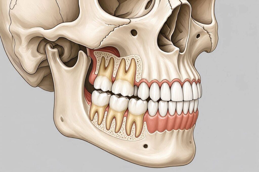
The wisdom teeth, like all teeth, are housed within the alveolar process of the maxilla and the mandible. In the maxilla, the third molar sits in the maxillary tuberosity, the posterior-most extension of the alveolar process, just anterior to the pterygomaxillary fissure. In the mandible, the third molar sits in the retromolar region of the alveolar process, just anterior to the ascending ramus. The relationship between the developing third molar and the surrounding alveolar bone is the anatomical stage upon which the drama of impaction unfolds.
The Impaction Problem: When the Jaw Is Too Small
The wisdom tooth is a normal, programmed tooth that develops in virtually all humans. It is not a pathological structure. The pathology arises because the modern human jaw is frequently too small to accommodate it. This is the evolutionary mismatch that defines the clinical significance of third molars.
The developing third molar germ begins to form around age 7–9, late in the sequence of tooth development. The crown calcifies, and the root begins to elongate in adolescence. The tooth attempts to erupt into the oral cavity, following the path of the previously erupted first and second molars. But in a significant percentage of the population, there is insufficient space in the dental arch. The retromolar space—the distance between the distal surface of the second molar and the anterior border of the ascending ramus of the mandible—is inadequate to accommodate the mesiodistal width of the third molar crown. The tooth becomes impacted: trapped, partially or completely, within the alveolar bone of the mandible or maxilla.
The impaction is a failure of the jaw to provide adequate housing. The tooth itself is normal. The jawbone is the limiting factor. This makes the wisdom tooth’s “part of the jaw” status clinically critical: the pathology of impaction is not a disease of the tooth but a structural inadequacy of the jawbone. The tooth is the innocent victim of its bony environment. The surgical removal of the impacted third molar is a procedure on the jawbone—osteotomy (bone removal), tooth sectioning, and elevation—as much as it is a procedure on the tooth.
The Relationship to Key Anatomical Structures: The Danger Zones
The wisdom tooth’s location within the jawbone places it in intimate proximity to several critical anatomical structures that are also “part of the jaw.” The surgical risk of third molar extraction is defined by these relationships.
In the mandible, the inferior alveolar nerve (IAN) runs through the mandibular canal, a bony tunnel within the body of the mandible. The roots of the mandibular third molar often extend inferiorly toward this canal, and in some cases, they are intimately associated with it—touching, grooving, or encircling the nerve. The lingual nerve, which supplies sensation to the anterior two-thirds of the tongue, runs along the lingual aspect of the mandibular third molar, just beneath the oral mucosa. Both nerves are part of the jaw’s neurovascular anatomy. The wisdom tooth is a resident of this anatomy, and its extraction involves navigating these nerves.
In the maxilla, the roots of the upper wisdom tooth are intimately related to the maxillary sinus floor. The sinus is an air-filled cavity within the maxillary bone. The roots of the third molar may protrude into the sinus, separated from the sinus airspace by only a thin layer of bone and the Schneiderian membrane. Extraction of the maxillary third molar can create an oro-antral communication—a hole between the oral cavity and the sinus—which may require surgical repair. The sinus is part of the maxillary jawbone, and the tooth’s roots are embedded in the bone that forms the sinus floor.
The Evolutionary Context: Why the Jaw Is Shrinking
The mismatch between tooth size and jaw size that produces impacted wisdom teeth is a relatively recent evolutionary phenomenon. The hominin fossil record shows that our ancestors—Australopithecus, early Homo erectus, Neanderthals—had larger, more prognathic jaws that comfortably accommodated a full complement of 32 teeth, including third molars that erupted into functional occlusion. The jaw was large. The teeth were in proportion. Impaction was rare.
The transition to agriculture and, later, to industrial food processing dramatically altered the mechanical demands on the developing jaw. A diet of tough, fibrous, unprocessed foods requires prolonged, forceful chewing, which stimulates mandibular growth. A diet of soft, processed, calorie-dense foods requires less chewing, reducing the mechanical stimulus for jaw development. The result, over generations, has been a reduction in jaw size—particularly in the retromolar space and the inter-canine width—without a corresponding reduction in tooth size. The teeth have remained roughly the same size. The jaw has shrunk. The third molars, as the last teeth to erupt, are squeezed out of the arch. They become impacted. They are still “part of the jaw,” but they are part of a jaw that no longer has room for them.
This evolutionary perspective answers the question “Are wisdom teeth part of your jaws?” in a deeper sense: they are part of an ancestral jaw that no longer exists in many modern humans. They are a vestigial component of a larger, more robust masticatory apparatus that has been gradually selected against by dietary and behavioral changes. They are part of the jaw’s evolutionary history, even when they are clinically problematic in the jaw’s modern form.
The Congenital Absence Phenomenon: When the Tooth Never Forms
A significant and growing percentage of the population is born without one or more wisdom teeth. This condition is called agenesis of the third molars, and it is the most common form of congenital tooth agenesis. Rates vary by population, but studies suggest that 20–35% of individuals worldwide are missing at least one third molar, with some populations showing rates exceeding 40%.
Agenesis represents the ultimate evolutionary response to the shrinking jaw: the tooth simply stops developing. The dental lamina fails to produce the third molar bud. The jaw develops without the tooth germ, and the alveolar process in the retromolar region remains undeveloped. The jaw is “missing” the tooth, and in this case, the tooth is clearly not part of the jaw—it never formed.
Patients with third molar agenesis are spared impaction, pericoronitis, and the need for surgical extraction. Their jaws are smaller, and the tooth count matches the available space. This is, from an evolutionary perspective, the direction in which the human species is headed: a jaw that houses 28 teeth, not 32. The wisdom tooth is becoming obsolete, a topic explored in depth in a companion article. Its status as “part of the jaw” is, in evolutionary time, temporary and fading.
The Periodontal Ligament: The Interface Between Tooth and Bone
The periodontal ligament (PDL) is the connective tissue that bridges the tooth root and the alveolar bone. It is a dense, fibrous, highly vascularized and innervated tissue that serves multiple functions: it anchors the tooth in the socket, it transmits occlusal forces to the bone, it provides proprioceptive feedback during chewing, and it serves as a reservoir of stem cells for tissue repair.
The PDL is part of the jaw. It is derived from the dental follicle, which is part of the tooth germ, but it is functionally integrated into the alveolar bone. The Sharpey’s fibers that embed into the cementum on one side and the alveolar bone on the other are continuous with the collagen matrix of the bone. When a tooth is extracted, the PDL is removed with the tooth, leaving a bony socket that fills with a blood clot and eventually remodels into new bone. The extraction severs the PDL, the tooth, and the alveolar bone from each other.
The PDL is the reason a tooth is not ankylosed—fused directly to the bone. Ankylosis is a pathological condition where the cementum fuses to the alveolar bone, obliterating the PDL space. An ankylosed tooth is literally part of the jawbone in a way that a normal tooth is not. It cannot be moved orthodontically. It cannot be extracted without removing the surrounding bone. Ankylosis is the exception that proves the rule: the normal tooth-bone relationship is a fibrous joint, not a fusion.
Conclusion
Wisdom teeth are anatomically part of the jaws, developing within the alveolar processes of the maxilla and mandible, erupting through them, and remaining connected to the surrounding bone via the periodontal ligament, but they are embryologically and histologically distinct organs—ectodermal and ectomesenchymal structures—housed within the mesodermally derived jawbone like a biological implant. The clinical pathology of impaction arises precisely because the tooth is trapped within a jaw that is evolutionarily too small to accommodate it, making the jawbone itself the limiting factor. The tooth is part of the jaw, but it is a part that is increasingly absent, increasingly impacted, and, over evolutionary time, increasingly obsolete.
FAQ
Q: Can you feel your wisdom teeth growing in your jaw?
A: Yes, particularly during the active eruption phase in late adolescence and early adulthood. The sensation is usually a dull, persistent pressure or aching in the back of the jaw, often radiating toward the ear or the temporomandibular joint. This is the pressure of the erupting tooth pushing against the overlying bone and soft tissue. If the tooth is impacted and cannot erupt, the pressure may be intermittent and recurrent over years. Sharp, severe, or throbbing pain usually indicates pericoronitis (infection of the gum over a partially erupted tooth) or a developing abscess, not normal eruption.
Q: If wisdom teeth are part of the jaw, does removing them weaken the jawbone?
A: In the short term, the extraction socket creates a localized defect in the alveolar bone that heals over 6–8 weeks with new bone formation. In the long term, the alveolar bone in the extraction site resorbs because the tooth is no longer present to stimulate it. This is a normal, expected remodeling process. The basal bone of the mandible—the structural foundation—is not compromised by routine third molar extraction. However, removing multiple large, deeply impacted third molars in an older patient with an already atrophic mandible can increase the risk of a post-operative mandibular fracture, a rare but serious complication that occurs when the bony defect from the extraction is so large that the remaining bone cannot withstand normal occlusal forces during the healing period.
Q: Are wisdom teeth connected to the rest of the body’s skeleton through the jaw?
A: Yes, indirectly. The mandible articulates with the temporal bone of the skull at the temporomandibular joint (TMJ). The maxilla is fused to the skull base. Forces applied to the wisdom teeth—during chewing, clenching, or surgical extraction—are transmitted through the jawbones to the TMJ and the skull base. Dental infections from wisdom teeth can spread through the fascial planes of the head and neck, which are continuous with the jaw’s soft tissue spaces. The wisdom tooth is a localized structure within the jaw, but the jaw is integrated into the cranial skeleton and the deep fascial compartments of the head and neck. Dental pathology is never truly isolated from the rest of the body.
Additional Resource
For a detailed anatomical atlas of the human jaw, including the relationship of the third molars to the inferior alveolar nerve, the maxillary sinus, and the surrounding skeletal structures, visit the online learning resources of the American Association of Anatomists at www.anatomy.org.