Are You Referring To Bone Grafting?
The phrase lands in the middle of a dental consultation like an unexpected bill. You came in to discuss replacing a missing tooth with an implant, and suddenly the dentist is talking about harvesting bone, membranes, and healing periods that stretch for months. “Are you referring to bone grafting?” you ask, your mind racing through images of orthopedic surgeons and hip replacements. Yes. That is exactly what they are referring to, and your immediate reaction—confusion, fear, skepticism about whether this is a necessary procedure or an upcharge—is entirely rational.
Dental bone grafting is one of the most misunderstood and unexpectedly frequent procedures in modern restorative dentistry. It is not a scam. It is not a luxury add-on. It is, for millions of patients who have lost teeth and the bone that supported them, the biological foundation without which a dental implant is doomed to fail. This article is a deep exploration of what bone grafting in the oral cavity actually entails, the cellular biology that makes it work, the different graft materials and their sources, the specific clinical scenarios that trigger a graft recommendation, the timeline of healing, and the honest risk-benefit calculus patients need to make an informed decision.
The Atrophy Problem: Why Bone Disappears After Tooth Loss
To understand why bone grafting is so frequently necessary, one must understand the cruel biological irony of tooth loss. The alveolar bone—the ridge of bone that houses the tooth sockets—exists for one purpose: to hold teeth. It is a use-it-or-lose-it tissue. The mechanical stimulation of chewing, transmitted through the periodontal ligament to the bone, signals to the osteoblasts (bone-building cells) to maintain that bone. When a tooth is extracted, that signal ceases. The body, in its relentless metabolic efficiency, resorbs the bone. Osteoclasts (bone-eating cells) dissolve the mineral matrix, and the calcium is recycled into the bloodstream.
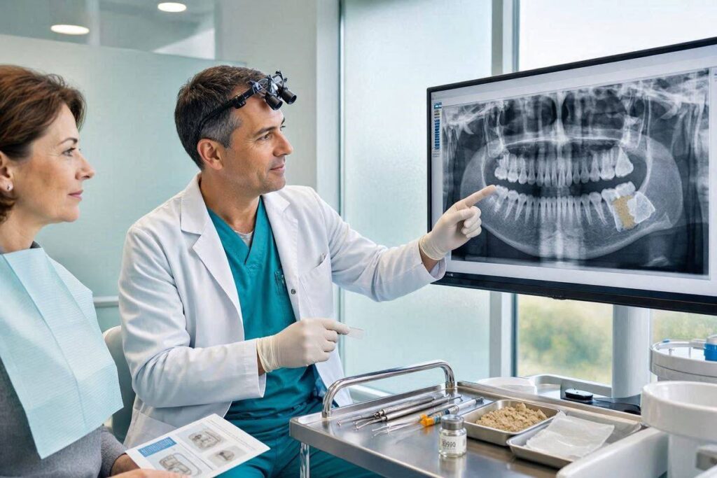
This resorption is dramatic and rapid. The majority of bone width loss occurs in the first 6–12 months post-extraction. Over a lifetime, an edentulous ridge can resorb to a thin, knife-edged sliver of bone that is structurally inadequate to support a dental implant. The implant requires a minimum volume of bone—roughly 1.5–2 millimeters of bone on all sides of the implant and a certain height above the inferior alveolar nerve and maxillary sinus—to osseointegrate successfully. If the bone has atrophied, the implant cannot be placed without first rebuilding the missing volume. This is the primary scenario where the dentist says, “We need to graft.” They are not inflating the treatment plan; they are responding to a biological deficit that, if ignored, will result in implant failure, nerve injury, or sinus perforation.
Socket Preservation: The Proactive Graft
The most common bone grafting procedure is not a massive reconstruction of a jaw that has been toothless for decades. It is a small, strategic, time-sensitive graft performed immediately after a tooth extraction. This is called socket preservation, or ridge preservation, and it is the dental equivalent of putting a placeholder in a parking spot.
When a tooth is extracted, the empty socket is a four-walled bony defect (assuming the buccal and lingual walls are intact). The body will naturally fill this socket with a blood clot, which will organize into granulation tissue and eventually woven bone. However, during this natural healing, the buccal wall—the thin outer plate of bone facing the cheek—often collapses inward, and the ridge shrinks in width. Socket preservation places a particulate bone graft material into the empty socket immediately after the tooth is removed. The graft acts as a scaffold, maintaining space and preventing the soft tissue and external pressure from collapsing the socket. A resorbable collagen membrane is often placed over the top to contain the graft particles and exclude gum tissue from growing into the bone compartment.
The purpose is to preserve the ridge dimensions for a future implant. Without socket preservation, the ridge may resorb 30–60% of its width, making implant placement later impossible without a more extensive, more expensive, and more morbid block graft. The socket graft is a minor procedure with a high return on investment. It is the single most cost-effective bone grafting intervention because it prevents the need for a major graft later.
Guided Bone Regeneration (GBR): The Membrane Principle
Socket preservation is a subset of a broader category called Guided Bone Regeneration (GBR). The principle of GBR is that different tissues heal at different speeds. Soft tissue (gum epithelium and connective tissue) grows rapidly and will invade a bony defect within days. Bone cells grow slowly. If you simply place bone graft material into a defect without a barrier, the fast-growing soft tissue will colonize the space and form scar tissue, not bone.
GBR uses a barrier membrane to exclude the unwanted soft tissue cells and create a protected space where only the slower-growing bone-forming cells (osteoblasts from the surrounding bony walls) can populate the graft. The membrane is the critical component. It can be a resorbable collagen membrane that dissolves over 4–6 months, or a non-resorbable PTFE (Teflon) membrane that requires a second minor surgery to remove. Resorbable membranes are the current standard for most routine cases. The membrane is tucked under the gum flaps, draped over the graft, and sutured into place. The gum is closed over the membrane, and the site heals for 4–9 months, after which an implant can be placed into the newly regenerated bone.
Graft Materials: Where the Bone Comes From
The term “bone graft” conjures images of a second surgical site, a hip or a rib being harvested in a painful, invasive procedure. For the vast majority of dental bone grafting, this is not the case. The graft material is typically a particulate powder or small granules, not a solid block, and the source is often not the patient’s own body. The materials fall into four categories.
- Autograft (Your Own Bone): This is bone harvested from the patient’s own body. In dentistry, the most common donor site is the mandibular ramus (the back of the lower jaw) or the chin (mental symphysis). Autograft is the gold standard because it is osteogenic—it contains live bone cells and growth factors. It is also osteoconductive (a scaffold for new bone) and osteoinductive (recruits stem cells to become bone). However, it requires a second surgical site, which adds morbidity, pain, and operative time. It is typically reserved for larger, more complex defects where its superior biological properties justify the donor site cost.
- Allograft (Human Donor Bone): This is bone from a human cadaver donor, processed and sterilized by tissue banks. Allograft is osteoconductive but not osteogenic—the processing kills the cells. It is the most common graft material used in socket preservation and GBR. It is available in abundance, avoids a donor site, and has an excellent safety record. Patients may initially recoil at the idea of cadaver bone, but the processing is rigorous, and the risk of disease transmission is vanishingly small.
- Xenograft (Animal Bone): The most common xenograft is deproteinized bovine bone mineral, derived from cow bone. The organic material is removed, leaving a porous, calcium-phosphate scaffold that is structurally very similar to human cancellous bone. Xenograft is very slow to resorb—some particles remain for years, acting as a permanent scaffold. This can be an advantage in sites where volume maintenance is critical. It is widely used, safe, and effective, but carries the psychological barrier of animal origin for some patients.
- Alloplast (Synthetic): These are laboratory-synthesized materials: hydroxyapatite, beta-tricalcium phosphate, bioactive glasses. Alloplasts are purely osteoconductive scaffolds with no biological growth factors. They are completely synthetic, eliminating any theoretical disease transmission risk and the psychological aversion to human or animal tissue. They resorb at varying rates depending on the formulation and are commonly used in periodontal defects and smaller grafting sites.
The choice of material is a clinical decision based on defect size, healing timeline, the need for resorption rate control, and patient preference. Most routine socket grafts use a combination of allograft and xenograft, or an alloplast/xenograft blend.
The Sinus Lift: The Upper Jaw Challenge
A specific and dramatically named bone grafting procedure deserves focused attention: the sinus lift, or sinus augmentation. The maxillary posterior teeth (upper molars and premolars) sit directly beneath the maxillary sinus, a large air-filled cavity. When these teeth are lost, the sinus floor often pneumatizes—it expands downward into the space formerly occupied by the roots. The remaining bone between the sinus and the oral cavity can be paper-thin, sometimes 1–3 millimeters.
A dental implant requires a minimum of 6–10 millimeters of bone height in the posterior maxilla. To create this height, the sinus membrane must be lifted upward, and bone graft material packed into the space created beneath it. This is the sinus lift. It can be performed via a lateral window approach (access through a small window created in the lateral wall of the sinus) or a crestal approach (access directly through the implant osteotomy site, using osteotomes to gently fracture the sinus floor upward). The lateral window approach is the classic, more invasive technique for cases with very little residual bone. The crestal approach is less invasive and can often be performed at the same time as implant placement if adequate primary stability is achieved.
The sinus lift is a predictable, well-documented procedure with success rates exceeding 90%. The primary complication is perforation of the Schneiderian membrane (the sinus lining), which occurs in a minority of cases. A small perforation can be patched with a collagen membrane; a large perforation may require aborting the procedure and allowing the membrane to heal before re-attempting. This is not a failure of the concept; it is a manageable intraoperative complication.
The Block Graft: The Major Reconstruction
For significant horizontal or vertical ridge defects—typically in a jaw that has been edentulous for many years—particulate graft alone is insufficient. The defect lacks the containing walls to hold the particulate material in place, and the volume required exceeds what can be achieved with loose granules. This is where the autogenous block graft enters the picture.
The block graft harvests a solid piece of cortical and cancellous bone, usually from the mandibular ramus or the chin, and secures it to the atrophic ridge with small titanium screws. The block is contoured to match the ridge anatomy, and any gaps are filled with particulate graft. The site is covered with a membrane, and the soft tissue is closed. The graft is allowed to heal for 4–6 months, integrating with the host bone. After this consolidation, the screws are removed, and implants can be placed into the now-voluminous ridge.
Block grafting is the most surgically involved dental bone graft. It has higher morbidity, a risk of temporary or permanent neurosensory disturbance at the donor site (numbness of the lower lip or chin if the mental nerve is traumatized during chin graft harvest), and a longer recovery. It is reserved for the most severely atrophic cases and is increasingly being supplanted by alternatives like distraction osteogenesis or the use of recombinant human bone morphogenetic protein (rhBMP-2) with a titanium mesh or resorbable scaffold, which can generate large volumes of bone without a donor site.
The Timeline Problem: Why Implants Take So Long
The most frequent patient frustration with bone grafting is the time. A patient wants a tooth replaced. They are told they need a graft, then 4–6 months of healing, then the implant, then 3–4 months of osseointegration, then the crown. The total timeline from extraction to final restoration can be 9–18 months. This is a psychologically brutal wait.
The timeline is dictated by the biology of bone healing and osseointegration. The graft must be revascularized—new blood vessels must grow into the graft material, bringing oxygen, nutrients, and osteoprogenitor cells. The graft is slowly replaced by living bone through a process called creeping substitution. This is slow. Cutting the healing time short to place an implant into immature, poorly vascularized graft risks implant failure due to lack of osseointegration. The implant must then undergo its own 3–4 month integration period.
There are protocols to accelerate or combine stages. Immediate implant placement into an extraction socket, with simultaneous grafting of any gap between the implant and the buccal wall, is a one-stage procedure that can save months. But this requires a favorable extraction socket with an intact buccal wall and adequate bone apical to the socket for primary implant stability. If these conditions are not met, the staged approach is necessary. The surgeon is not trying to extend the treatment for financial gain; they are respecting the biological speed limits of hard tissue healing.
Risks and Complications: The Honest Reckoning
Bone grafting, like all surgical procedures, carries a defined set of risks. Patients deserve to hear them unvarnished.
- Infection: The oral cavity is a contaminated field. A graft site can become infected, leading to graft loss, sequestration (the graft material dying and being extruded as small particles), and failure of the bone regeneration. Perioperative antibiotics, chlorhexidine mouth rinse, and meticulous surgical sterility minimize this risk.
- Membrane Exposure: A common complication of GBR is premature exposure of the membrane. The gum tissue over the graft can break down, exposing the membrane to the oral environment. This is a wound dehiscence. If the membrane becomes exposed, it is rapidly colonized by oral bacteria, and the graft may become infected. Management ranges from frequent chlorhexidine rinses and monitoring to early membrane removal and graft salvage, depending on the extent and timing.
- Nerve Injury: Grafting in the posterior mandible near the inferior alveolar nerve, or harvesting a chin graft near the mental nerve, carries a risk of paresthesia (altered sensation) or anesthesia of the lower lip and chin. A cone beam CT scan is mandatory for pre-operative planning to map the nerve’s position. The risk of permanent nerve injury from a routine socket graft is very low; the risk from a chin block graft harvest is higher and must be explicitly disclosed.
- Sinus Complications: Sinus lift can cause sinus membrane perforation, post-operative sinusitis, or, rarely, displacement of graft material into the sinus cavity. A skilled surgeon can manage most membrane perforations intraoperatively. Persistent post-operative sinus congestion or infection should be evaluated by an ENT specialist.
- Graft Failure: A graft can simply fail to consolidate—the material resorbs, is replaced by fibrous scar tissue rather than bone, and the implant site is lost. This is more common in smokers, patients with uncontrolled diabetes, or sites of previous radiation therapy. Smoking is a strong relative contraindication to bone grafting; smokers have significantly higher graft failure rates due to vasoconstriction and impaired wound healing.
Comparative Table: Graft Material Properties
| Material | Source | Osteogenic? | Resorption Rate | Cost | Best For |
|---|---|---|---|---|---|
| Autograft | Patient’s own body. | Yes (gold standard). | Moderate (replaced by living bone). | High (second surgical site). | Large defects, block grafts. |
| Allograft | Human cadaver donor. | No (osteoconductive). | Slow to moderate. | Moderate. | Socket preservation, GBR. |
| Xenograft | Bovine bone. | No. | Very slow (years). | Moderate. | Sites requiring long-term volume maintenance. |
| Alloplast | Synthetic (HA, TCP). | No. | Variable (controlled). | Moderate. | Periodontal defects, small sockets. |
The Non-Grafting Alternatives: When Bone Grafting is Not the Only Answer
In selected cases, bone grafting can be avoided. Short implants—implants less than 8 millimeters in length—can be placed in areas of limited vertical bone height, avoiding the need for a sinus lift. Zygomatic implants bypass the maxillary bone entirely and anchor into the zygomatic (cheek) bone, eliminating the need for grafting in the severely atrophic maxilla. Subperiosteal implants, though largely historical, sit on top of the bone under the periosteum and are custom-fabricated for the patient’s ridge anatomy.
These alternatives have their own risk profiles, success rates, and limitations. Short implants have higher failure rates in poor quality bone. Zygomatic implants require an advanced surgical skill set and carry risks of orbital penetration and sinus complications. The alternative to a graft is not always a simpler path; it is often a technically more demanding one. But for the patient who is adamantly opposed to bone grafting, a discussion of these alternatives is appropriate.
Important Note: The “Wait and See” Deception
A destructive myth in dental implant conversations is that grafting can be deferred: “Let’s just pull the tooth and see how the bone heals.” This is not a neutral strategy. It is a decision to allow resorption to occur, and it converts a simple socket graft (performed at the time of extraction) into a more complex, costly block graft (performed months or years later). If an implant is being considered as a future replacement option, the time to graft is at the moment of extraction, not after the ridge has collapsed. “Wait and see” is a choice to lose bone volume, and the patient should understand that it is an irreversible choice with significant downstream consequences.
Conclusion
Dental bone grafting is the surgical placement of bone or a bone substitute material into a deficient jaw site to regenerate the volume and density required for successful dental implant osseointegration, most commonly performed immediately after extraction as socket preservation. Graft materials range from the patient’s own bone (autograft, the gold standard) to cadaver, bovine, and synthetic sources, each with distinct biological activity and resorption profiles, selected based on defect size and healing timeline. The grafting process extends the total implant treatment timeline by 4–9 months but is a biologically necessary foundation; skipping it when bone volume is inadequate results in implant failure, nerve injury, or sinus perforation.
FAQ
Q: Is bone grafting painful?
A: The grafting procedure itself is performed under local anesthesia (or IV sedation if combined with other surgeries) and is painless. Post-operative discomfort is typically mild to moderate, managed with over-the-counter NSAIDs or prescribed analgesics for 2–3 days. Swelling and bruising are common, particularly with larger grafts or block harvests. Socket preservation grafts are minimally uncomfortable. A chin block graft has more significant post-operative pain and a risk of temporary chin numbness. Most patients report that the procedure was far less painful than they anticipated.
Q: Can I get a bone graft if I smoke?
A: Smoking significantly impairs graft healing due to nicotine-induced vasoconstriction and carbon monoxide-induced hypoxia. Smokers have a higher rate of graft failure, wound dehiscence, and infection. Many surgeons will not perform elective bone grafting on heavy smokers or will require a smoking cessation period of 2–4 weeks before and after the procedure. If you smoke, be honest with your surgeon. The risk calculus changes, and a staged, cautious approach with a lower success expectation is appropriate.
Q: Will my dental insurance cover bone grafting?
A: Bone grafting is a medical procedure but is billed under dental insurance. Coverage varies wildly. Some dental plans exclude grafting entirely as “experimental” or “not a covered benefit,” despite its well-established clinical evidence base. Others cover a portion under the surgical benefit, particularly if the graft is related to a traumatic extraction or cyst removal. Socket preservation is often excluded. The implant itself is rarely covered, and the grafting may be denied on the basis that it is part of an uncovered implant treatment plan. A pre-treatment estimate submitted to the insurer is essential.
Additional Resource
For an in-depth, patient-friendly explanation of bone biology, graft material science, and implant site development protocols, visit the American Academy of Periodontology’s patient education portal at www.perio.org.