Can Dental Implants Affect Mri?
You are scheduled for an MRI. Perhaps it is for your knee, your spine, or, more concerningly, your brain. The pre-screening questionnaire lands in your hands, and it asks a series of ominous questions. Do you have any metal in your body? A surgical clip? A pacemaker? Shrapnel? You remember the dental implant in your jaw. A titanium post, surgically placed, now fused to your bone. A cold wave of anxiety washes over you. Can this dental implant affect the MRI? Will it heat up, move, or tear itself from my jaw? Will it ruin the images my doctor desperately needs?
This article is a definitive, science-based guide to the interaction between dental implants and magnetic resonance imaging. We will explain the physics of what happens to titanium in a magnetic field, the safety data that has been gathered over decades, and the real-world effect on the diagnostic quality of the scan. The goal is to replace your anxiety with calm, factual knowledge.
The Metal Question: Ferrous vs. Non-Ferrous
The MRI machine is a giant, incredibly powerful magnet. Its magnetic field is measured in Tesla, with typical clinical scanners operating at 1.5T or 3.0T. The danger is not with all metals, but specifically with ferromagnetic metals. Ferromagnetic materials, such as iron, nickel, and cobalt, are strongly attracted to a magnet. An object made of ferromagnetic metal, if brought into the MRI room, can become a dangerous projectile, accelerating into the bore of the magnet with lethal force. An implanted ferromagnetic object, such as an old aneurysm clip of certain designs, can twist or migrate in the tissue under the influence of the strong magnetic field, causing catastrophic internal injury.
Dental implant posts are not made of ferromagnetic metals. The global standard for dental implants is medical-grade titanium alloy, specifically Ti-6Al-4V ELI. Titanium is a paramagnetic metal. This is a fundamentally different category. A paramagnetic material is very weakly attracted to a magnetic field, but this attraction is so slight that it has no clinical significance. It will not move. It will not heat up to a dangerous degree. It will not become a projectile. Titanium is one of the most MRI-compatible materials in existence. It is the same material used for many orthopedic implants, plates, and screws that routinely and safely go through MRI scanners every single day.
Zirconia dental implants, the white, metal-free alternative, are even more inert. Zirconia is a ceramic, completely non-magnetic. It is entirely invisible to the MRI’s magnetic field. A patient with a zirconia implant can enter an MRI scanner with the same magnetic profile as a patient with no implant at all.
The Safety Data: Decades of Safe Scanning
The safety of dental implants in MRI is not a theoretical assumption. It is a documented clinical reality with decades of experience. Millions of patients with titanium dental implants have undergone MRI scans of every body part without a single reported case of an implant being displaced by the magnetic field or causing a significant thermal injury. The American College of Radiology, the governing body for imaging safety, explicitly lists dental implants and dental fillings as items that are safe for MRI and do not require a period of waiting or a waiver.
The concern about heating is theoretically plausible. The rapidly switching radiofrequency pulses and magnetic gradients of the MRI machine can induce small electrical currents in a conductive metal loop. This can, in large, loop-shaped implants like some hip prostheses, cause a slight warming of the surrounding tissue. A dental implant is a tiny, single post. It does not form a conductive loop. The amount of radiofrequency energy it can absorb is minuscule, and the heat is immediately dissipated by the high blood flow in the jaw. The temperature rise at the implant site during a clinical MRI is negligible and completely harmless to the surrounding bone and gum.
Your dental implant is not a threat to your safety in the MRI scanner. It is a passive, inert piece of metal that the magnet will essentially ignore. You can be scanned with full confidence.
The Real-World Effect: Image Distortion and Artifact
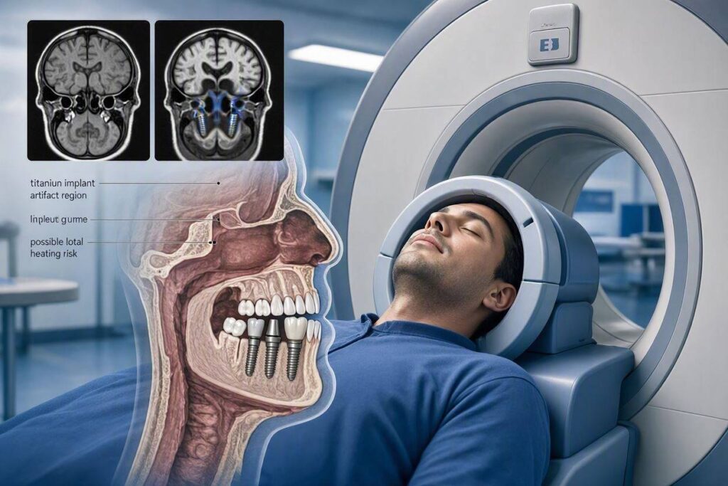
The significant effect of a dental implant on an MRI is not a safety risk. It is a diagnostic nuisance. It can affect the quality of the images. This phenomenon is called a susceptibility artifact. The titanium implant, while non-ferromagnetic, has a different magnetic susceptibility than the surrounding soft tissue and bone. It distorts the local magnetic field homogeneity. The scanner’s imaging algorithms rely on a perfectly uniform magnetic field to assign spatial positions. The implant creates a local field distortion, and the scanner mis-maps the signal from the tissues immediately adjacent to the implant.
The result is a signal void, a black hole in the image, surrounded by a rim of bright signal and geometric warping of the anatomy. The artifact is much larger than the implant itself. A dental implant can obscure a region of the jaw several times the diameter of the implant post. This is a significant problem if the MRI is specifically designed to evaluate the jaw, the floor of the mouth, or the tongue. A tongue cancer study, an evaluation for a jaw cyst, or a TMJ protocol can be severely compromised by the presence of multiple dental implants.
However, the critical point for most patients is one of anatomical distance. If your dental implant is in your jaw and you are undergoing an MRI of your brain, your cervical spine, or your knee, the artifact zone in your jaw is physically distant from the anatomy being imaged. It will not affect the diagnostic quality of those distant scans. The implant’s black hole is confined to the mouth. The brain, centimeters away, is imaged perfectly. The artifact will be visible on the localizer images, the initial scout views, and the radiologist will note the dental artifact. But the clinical question, the reason for the MRI, will be answered without interference. The only scenario where a dental implant truly impairs an MRI is when the scan is focused on the implant’s immediate neighborhood.
A Crucial Pre-Scan Action: Tell your MRI technologist and radiologist about your dental implant. Do not assume they will see it on the form. Describe the location and the number of implants. If the scan is of your head, neck, or face, the technologist may adjust the pulse sequence parameters, such as the bandwidth or the echo time, to minimize the size of the artifact. There are specific “metal artifact reduction sequences” (MARS) that can be employed to significantly improve image quality around metal implants. This is a conversation that should happen before the scan begins, not after.
The Absolute Contraindication: Removable Implant Overdentures
There is one scenario related to dental implants that does create a safety hazard. It is not the implant itself, but the removable prosthesis that attaches to it. Many implant-supported overdentures, like a “snap-on” denture, contain a metal housing with a ferromagnetic magnet used to retain the denture onto the implant abutments. This magnet can be strongly ferromagnetic. If a patient enters the MRI scanner with this removable denture in their mouth, the magnetic retention component can be forcefully pulled by the MRI’s magnetic field. This could damage the denture, cause injury to the patient’s mouth, or, in a worst-case scenario, become a projectile.
All removable dental prostheses, including implant-retained dentures, must be removed before you enter the MRI scanner room. This is a non-negotiable safety rule, the same as removing jewelry, hearing aids, and eyeglasses. The titanium implant posts in your jaw are safe. The removable appliance with the magnet is not. Confirm with your restorative dentist whether your overdenture contains magnetic attachments. If it does, it comes out before the MRI.
Conclusion
Dental implants, made of paramagnetic titanium or non-magnetic zirconia, are safe for MRI. They will not move, heat up dangerously, or become projectiles. The only significant effect is a localized image artifact, a dark distortion around the implant, which can obscure diagnostic detail in the jaw and mouth. For scans of any other body part, this artifact is irrelevant. Always inform your radiologist about your implant, and always remove any removable, implant-supported dentures that may contain magnetic components before entering the scanner.
Frequently Asked Questions
Will my dental implant set off the metal detector before the MRI?
No. The metal detectors used in MRI facilities are calibrated to detect ferromagnetic metals. A titanium dental implant is non-ferromagnetic and will not trigger the alarm. However, you must still declare your implant on the screening form and verbally to the technologist.
Can I have an MRI with a newly placed dental implant?
Yes. There is no biological or physical reason to wait. The titanium post is stable the moment it is placed. The osseointegration process is not affected by the MRI’s magnetic field or radiofrequency pulses. You can have an MRI the day after implant surgery, although the post-operative swelling and discomfort might make lying still for a prolonged scan unpleasant for other reasons.
Is a 3T MRI safe for dental implants?
Yes. A 3.0 Tesla MRI has a much stronger magnetic field than a 1.5 Tesla machine. The artifact will be larger and more pronounced, potentially more distorting to head and neck images. But the safety profile remains unchanged. The titanium implant is still non-ferromagnetic and will not pose a movement or heating risk at the higher field strength.
Additional Resource:
For the official safety guidelines on medical implants and MRI from the American College of Radiology, visit the ACR Manual on MR Safety: https://www.acr.org/Clinical-Resources/Radiology-Safety/MR-Safety