Can Dental Implants Close Gaps?
You have a gap in your smile. Perhaps it is a single dark space where a tooth once resided, a visual interruption in an otherwise even row of teeth. Perhaps it is multiple gaps, the legacy of extractions, trauma, or congenitally missing teeth that never developed. The gap bothers you. It catches food, it makes you self-conscious when you smile, and it feels like a flaw in an otherwise functional mouth. You have heard that dental implants are the gold standard for replacing missing teeth, and the question forms naturally: can an implant close this gap, restoring my smile to its original, uninterrupted wholeness?
The answer is a confident yes. Closing the gap left by a missing tooth is the fundamental purpose of a single-tooth dental implant. The implant replaces the absent root, and the crown fills the space in the dental arch. However, the simplicity of this answer belies a deeper layer of clinical nuance. An implant does not “close” a gap in the way that orthodontics closes a gap, by sliding adjacent teeth together. It fills the gap with a prosthetic tooth. This distinction has profound implications for the size of the gap, the health of the adjacent teeth, the condition of the underlying bone, and the final aesthetic outcome. Not every gap is suited to a single implant. Some gaps are too small, too large, or complicated by the drifting of adjacent teeth.
This guide explores the use of dental implants to restore the spaces left by missing teeth. We will explain the difference between prosthetic gap filling and orthodontic gap closure, the critical factors that determine whether an implant is the right solution for your specific gap, and the cosmetic and functional considerations that produce a result that looks and feels as though the gap was never there.
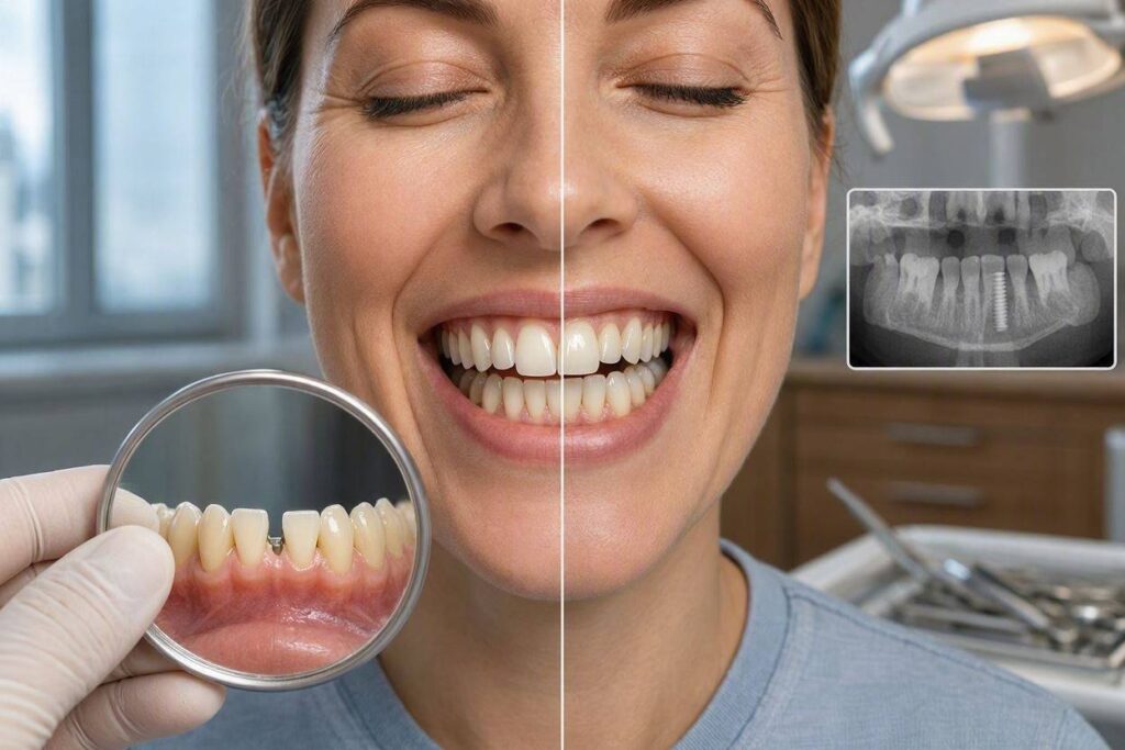
The Difference Between Filling a Gap and Closing a Gap
In dentistry, the words “fill” and “close” describe fundamentally different approaches to managing a space between teeth. Understanding this distinction is the first step in making an informed decision about your gap.
Orthodontic gap closure uses braces or clear aligners to physically move the adjacent teeth into the empty space. The teeth on either side of the gap are pulled together until they touch, and the space disappears. No prosthetic tooth is placed. The gap is eliminated by repositioning the natural teeth. This approach works well for small to moderate gaps, particularly when the adjacent teeth could benefit from orthodontic alignment anyway. The advantage is that no foreign material is introduced, and the result is entirely natural. The disadvantage is that the treatment takes months to years, and the space must be suitable for closure without creating an unbalanced bite.
Prosthetic gap filling, the implant approach, leaves the adjacent teeth where they are and places an artificial tooth in the space. The implant is drilled into the bone at the center of the gap. A custom crown is fabricated to occupy the exact dimensions of the missing tooth, touching the adjacent teeth on either side with light, hygienic contact points. The gap is visually and functionally filled, but the adjacent teeth have not moved. The advantage is that the adjacent teeth are untouched, preserved in their natural state. The disadvantage is that a surgical procedure is required, and the treatment involves a foreign body and a higher upfront cost.
The choice between these two approaches depends on the specific clinical situation. A patient with a missing tooth and perfectly aligned adjacent teeth is often best served by an implant, which preserves the alignment without disturbing the healthy teeth. A patient with a missing tooth and crowded, misaligned adjacent teeth may be better served by orthodontic treatment that simultaneously closes the gap and straightens the teeth, avoiding the need for both an implant and orthodontics.
When the Gap Is Too Small or Too Large
The size of the gap is a critical determinant of whether a single implant is feasible. A standard dental implant has a platform diameter of roughly 3.5 to 5.0 millimeters, and the crown that sits on top is wider than the implant platform. The crown must have enough space to look like a natural tooth and to establish proper contact with the adjacent teeth. If the gap is too narrow, the implant crown will look like a sliver, or it may not be possible to place an implant at all without encroaching on the roots of the adjacent teeth.
The minimum mesiodistal space—the distance between the adjacent teeth at the level of the crown—for a single implant restoration is approximately 6 to 7 millimeters for a lower incisor and 7 to 8 millimeters for a molar. If the space is smaller than this, a single implant may not be feasible. The options in a narrow gap include orthodontic closure of the space, a resin-bonded bridge that does not require an implant, or accepting a small residual space.
If the gap is too large—significantly wider than the original tooth—placing a single standard-diameter implant will leave excess space that results in an unnaturally wide crown or open spaces between the crown and the adjacent teeth. An oversized crown is aesthetically displeasing and functionally compromised because food will pack into the open contacts. The options for a large gap include placing a wider-diameter implant, placing two implants with two smaller crowns, or fabricating an implant-supported bridge where two implants support a prosthetic tooth or teeth that span the space.
The patient who has been missing a tooth for many years may find that the gap is larger than they remember. The adjacent teeth may have drifted apart, widening the space. The opposing tooth may have supra-erupted, intruding into the gap from the opposite arch. These secondary changes complicate the implant treatment plan. The gap may need to be orthodontically reduced to its original dimensions before an implant can be placed. The overerupted opposing tooth may need to be reduced or extracted. A gap that appears simple on the surface may, upon examination and CBCT analysis, reveal a complex restorative problem.
The Implant Gap Restoration Process
Restoring a gap with a dental implant follows the standard implant treatment sequence, with specific attention to the spatial and aesthetic dimensions of the edentulous space. The process begins with a thorough diagnostic workup. The dentist measures the gap with calipers or a digital scan, assessing the mesiodistal width, the buccolingual depth, and the vertical distance from the bone to the opposing tooth. A CBCT scan reveals the underlying bone volume, the position of the adjacent tooth roots, and the proximity of nerves and sinuses.
If the gap dimensions are suitable, the implant is surgically placed at the center of the edentulous space. The position is critical: the implant must be centered mesiodistally and located sufficiently lingually or palatally so that the screw access hole emerges on the biting surface or the lingual surface of the crown, not on the facial surface. An implant placed too far facially will create an aesthetic disaster, with the gray titanium or the screw access visible through the crown or the gum.
After osseointegration, the restorative phase begins. The dentist takes an impression or a digital scan of the implant position and the surrounding teeth. The dental laboratory fabricates a custom abutment and crown. The crown is designed to fill the gap precisely, with light contact on the adjacent teeth. The contact is tight enough to prevent food impaction but light enough to allow the patient to floss comfortably. The emergence profile—the contour of the crown as it emerges from the gum—is sculpted to mimic the natural tooth that was lost. In the anterior region, particular attention is paid to the incisal edge position, the color, the translucency, and the surface texture.
Cosmetic Considerations for Anterior Gaps
A gap in the anterior region, the smile zone, presents the highest cosmetic stakes. The implant crown must not only fill the space but also blend imperceptibly with the adjacent natural teeth. The color match must be exact. The shape, the length, the width, and the position of the zenith—the highest point of the gum contour—must harmonize with the neighboring teeth.
The cosmetic success of an anterior implant depends heavily on the condition of the bone and gum tissue at the time of implant placement. If the tooth was extracted traumatically, or if the gap has been present for years, the buccal bone plate on the facial side of the ridge may be deficient. The gum tissue may have collapsed into the defect, creating a concavity. An implant placed into this deficient ridge will not have the proper soft tissue support, and the crown will appear too long, with a dark shadow at the gum line.
Bone grafting and soft tissue grafting may be necessary to rebuild the ridge to a contour that supports a natural-looking crown. A connective tissue graft, harvested from the patient’s palate, can thicken the gum tissue and improve the emergence profile. These grafting procedures add time and cost to the treatment but are often the difference between a visible prosthetic restoration and an invisible one.
Multiple Gaps and Implant-Supported Bridges
A patient with several adjacent missing teeth faces a larger restorative challenge. Three adjacent missing teeth, for example, cannot necessarily be replaced with three individual implants. The space may not accommodate three implants without crowding the roots or compromising the bone between them. The restorative solution is often an implant-supported bridge.
Two implants, placed at the terminal positions of the edentulous span, can support a three-unit bridge that replaces three missing teeth. The bridge consists of two implant crowns, on the implants, connected by one or more pontics—prosthetic teeth that span the gap and rest on the gum tissue without underlying implants. The bridge fills the entire gap with a single, rigid restoration. This approach reduces the number of implants required, lowers the surgical burden, and can be more cost-effective than three individual implants.
The trade-off is that the bridge is more difficult to clean than individual implant crowns. The pontic rests on the gum, creating a surface that must be cleaned with a floss threader or a water flosser. The connection between the implant crowns and the pontics is a potential site for fracture or debonding. The entire bridge is a single unit, so if one implant fails, the entire restoration is compromised. These considerations must be weighed against the benefits of a fixed, implant-supported solution.
Conclusion
Dental implants are an excellent solution for filling the gaps left by missing teeth, with a single implant and crown capable of restoring a single tooth space to full function and aesthetics, provided the gap dimensions and bone volume are adequate. The implant approach fills the gap with a prosthetic tooth, a fundamentally different process from orthodontic closure, which moves natural teeth together. Gaps that are too narrow or too wide require modification through orthodontics or alternative restorative designs such as bridges.
Frequently Asked Questions
Q: How small can a gap be and still fit a dental implant?
A: The minimum mesiodistal space for a single implant and crown is approximately 6 to 7 millimeters for a lower incisor and 7 to 8 millimeters for a posterior tooth, measured at the level of the crown. If the space is smaller, the implant crown may not have a natural shape, or the implant may risk damaging the roots of the adjacent teeth. A CBCT scan provides the definitive measurement of the available space and the root positions.
Q: Can I get an implant if my teeth have shifted into the gap over time?
A: If the adjacent teeth have drifted into the gap, the available space may be insufficient for an implant. Orthodontic treatment can reopen the space to its original dimensions by moving the drifted teeth back into their proper positions. Once the space is restored, the implant can be placed. This combined orthodontic-implant approach is common and effective.
Q: Will an implant close a gap between my front teeth that I have always had?
A: An implant replaces a missing tooth. It does not close a diastema, which is a gap between existing natural teeth. If you have a gap between your two upper central incisors but both teeth are present, an implant is not the solution. Orthodontics or cosmetic bonding are the appropriate treatments for closing a diastema between natural teeth.
Q: How long after tooth extraction should I wait to get an implant to close the gap?
A: The timing depends on the condition of the extraction socket and the presence of infection. In ideal circumstances, an immediate implant can be placed at the same appointment as the extraction, and a temporary crown can fill the gap immediately. If infection is present, or if the bone is insufficient, a delayed approach is preferred, with the extraction site healing for two to four months before implant placement. A temporary removable partial denture or a bonded bridge can fill the gap during the healing period.
Additional Resource:
For information on tooth replacement options, visit the American College of Prosthodontists: https://www.gotoapro.org/
Ready for the next article? Continue to Part 35: Can Dental Implants Cause Nerve Damage? for a detailed examination of nerve injury risk, prevention, and management in implant dentistry.
continue
Meta Description: Can dental implants cause nerve damage? We examine the risks to the inferior alveolar, lingual, and mental nerves, how to prevent injury with CBCT planning, and what to do if nerve damage occurs.
Can Dental Implants Cause Nerve Damage?
You are considering dental implant surgery, and you have done your research. You understand the impressive success rates, the transformative benefits, and the general safety of the procedure. Then you encounter a forum post, a news article, or a friend-of-a-friend story describing a devastating complication: permanent numbness of the lip, chin, or tongue following implant placement. The implant, intended to restore function and aesthetics, instead caused a nerve injury that altered sensation and diminished quality of life. The question that follows is natural and urgent: can this happen to me? Is nerve damage a real risk of dental implant surgery?
The honest answer is yes. Nerve damage is a recognized, documented, and serious complication of dental implant surgery. It is not a myth or an exaggeration. However, it is also a complication that can be almost entirely prevented with modern diagnostic imaging, careful surgical planning, and precise surgical technique. The critical distinction is between nerve injuries caused by inadequate preoperative assessment—which are largely avoidable—and nerve injuries caused by rare anatomical variations or unpredictable surgical events, which are far less common. The vast majority of dental implant procedures are completed without any nerve involvement whatsoever. Understanding the specific nerves at risk, how they are protected, and what to do if an injury occurs transforms fear into informed vigilance.
This guide provides a comprehensive, anatomically precise examination of nerve injury risk in dental implant surgery. We will describe the three major nerves of concern: the inferior alveolar nerve, the lingual nerve, and the mental nerve. We will explain how preoperative CBCT imaging identifies and maps these nerves, how the surgeon designs the implant plan to maintain a safe distance, and the intraoperative techniques that minimize the risk of injury. We will also address the spectrum of nerve injuries, from transient paresthesia to permanent anesthesia, and the treatment options available if damage occurs.
The Nerves at Risk: Anatomy of the Surgical Danger Zones
The placement of dental implants in the lower jaw involves navigating a landscape of critical sensory nerves. The upper jaw, the maxilla, has fewer major nerve trunks in the implant-relevant zones, though the nasopalatine nerve and the posterior superior alveolar nerve can be encountered. The lower jaw, the mandible, is the primary locus of nerve injury risk because it houses the inferior alveolar nerve, the mental nerve, and the lingual nerve in close proximity to the implant osteotomy sites.
The Inferior Alveolar Nerve: This is the nerve of greatest concern in implant dentistry. The inferior alveolar nerve is a branch of the mandibular division of the trigeminal nerve, the fifth cranial nerve. It enters the mandible through the mandibular foramen on the inner aspect of the ramus and travels within the mandibular canal, a bony tunnel that runs through the body of the mandible. The nerve provides sensory innervation to the lower teeth, the lower lip, and the chin on that side.
The mandibular canal is the critical anatomical structure that every implant surgeon must visualize and respect. In the posterior mandible, where implants are placed to replace molars and premolars, the available bone height above the canal is often limited. The implant osteotomy must not violate the canal. Contact with the nerve within the canal can cause direct mechanical trauma, compression from an implant that encroaches on the canal, or thermal injury from the heat generated during drilling.
The consequences of inferior alveolar nerve injury are profound. The patient experiences paresthesia, which is an abnormal sensation such as tingling or numbness; dysesthesia, which is an unpleasant, often burning sensation; or anesthesia, which is complete loss of sensation in the lower lip and chin on the affected side. This sensory loss affects speech, eating, drinking, and kissing. Saliva and food can go unnoticed on the chin. The psychological impact of a numb, insensate lip is significant and can be permanent if the nerve damage is severe.
The Mental Nerve: The mental nerve is the terminal branch of the inferior alveolar nerve. It exits the mandible through the mental foramen, an opening on the facial surface of the mandible, typically located between the roots of the lower premolars. The mental nerve provides sensation to the lower lip, the chin, and the buccal gingiva of the lower anterior teeth.
The mental foramen is a key landmark for implant planning in the premolar region. An implant placed too close to the foramen, or a surgical flap that traumatically retracts the nerve, can cause mental nerve injury. The mental foramen is usually identifiable on a panoramic radiograph and on a CBCT scan, but its exact position varies among individuals and can be located more anteriorly or posteriorly than expected. The mental nerve can also form an anterior loop, where the nerve courses forward beyond the mental foramen before looping back to exit. An implant placed anterior to the mental foramen, thinking it is safely away from the nerve, can still injure this anterior loop if its presence is not recognized on the CBCT scan.
The Lingual Nerve: The lingual nerve is a branch of the mandibular division of the trigeminal nerve that provides general sensation and taste to the anterior two-thirds of the tongue. It descends near the inner surface of the mandible in the third molar region, running close to the lingual cortical plate. The lingual nerve is at risk during implant surgery in the posterior mandible, particularly when a lingual flap is elevated or when the implant osteotomy perforates the lingual cortical plate.
Lingual nerve injury results in numbness, tingling, or loss of taste on the affected side of the tongue. The patient may bite their tongue without realizing it. The loss of taste can significantly reduce the enjoyment of food. Lingual nerve injury is less common than inferior alveolar nerve injury in implant surgery, but it is a recognized and serious complication.
The CBCT Mandate: Preoperative Nerve Mapping
The single most important tool for preventing nerve injury in implant surgery is the Cone Beam CT scan. A two-dimensional panoramic radiograph, while useful for screening, does not show the buccolingual position of the mandibular canal, the exact location of the mental foramen, or the presence of an anterior loop of the mental nerve. Placing implants based solely on a panoramic radiograph is an outdated practice that exposes the patient to an unnecessary risk of nerve injury.
The CBCT scan provides a three-dimensional, sub-millimeter-resolution map of the mandibular anatomy. The surgeon scrolls through the axial, coronal, and sagittal slices and generates oblique cross-sectional views that are perpendicular to the curve of the mandibular arch. On these cross-sectional views, the mandibular canal appears as a round or oval radiolucency. The surgeon measures the vertical distance from the crest of the alveolar ridge to the superior border of the canal. This measurement determines the maximum safe length of the implant.
The surgeon also traces the mental foramen and looks for the anterior loop of the mental nerve. The CBCT software allows the surgeon to place a virtual implant in the bone and to measure the distance from the implant to the nerve in three dimensions. The safety zone is generally considered to be a minimum of 1.5 to 2.0 millimeters of bone between the implant apex and the superior border of the mandibular canal. This margin accounts for minor inaccuracies in the CBCT measurement and for the surgical reality that the drill may advance slightly beyond the planned depth.
The CBCT scan also reveals the lingual concavity in the posterior mandible. The submandibular fossa, a depression on the lingual side of the mandible, can be deep, and the lingual cortical plate can be thin or perforated. An implant that is placed too far lingually, or that is angled such that the apex perforates the lingual plate, can injure the lingual nerve. The CBCT shows the anatomy of the lingual plate, and the surgeon can plan the implant position to avoid perforation.<div style=”border-left: 4px solid #e76f51; padding: 15px; margin: 25px 0; background-color: #fff5f3;”> <p style=”font-weight: bold; margin-bottom: 5px;”>⚠️ The Non-Negotiable Standard of Care</p> <p style=”margin: 0;”>If an implant surgeon does not obtain a CBCT scan before placing implants in the posterior mandible, they are not adhering to the modern standard of care. The CBCT is not a luxury or an upsell; it is a patient safety imperative. A patient considering mandibular implant surgery should verify that a CBCT will be taken and reviewed as part of the treatment planning process.</p> </div>
Intraoperative Techniques for Nerve Protection
Even with a perfect CBCT plan, the surgeon must employ meticulous surgical technique to protect the nerves during the procedure. The CBCT plan is a map; the surgery is the actual journey through the terrain.
The surgeon uses a surgical guide, fabricated from the CBCT data, that controls the position, angle, and depth of the implant osteotomy. The guide fits onto the patient’s teeth or bone and has metal sleeves that direct the drills. The drills have depth markings or physical stops that correspond to the planned depth. Using a surgical guide and depth-controlled drills significantly reduces the risk of inadvertent nerve violation compared to freehand drilling.
The surgeon must be aware of the heat generated during drilling. Bone necrosis from excessive heat can occur at temperatures above 47 degrees Celsius sustained for more than one minute. Sharp, new drills, copious irrigation with chilled saline, and an intermittent drilling technique that allows the bone to cool between passes are the standard measures to prevent thermal injury. Thermal injury to the bone surrounding the nerve can cause nerve damage even if the drill does not directly contact the nerve.
The surgeon must also exercise caution when elevating the surgical flap in the posterior mandible. The lingual flap should be reflected gently, and retraction should avoid excessive pressure on the lingual tissues where the lingual nerve courses. The mental nerve should be identified and protected during flap elevation in the premolar region. A releasing incision that is placed too close to the mental foramen can transect the mental nerve.
Recognizing and Responding to an Intraoperative Nerve Event
Despite the best planning and technique, nerve events can occur during implant surgery. The patient, if under local anesthesia without heavy sedation, may report a sudden, sharp, electric-shock sensation during drilling or implant placement. This is a critical warning sign. It suggests that the drill or the implant has contacted the nerve.
If the patient reports this sensation, the surgeon should immediately stop the procedure and reassess. The drill or implant may be retracted slightly. The depth, angulation, and position should be re-evaluated relative to the CBCT plan. In some cases, the implant can be placed in a slightly modified position or a shorter implant can be selected. If the nerve has been directly traumatized, the surgeon may decide to abort the procedure, remove the implant, and allow the nerve to heal before attempting a different approach.
The worst response to a patient’s report of intraoperative nerve pain is to dismiss it, to attribute it to anxiety, or to proceed without modification. Ignoring the patient’s warning signal is a recipe for a permanent nerve injury and a medicolegal disaster.
The Spectrum of Nerve Injuries and Recovery
Nerve injuries from dental implant surgery range from transient, mild paresthesia to permanent, complete anesthesia. The classification of nerve injuries, originally developed by Seddon and Sunderland for peripheral nerve trauma, is applied to implant-related nerve injuries.
Neuropraxia is the mildest form of nerve injury. The nerve is compressed or mildly stretched, but the axon and the connective tissue sheaths remain intact. The conduction of nerve impulses is temporarily blocked. The patient experiences numbness or tingling. Recovery is typically complete and occurs within days to weeks.
Axonotmesis is a more severe injury. The axon is disrupted, but the surrounding connective tissue sheaths, including the endoneurial tubes that guide regenerating axons, remain intact. Wallerian degeneration occurs in the distal segment of the nerve. The nerve can regenerate, with the axons growing down the preserved endoneurial tubes at a rate of approximately one millimeter per day. Recovery is possible but takes months to years, and it may be incomplete.
Neurotmesis is the most severe injury. The nerve is completely severed or so severely disrupted that the connective tissue sheaths are destroyed. Without the guidance of the endoneurial tubes, regenerating axons may form a painful neuroma, a tangled mass of nerve fibers, rather than reaching their intended targets. Spontaneous recovery is unlikely. Surgical repair, such as direct nerve anastomosis or nerve grafting, may be attempted, but the results are unpredictable and often incomplete.
The prognosis for nerve recovery depends on the severity of the injury, the time elapsed since the injury, and the age and general health of the patient. Early intervention for severe nerve injuries offers the best chance of recovery. A patient who experiences numbness or altered sensation following implant surgery should be evaluated promptly. The surgeon may prescribe a course of oral steroids to reduce inflammation around the nerve. If the sensory deficit persists beyond a few weeks, referral to an orofacial pain specialist, a neurologist, or a micro-neurosurgeon is indicated. Sensory testing, including two-point discrimination, directional discrimination, and thermal testing, maps the extent of the deficit.
Prevention Is the Only True Cure
The most effective treatment for implant-related nerve injury is prevention. The combination of a high-quality CBCT scan, restoratively driven implant planning with a safety zone of at least 1.5 to 2.0 millimeters from the nerve, the use of a surgical guide, depth-controlled drilling, copious irrigation, and careful flap management reduces the risk of nerve injury to an extremely low level. A patient who chooses a well-trained, experienced implant surgeon who embraces these technologies and techniques is choosing the best possible protection for their nerves.
Conclusion
Dental implant surgery carries a real but largely preventable risk of nerve damage, with the inferior alveolar, mental, and lingual nerves being the primary structures of concern in the mandible. Modern CBCT imaging, meticulous three-dimensional treatment planning, surgical guides, and careful intraoperative technique are the pillars of nerve injury prevention. When nerve injury does occur, prompt recognition, honest communication with the patient, and timely referral for specialist evaluation offer the best chance of maximizing recovery.
Frequently Asked Questions
Q: How common is permanent nerve damage from dental implants?
A: The reported incidence of permanent inferior alveolar nerve injury from implant surgery varies in the literature but is generally considered to be less than 1% when modern diagnostic and surgical protocols are followed. The incidence is higher when implants are placed without CBCT guidance or by less experienced surgeons. Transient sensory disturbances are more common but typically resolve within weeks.
Q: Can nerve damage from an implant be repaired surgically?
A: In cases of severe nerve injury, microsurgical repair may be attempted. This can involve direct suturing of the severed nerve ends, nerve grafting using a donor nerve from elsewhere in the body, or neurolysis, which is the removal of scar tissue compressing the nerve. The success rates for these procedures decline with time, making early referral essential. Even with optimal surgical repair, full return of normal sensation is not guaranteed.
Q: What are the first signs that an implant has damaged a nerve?
A: The most immediate sign is a sharp, electric-shock sensation during the surgery, reported by the patient if they are not heavily sedated. After the surgery, the signs include persistent numbness, tingling, a “pins and needles” sensation, a burning sensation, or complete loss of feeling in the lower lip, chin, gums, or tongue on the side of the implant. Any altered sensation that persists beyond the expected duration of the local anesthetic warrants immediate evaluation by the surgeon.
Q: Can a CBCT scan guarantee that nerve damage will not occur?
A: A CBCT scan dramatically reduces the risk but does not eliminate it entirely. Anatomical variations, such as a bifid mandibular canal with a smaller, accessory canal that is not visible on the scan, can result in nerve injury despite careful planning. Surgical error, such as drill slippage or exceeding the planned depth, can also occur. The CBCT is the most powerful preventive tool available, but it must be combined with careful surgical execution.
Additional Resource:
For information on nerve injury management in dentistry, visit the American Association of Oral and Maxillofacial Surgeons: https://www.aaoms.org/
Ready for the next article? Continue to Part 36: Can Dental Implants Cause Loss Of Taste? for an exploration of gustatory function and implant-related taste disturbances.
continue
Meta Description: Can dental implants cause loss of taste? We examine the relationship between implant surgery and gustatory dysfunction, explaining how nerve proximity, surgical trauma, and medications can affect your sense of taste.
Can Dental Implants Cause Loss Of Taste?
You have undergone dental implant surgery, and as the anesthesia wears off and the surgical site heals, you notice something unexpected. Food does not taste quite right. Your morning coffee seems muted. The citrus brightness of an orange is dulled. A metallic flavor lingers in your mouth that no amount of brushing can banish. You recall that taste is a function of nerves, and you know that implant surgery involves drilling near nerves. An unsettling question forms: has the implant surgery damaged my ability to taste?
The answer is that a direct, permanent loss of taste from dental implant surgery is a rare but possible complication, specifically associated with injury to the chorda tympani branch of the facial nerve or the lingual nerve during surgery in the posterior mandible. Far more common are temporary taste disturbances caused by surgical trauma, inflammation, medications, and the presence of metallic components in the mouth. The vast majority of post-implant taste alterations are transient and resolve as the tissues heal. Distinguishing between the rare, serious nerve injury and the common, self-limiting taste disturbance is essential for appropriate management and peace of mind.
This guide explores the anatomy and physiology of taste, the mechanisms by which implant surgery can temporarily or permanently alter gustatory function, and the diagnostic steps to take if you experience a change in your sense of taste following implant placement. Understanding the specific nerves involved and the typical recovery timeline will help you navigate this unsettling but usually temporary postoperative experience.
The Anatomy of Taste: A Delicate Sensory Network
The sense of taste, or gustation, is mediated by specialized sensory cells clustered in taste buds located primarily on the tongue, but also on the soft palate, the pharynx, and the epiglottis. These taste buds detect five basic taste qualities: sweet, salty, sour, bitter, and umami. The taste information is transmitted from the taste buds to the brain via three cranial nerves, each serving a different region of the tongue and oral cavity.
The facial nerve, cranial nerve VII, carries taste sensation from the anterior two-thirds of the tongue via a small branch called the chorda tympani. The chorda tympani joins the lingual nerve, a branch of the mandibular division of the trigeminal nerve, in the infratemporal fossa and travels with it to the tongue. This anatomical proximity is the reason lingual nerve injury, discussed in the previous article, can also affect taste. The chorda tympani is exquisitely sensitive to stretch, compression, and transection.
The glossopharyngeal nerve, cranial nerve IX, carries taste from the posterior one-third of the tongue. This nerve is rarely involved in dental implant surgery because the surgical field for implants does not extend this far posteriorly. The vagus nerve, cranial nerve X, carries taste from the epiglottis and the extreme posterior pharynx, also well outside the implant surgical field.
The important clinical point is that an implant placed in the posterior mandible, particularly in the molar region, is in close proximity to the lingual nerve and its accompanying chorda tympani fibers. Surgical trauma to these structures can affect both general sensation, the feeling of touch and temperature on the tongue, and the special sensation of taste. An implant placed in the anterior mandible or in the maxilla is far less likely to affect taste.
Mechanisms of Taste Disturbance After Implant Surgery
The causes of taste alteration following implant surgery can be categorized into direct nerve injury, indirect inflammatory effects, medication side effects, and the taste of metal.
Direct Nerve Injury: As described in the article on nerve damage, the lingual nerve and the chorda tympani can be injured during implant surgery in the posterior mandible. A lingual flap that is aggressively retracted can stretch the lingual nerve. An implant osteotomy that perforates the lingual cortical plate can directly traumatize the nerve. A suture placed too deeply in the lingual flap can entrap the nerve. A hematoma that forms in the floor of the mouth can compress the nerve. Depending on the severity of the injury, the taste disturbance can range from a mild, transient hypogeusia, which is reduced taste sensitivity, to a complete and permanent ageusia, which is loss of taste, on the affected side of the tongue.
It is important to note that even a complete unilateral chorda tympani injury rarely results in a subjective complaint of total taste loss. The taste buds on the other side of the tongue, the posterior tongue served by the glossopharyngeal nerve, and the palate continue to function. The brain integrates taste information from all these sources, and the patient may only notice a subtle reduction in taste intensity on the affected side. The most common complaint following unilateral chorda tympani injury is a persistent metallic or phantom taste, not a complete absence of taste.
Inflammatory and Traumatic Effects: The surgical procedure itself creates a significant inflammatory response in the tissues surrounding the implant site. This inflammation can spread to involve the lingual nerve and the chorda tympani without causing permanent structural damage. The inflammatory mediators sensitize the nerve endings and alter their signaling properties. The patient experiences a distorted taste, known as dysgeusia, or a reduced taste, known as hypogeusia, that resolves as the inflammation subsides over days to weeks.
The surgical trauma can also cause localized swelling that physically compresses the taste nerves. The tongue itself may be swollen or bruised if it was retracted during the surgery. The simple mechanical disruption of the oral tissues can temporarily alter the oral environment and the exposure of the taste buds to tastants.
Medication Side Effects: A wide range of medications prescribed in the perioperative period can alter taste sensation. Antibiotics, particularly metronidazole and clarithromycin, are notorious for causing a metallic taste. Chlorhexidine mouthwash, the gold standard antiseptic rinse prescribed after implant surgery, frequently causes a temporary alteration of taste perception. Analgesics, including non-steroidal anti-inflammatory drugs like ibuprofen, can cause taste disturbances. The taste alteration from medications is systemic, affecting the entire mouth rather than just the side of the implant, and it resolves when the medication is discontinued.
Metallic Taste from the Implant or Restorations: Some patients report a persistent metallic taste that they attribute to the titanium implant itself. The evidence for this phenomenon is mixed. Titanium is considered biologically inert and does not corrode significantly in the oral environment. However, the presence of a metal object in the jaw, or the galvanic interaction between the titanium implant and other metal restorations in the mouth, as discussed in the article on implant-related illness, can theoretically generate a metallic taste in some patients. The metallic taste may also originate from the abutment, the healing cap, or the temporary crown materials rather than from the implant fixture itself. The definitive all-ceramic crown, once placed, eliminates most of these potential sources.
Distinguishing Between Causes: A Diagnostic Approach
If you experience a change in your sense of taste following implant surgery, a systematic approach can help identify the likely cause and guide your expectations for recovery.
First, localize the taste disturbance. Is it limited to one side of the tongue, the side of the implant? If so, a local nerve issue is more likely. Is the taste alteration present throughout the entire mouth? This suggests a medication side effect or a systemic issue. Is the taste alteration specifically a metallic taste that you notice even when not eating? This is a common dysgeusia associated with medications, chlorhexidine, or the presence of metal components.
Second, note the timeline. Did the taste disturbance begin immediately after surgery, as soon as the local anesthetic wore off? This suggests a direct surgical effect on the nerve. Did it begin several days after surgery, coinciding with the start of antibiotics or chlorhexidine rinses? This strongly implicates the medication. Did it begin weeks or months after surgery, after the final crown was placed? This may relate to the restoration rather than the surgery.
Third, monitor the trajectory. A taste disturbance that is gradually improving over days to weeks is consistent with resolving inflammation or the discontinuation of a medication. A taste disturbance that is static or worsening over weeks to months is more concerning for a significant nerve injury and warrants specialist evaluation.
When to Seek Specialist Evaluation
A mild, generalized alteration in taste that improves over the first few weeks after implant surgery is common and generally does not require specialist intervention beyond reassurance. However, the following scenarios should prompt a referral to an orofacial pain specialist, an oral medicine specialist, or a neurologist:
- Complete loss of taste on one side of the tongue that persists beyond one month.
- A persistent, unpleasant taste—cacogeusia—localized to one side of the tongue.
- Altered taste accompanied by other signs of lingual nerve injury, such as numbness, tingling, or burning of the tongue on the affected side.
- Taste disturbance that is worsening rather than improving over time.
Specialist evaluation may include quantitative gustatory testing, where the patient is asked to identify and rate the intensity of standardized taste solutions applied to different regions of the tongue. This testing objectively maps the extent of the taste deficit. Electrophysiologic testing of the lingual and facial nerves may be performed in select cases. A CBCT scan can assess the proximity of the implant to the lingual nerve and the mandibular canal.
Recovery and Prognosis
The prognosis for implant-related taste disturbance depends on the cause. Medication-related and inflammation-related taste alterations almost always resolve completely when the offending medication is stopped and the surgical site heals. This typically occurs within two to four weeks.
Lingual nerve and chorda tympani injuries classified as neuropraxia, the mildest form, also carry an excellent prognosis, with recovery typically occurring within weeks to a few months. Axonotmesis injuries, where the axon is disrupted but the nerve sheath is intact, can recover over many months as the axon regenerates. The taste system has a remarkable capacity for plasticity, and the brain adapts to altered sensory input over time.
Neurotmesis, the complete severing of the nerve, carries a poor prognosis for spontaneous recovery. Microsurgical repair of the lingual nerve is possible but technically challenging, and the results for taste recovery are less predictable than for sensory recovery. The chorda tympani, being small and delicate, is particularly difficult to repair surgically. Prevention, through careful surgical planning and technique, remains the most effective strategy for preserving taste function.
Conclusion
Dental implant surgery can cause a temporary alteration in taste due to surgical inflammation, medications like antibiotics and chlorhexidine, and the presence of metal components in the mouth, with these disturbances typically resolving within weeks. Permanent, localized loss of taste is a rare complication associated with injury to the chorda tympani or lingual nerve during posterior mandibular implant surgery. Distinguishing between the common, self-limiting causes and the rare, serious nerve injury through careful attention to the localization, timeline, and trajectory of the taste disturbance guides appropriate management and reassurance.
Frequently Asked Questions
Q: How long will the metallic taste last after implant surgery?
A: A metallic taste caused by postoperative medications or chlorhexidine mouthwash typically resolves within a few days to a week after you stop using those products. A metallic taste associated with a temporary healing abutment may persist until the final crown is placed. A metallic taste associated with nerve trauma may persist for weeks to months as the nerve heals. If a metallic taste persists beyond three months, seek evaluation from your implant surgeon.
Q: Can a dental implant on the upper jaw affect my sense of taste?
A: It is significantly less likely. The nerves responsible for taste to the tongue travel in the lower jaw and the floor of the mouth. An upper jaw implant, particularly in the anterior maxilla, is far from these nerves. A taste disturbance following an upper implant is more likely due to medications, postoperative nasal or sinus congestion affecting the sense of smell, which heavily influences taste, or a coincidental unrelated condition.
Q: Will my sense of smell be affected by dental implant surgery?
A: Direct injury to the olfactory nerve from implant surgery is essentially impossible because the olfactory nerve is located high in the nasal cavity, far from the oral surgical sites. However, an upper posterior implant that encroaches on the maxillary sinus can cause sinus inflammation or infection, and the resulting nasal congestion can temporarily impair the sense of smell, and thus the perception of flavor, which is the combination of taste and smell.
Q: Can a zirconia implant prevent taste disturbances?
A: A zirconia implant eliminates the metal component, which may reduce the likelihood of a metallic taste associated with galvanic interactions or metal sensitivity. However, the primary risk to the taste nerves is surgical trauma, which is independent of the implant material. A zirconia implant placed with the same careful surgical technique as a titanium implant carries the same low risk of nerve injury and the same high probability of preserving taste function.
Additional Resource:
For information on taste disorders and oral sensory function, visit the National Institute on Deafness and Other Communication Disorders: https://www.nidcd.nih.gov/
Ready for the next article? Continue to Part 37: Can Dental Implants Cause Liver Cyst? for an evidence-based examination of the alleged link between dental implants and hepatic lesions.
continue
Meta Description: Can dental implants cause liver cysts? We investigate the scientific evidence behind this alarming claim, examining titanium particle dispersion, systemic effects, and what the research actually shows.
Can Dental Implants Cause Liver Cyst?
You have dental implants, or you are considering them, and you have encountered a disturbing claim in your research. Some online sources, alternative health practitioners, or well-meaning acquaintances have suggested that dental implants, particularly titanium implants, can cause liver cysts. The proposed mechanism sounds plausible enough to be alarming: the implant corrodes over time, releasing titanium particles into the bloodstream, which accumulate in the liver and trigger the formation of cysts. This claim, if true, would fundamentally alter the risk-benefit calculus of implant dentistry. A replacement tooth is not worth a hepatic lesion. The question demands a rigorous, evidence-based answer.
The answer, grounded in the current body of scientific literature and clinical epidemiology, is that there is no credible evidence establishing a causal link between dental implants and liver cysts. The claim is not supported by large-scale epidemiological studies, by the known toxicology of titanium, or by the clinical experience of implant dentistry over more than five decades. Titanium particles can be detected in the tissues surrounding implants and, in trace amounts, in distant organs including the liver. This is an established fact. However, the presence of particles does not equate to the causation of disease. The leap from “titanium particles are detectable” to “titanium particles cause liver cysts” is a speculation that lacks scientific validation and contradicts the vast body of safety data on titanium medical implants.
This guide examines the claim with scientific rigor and clinical honesty. We will explore the evidence for titanium particle release from dental implants, the fate of those particles in the body, the known causes of liver cysts, and the reasons why a causal link between the two is implausible based on current evidence. The goal is not to dismiss patient concerns but to provide the factual foundation upon which an informed decision can be made.
Titanium Particles and Systemic Distribution: What the Research Shows
Titanium and its alloys are widely regarded as biocompatible, corrosion-resistant materials. This is why they have been used for decades in dental implants, orthopedic joint replacements, pacemaker housings, and other permanent medical implants. However, “corrosion-resistant” is not synonymous with “corrosion-proof.” Titanium implants do release particles into the surrounding environment, a fact that the scientific literature has documented extensively.
The surface of a titanium implant is covered by a thin, stable oxide layer, primarily titanium dioxide, that forms spontaneously on contact with air or water. This oxide layer is what gives titanium its excellent biocompatibility. However, under certain conditions—mechanical wear from chewing forces, the acidic and enzymatic environment of inflammation, and the electrochemical conditions created by the presence of dissimilar metals—the oxide layer can be disrupted. Microscopic particles of titanium and titanium dioxide can be released from the implant surface through processes of fretting, corrosion, and wear.
These particles are found in the peri-implant tissues, the gum and bone immediately surrounding the implant. This is expected and well-documented. The more controversial question is whether these particles can enter the systemic circulation and accumulate in distant organs. The answer, based on autopsy studies and animal experiments, is yes. Titanium particles have been detected in the lymph nodes, spleen, liver, and lungs of patients with titanium implants. The lymphatic system drains the peri-implant tissues and transports particles to regional lymph nodes. From there, particles can enter the venous circulation and be distributed systemically. The liver, as the body’s primary filtration organ, is a natural site of particle accumulation.
The critical point, however, is that the detection of particles does not imply the causation of disease. The body has a remarkable capacity to sequester inert particles within tissues without triggering a pathological response. Titanium dioxide particles, in particular, are considered to have low toxicity. The mere presence of titanium in the liver of a person with liver cysts does not establish that the titanium caused the cysts. Correlation is not causation, and in the case of titanium and liver cysts, even a compelling correlation has not been demonstrated.
The Known Causes of Liver Cysts
Liver cysts are fluid-filled sacs that occur within the liver parenchyma. They are extremely common, particularly with advancing age. Simple hepatic cysts are benign, usually asymptomatic, and discovered incidentally on abdominal imaging performed for unrelated reasons. They are estimated to occur in approximately 5% to 18% of the population, with prevalence increasing with age.
The vast majority of liver cysts are congenital, meaning the person is born with a predisposition to develop them. They arise from biliary ducts that fail to connect properly with the main biliary tree during embryonic development and slowly expand over decades. They are not caused by an external agent. Polycystic liver disease, a genetic condition often associated with polycystic kidney disease, leads to the development of multiple cysts throughout the liver. This condition is hereditary, not acquired from environmental exposures.
Other types of hepatic cysts include parasitic cysts, caused by Echinococcus tapeworm infection; neoplastic cysts, which are cystic tumors that can be benign or malignant; and cysts associated with biliary tract diseases. None of these conditions have an established link to titanium or other metallic particles. The known causes of liver cysts are genetic, congenital, infectious, or neoplastic. Metal particle deposition is not recognized as a cause of hepatic cyst formation by hepatology or toxicology.
The Flawed Logic of the Implant-Cyst Connection
The claim that dental implants cause liver cysts typically follows a chain of reasoning that contains multiple logical gaps. The reasoning goes: implants release titanium particles, titanium particles travel to the liver, titanium is a foreign substance, foreign substances cause inflammation, inflammation causes cysts. Each step in this chain is problematic.
First, the release of titanium particles from a well-integrated, healthy implant is extremely low, often at levels that are difficult to distinguish from the background titanium exposure that all humans receive from dietary sources, cosmetics, and environmental pollution. Titanium dioxide is a ubiquitous compound used as a whitening agent in food, toothpaste, sunscreen, and countless consumer products. The contribution of a dental implant to the total body burden of titanium is minor compared to these everyday exposures.
Second, the liver’s response to inert, insoluble particles is typically sequestration, not cyst formation. Macrophages engulf the particles and store them within the liver sinusoids without causing significant architectural disruption or cyst formation. The formation of a cyst requires a specific pathological process: a fluid-filled cavity lined by epithelium. Metal particles do not create this structure.
Third, epidemiological evidence for an association between dental implants and liver cysts is absent. If dental implants caused liver cysts with any meaningful frequency, the millions of people with dental implants, many of whom undergo abdominal imaging for other reasons, would show a higher prevalence of liver cysts than the non-implant population. This signal has not been detected. No large-scale study has demonstrated an increased incidence of hepatic cysts in patients with titanium dental implants. The claim is based on isolated anecdotes, case reports, and theoretical speculation, not on the kind of controlled, population-level evidence that would be required to establish causality.<div style=”border-left: 4px solid #2a9d8f; padding: 15px; margin: 25px 0; background-color: #f0fdfa;”> <p style=”font-weight: bold; margin-bottom: 5px;”>💡 A Note on Online Health Information</p> <p style=”margin: 0;”>The claim that dental implants cause liver cysts circulates primarily in online forums, social media, and websites promoting alternative health philosophies. These sources often cite case reports or anecdotal patient testimonials as proof, while ignoring the vast body of peer-reviewed safety data. When evaluating a health claim, consider the source, the quality of the evidence, and whether the claim is consistent with the broader scientific consensus. Extraordinary claims require extraordinary evidence, and the evidence for an implant-cyst connection is far from extraordinary.</p> </div>
When Patient Concern Is Warranted
While the specific claim linking dental implants to liver cysts lacks evidence, the broader phenomenon of systemic symptoms attributed to implants is a legitimate area of patient concern and ongoing research. As discussed in a previous article on implant-related illness, some patients do experience systemic symptoms that they associate with their implants. These symptoms may include fatigue, cognitive difficulties, joint pain, and skin rashes. Liver cysts are not part of this symptom complex, but the concern about the implant affecting systemic health is shared.
The appropriate response to a patient who is worried about the systemic effects of their implant is not dismissiveness but a thoughtful, evidence-informed conversation. The patient should be encouraged to discuss their concerns with their primary care physician, who can evaluate any systemic symptoms on their own merits, independent of assumptions about the implant. If an abdominal ultrasound or CT scan incidentally reveals a liver cyst, the cyst should be evaluated by a hepatologist or gastroenterologist according to standard medical guidelines, which do not include implant removal as a treatment for hepatic cysts.
If a patient with liver cysts insists that their implants are the cause and demands removal, the clinician should acknowledge the patient’s conviction while providing the best available scientific evidence. A decision to remove a well-integrated, functional implant should be based on a clear, documented indication—peri-implantitis, implant fracture, nerve injury, verified allergy—not on an unsubstantiated theory of systemic particle toxicity.
Conclusion
There is no credible scientific evidence that dental implants cause liver cysts. While titanium particles from implants can be detected in distant organs including the liver, the presence of these particles does not establish a causal link to hepatic cyst formation, a condition that is common, usually congenital, and not associated with metal exposure in the medical literature. The claim circulates primarily in non-scientific venues and lacks the epidemiological, toxicological, and pathological support required to be taken seriously as a clinical risk.
Frequently Asked Questions
Q: Should I have my liver checked if I have dental implants?
A: There is no medical indication for routine liver imaging or liver function testing solely because you have dental implants. Standard medical care, including age-appropriate screening and evaluation of any new symptoms, should be followed. If you have symptoms such as abdominal pain, jaundice, or unexplained weight loss, consult your physician for a medical workup, not an implant surgeon.
Q: Can removing my dental implants resolve existing liver cysts?
A: There is no evidence that removing dental implants has any effect on pre-existing liver cysts. Liver cysts are fluid-filled structures that, once formed, do not typically resolve with the removal of a distant foreign body. Explantation of a well-functioning implant in the hope of shrinking a liver cyst is not a medically justified procedure.
Q: Are zirconia implants safer for the liver than titanium implants?
A: Zirconia implants eliminate the exposure to titanium particles, which may be a consideration for patients who are concerned about metal exposure or who have a documented titanium allergy. However, because the link between titanium particles and liver cysts is unsubstantiated, there is no evidence that zirconia implants confer a specific hepatic health benefit. The choice between titanium and zirconia should be based on well-established factors: aesthetics, the clinical track record, the specific implant system, and the surgeon’s experience.
Q: What about other systemic diseases? Can implants cause autoimmune disease or cancer?
A: Large-scale, long-term studies have not demonstrated an increased risk of autoimmune disease or cancer in patients with dental implants. Titanium is not classified as a carcinogen. The safety record of titanium implants over more than fifty years, with millions of patients, is robust. Ongoing research into the systemic effects of implant-derived particles is appropriate and necessary, but the current evidence does not support the removal of implants to prevent or treat systemic disease.
Additional Resource:
For information on liver cysts and hepatic health, visit the American Liver Foundation: https://liverfoundation.org/
Ready for the next article? Continue to Part 38: Can Dental Implants Cause Dry Mouth? for an exploration of xerostomia and its relationship to implant surgery and restoration.
continue
Meta Description: Can dental implants cause dry mouth? We explore the potential connections between implant surgery, medications, nerve function, and salivary flow, explaining when dry mouth is temporary and when it signals a deeper issue.
Can Dental Implants Cause Dry Mouth?
You have noticed a persistent dryness in your mouth since your dental implant surgery. Your tongue sticks to the roof of your mouth. You reach for a glass of water more frequently than before. Speaking for extended periods leaves your mouth feeling parched. The dryness is uncomfortable, and it worries you because you know that saliva is essential for protecting your teeth and your implant from decay and infection. You trace the onset of this dryness back to the implant procedure, and the question forms logically: did the implant surgery cause my dry mouth?
The answer is that dental implant surgery itself is rarely the direct, long-term cause of dry mouth, known medically as xerostomia. However, several factors directly related to the surgical experience can trigger temporary dry mouth, and the implant restoration can, in specific circumstances, contribute to altered salivary function or oral comfort. More commonly, the implant surgery coincides with other causes of dry mouth—medications, stress, dehydration, or underlying medical conditions—and the temporal association is mistaken for causation. Unraveling these overlapping factors is essential for identifying the true cause and finding effective relief.
This guide explores the relationship between dental implants and dry mouth. We will explain the normal function of the salivary glands, the mechanisms by which surgery and medications can temporarily reduce salivary flow, the rare instances where implant position can affect salivary ducts or nerves, and the diagnostic steps to determine whether your implant is the culprit or an innocent bystander. Understanding the salivary system empowers you to address dry mouth at its source.
The Salivary System: A Vital Oral Defense
Saliva is far more than water. It is a complex, biologically active fluid produced by three pairs of major salivary glands—the parotid glands in the cheeks, the submandibular glands under the jaw, and the sublingual glands under the tongue—as well as hundreds of minor salivary glands distributed throughout the oral mucosa. Saliva performs multiple essential functions: it lubricates the oral tissues for comfortable speech and swallowing, it initiates the digestion of starches, it buffers acids produced by plaque bacteria to prevent tooth decay, and it contains antimicrobial enzymes and antibodies that control the oral microbiome.
The salivary glands are innervated by the autonomic nervous system. Parasympathetic stimulation, the “rest and digest” state, produces a copious, watery saliva. Sympathetic stimulation, the “fight or flight” state, produces a smaller volume of thicker, more mucinous saliva. Medications that interfere with autonomic nerve signaling, particularly anticholinergic drugs, can dramatically reduce salivary flow. The glands are also sensitive to hormonal changes, systemic hydration status, and radiation exposure.
The implant surgical sites in the maxilla and mandible are located near, but generally do not directly involve, the major salivary glands. The parotid gland is located on the side of the face, near the ear, and its duct opens into the cheek opposite the upper second molar. The submandibular and sublingual glands are located in the floor of the mouth. Implant surgery in the posterior mandible approaches the floor of the mouth and the sublingual space, and a perforation of the lingual cortical plate can create a hematoma that compresses the sublingual gland. This is a rare but recognized complication that can transiently affect salivary flow from that gland.
Medications: The Most Common Postoperative Culprit
The most common cause of dry mouth in the period following implant surgery is medication, not the surgery itself. The medications prescribed or recommended in the perioperative period are a pharmacological assault on the salivary glands.
Antibiotics: Broad-spectrum antibiotics, such as amoxicillin or clindamycin, are frequently prescribed after implant surgery to prevent infection. While dry mouth is not the most prominent side effect of these drugs, they can alter the oral microbiome and contribute to a sensation of oral dryness or altered taste.
Analgesics: Non-steroidal anti-inflammatory drugs, such as ibuprofen, and opioid pain relievers, such as codeine or hydrocodone, can both cause dry mouth. Opioids, in particular, are known to reduce salivary flow through central nervous system mechanisms.
Chlorhexidine Mouthwash: The gold standard antiseptic rinse prescribed after implant surgery, chlorhexidine, has a well-documented side effect of causing a temporary alteration in taste and a sensation of dryness. The rinse can also cause a slight burning sensation of the oral mucosa. These side effects resolve when the rinse is discontinued, typically after one to two weeks of use.
Anesthetic Agents: The local anesthetic used during the surgery, particularly when a nerve block is administered, temporarily disrupts the autonomic nerve supply to the salivary glands on that side. The patient may notice a dry mouth on the side of the surgery for several hours until the anesthetic fully wears off. This effect is self-limiting.
If you experience dry mouth after implant surgery, review your medication list with your surgeon or pharmacist. The dry mouth often resolves when the postoperative medications are completed. If the dryness persists beyond the medication period, other causes must be investigated.
Surgical Trauma and Salivary Function
Direct surgical trauma to the salivary glands or their ducts is an uncommon but possible complication of implant surgery, particularly in the posterior mandible. The sublingual gland and the submandibular duct, known as Wharton’s duct, are located in the floor of the mouth, beneath the thin mucosa lingual to the lower molars. An implant drill that perforates the lingual cortical plate can enter the sublingual space, causing bleeding, hematoma formation, and potential compression or injury to the sublingual gland or Wharton’s duct.
If the duct is damaged or obstructed, saliva produced by the submandibular gland cannot empty into the mouth. The gland may swell, a condition called sialadenitis, and the patient experiences a dry mouth sensation, particularly under the tongue. This is a rare complication, and the implant surgeon takes specific precautions to avoid perforating the lingual plate. The lingual concavity is assessed on the preoperative CBCT scan, and the implant is planned with a safe zone of bone on the lingual side.
Nerve injury can also indirectly affect salivary function. The chorda tympani, which carries taste fibers and parasympathetic secretomotor fibers to the submandibular and sublingual glands, travels with the lingual nerve. An injury to the lingual nerve, as discussed in previous articles, can therefore affect not only taste and general sensation but also the neural stimulation of salivary secretion from these glands. The result is a reduced salivary flow on the affected side, contributing to a sensation of oral dryness. This is a rare and usually partial effect, as the other salivary glands continue to function.
The Implant Prosthesis and Oral Sensation
In some patients, the sensation of dry mouth is not due to a true reduction in salivary flow but to an altered oral perception created by the implant restoration. The implant crown, abutment, and the associated changes in the oral contours create a new sensory environment. The tongue and the oral mucosa explore these new surfaces, and the sensation can be interpreted as dryness, roughness, or a foreign body awareness.
A full-arch implant bridge, such as an All-on-4 restoration, occupies significant space in the oral cavity. The acrylic or zirconia prosthesis covers a portion of the palate or the lingual surfaces of the ridge. This can alter the normal wetting of the oral tissues and the distribution of saliva. The patient may feel that their mouth is drier because the prosthesis feels different from natural teeth and gums. This sensation typically diminishes over weeks to months as the brain adapts to the new oral environment, a process called sensory adaptation.
Unrelated Causes That Coincide with Implant Treatment
The population of patients seeking dental implants overlaps significantly with the population at risk for dry mouth from other causes. Age is a risk factor for both tooth loss and xerostomia. As people age, the salivary glands undergo degenerative changes, and the prevalence of systemic diseases and medications that cause dry mouth increases.
Medications for Chronic Conditions: Many adults take one or more medications that list dry mouth as a side effect. Antihypertensives, antidepressants, anti-anxiety medications, antihistamines, and proton pump inhibitors are among the most common. A patient who starts a new medication around the same time as their implant surgery may attribute the resulting dry mouth to the surgery rather than the medication.
Systemic Diseases: Sjögren’s syndrome, an autoimmune disease that attacks the salivary and lacrimal glands, causes severe dry mouth and dry eyes. Diabetes mellitus, thyroid disorders, and HIV/AIDS can also affect salivary function. A patient with undiagnosed or progressing Sjögren’s syndrome may notice an increase in dry mouth symptoms coincident with their implant treatment.
Dehydration and Lifestyle Factors: Simple dehydration, often from inadequate water intake, excessive caffeine or alcohol consumption, or mouth breathing, particularly at night, are common causes of dry mouth that can easily be mistaken for a surgical complication.
Diagnostic Steps for Persistent Dry Mouth
If dry mouth persists beyond the initial postoperative period and after the discontinuation of short-term medications, a systematic diagnostic approach is warranted. Your implant surgeon or general dentist can perform an initial evaluation, which may include measuring your salivary flow rate by collecting saliva over a timed period. A significantly reduced flow rate confirms true hyposalivation, as opposed to a subjective sensation of dryness with normal flow.
A thorough medication review with your physician is essential. A change in medication or a dose adjustment may resolve the dry mouth without any intervention related to the implant. A medical evaluation for underlying systemic conditions, including blood tests for autoimmune markers and diabetes screening, may be indicated.
If the implant is suspected of impinging on a salivary duct or gland, a CBCT scan can evaluate the implant’s position relative to the floor of the mouth and the submandibular duct. If a duct obstruction is identified, referral to an oral and maxillofacial surgeon or an ENT specialist for management is appropriate. In the vast majority of cases, however, the implant is found to be well clear of the salivary structures, and the cause of the dry mouth lies elsewhere.
Conclusion
Dental implant surgery is rarely the direct, permanent cause of dry mouth; the more common culprits are temporary postoperative medications, the lingering effects of anesthesia, and the normal sensory adaptation to a new oral prosthesis. In rare cases, surgical trauma to the salivary glands or ducts in the floor of the mouth, or nerve injury affecting salivary stimulation, can contribute to reduced salivary flow on the affected side. Persistent dry mouth warrants a comprehensive evaluation of medications, systemic conditions, and hydration status, as the implant is most often an innocent bystander rather than the cause.
Frequently Asked Questions
Q: How long does dry mouth last after implant surgery?
A: Dry mouth caused by the local anesthetic typically resolves within hours. Dry mouth caused by postoperative medications resolves within days to a week after those medications are discontinued. The sensation of dryness related to adapting to a new implant crown or bridge usually fades over several weeks. Dry mouth that persists beyond a month should be investigated for other causes.
Q: Can a dental implant cause Sjögren’s syndrome?
A: There is no evidence that dental implants cause or trigger Sjögren’s syndrome, which is a systemic autoimmune disease. The onset of Sjögren’s symptoms around the time of implant surgery is coincidental, not causal. The presence of a dental implant does not affect the progression or management of Sjögren’s syndrome, though the dry mouth associated with the disease increases the risk of peri-implantitis and requires meticulous oral hygiene.
Q: Will a zirconia implant reduce dry mouth compared to titanium?
A: The material of the implant fixture has no known effect on salivary gland function or salivary flow. Dry mouth is caused by factors affecting the salivary glands or their nerve supply, not by the chemical composition of the implant. Zirconia and titanium implants have an equivalent relationship to salivary function, which is essentially neutral.
Q: What can I do to manage dry mouth while I wait for it to resolve?
A: Increase your water intake and sip water frequently throughout the day. Use alcohol-free artificial saliva sprays or lozenges. Avoid caffeine, alcohol, and tobacco, which are drying. Chew sugar-free gum or suck on sugar-free candies to stimulate salivary flow. Use a humidifier in your bedroom at night. Maintain meticulous oral hygiene, as reduced saliva increases the risk of decay around natural teeth and peri-implant disease around implants.
Additional Resource:
For information on dry mouth causes and management, visit the American Dental Association: https://www.ada.org/
Ready for the next article? Continue to Part 39: Can Dental Implants Cause Drooling? for an exploration of sialorrhea and its relationship to implant restorations.
This response is AI-generated, for reference only.
continue
Meta Description: Can dental implants cause drooling? We examine the temporary increase in salivation after implant surgery, how new restorations affect oral sensation, and when excessive drooling signals a problem.
Can Dental Implants Cause Drooling?
You have recently received a new dental implant crown or a full-arch implant restoration. You expected some soreness and an adjustment period, but you did not anticipate this. You find yourself swallowing more frequently. You wake up with a damp spot on your pillow. You are more conscious of the saliva pooling in your mouth, and on occasion, a small droplet escapes the corner of your lips. The experience is embarrassing and disconcerting. You wonder: is the implant causing me to drool?
The answer is that a temporary increase in salivation, and in some cases a sensation of excessive saliva that can lead to minor drooling, is a normal and expected response to the introduction of a new dental restoration. The implant itself does not cause the salivary glands to overproduce fluid, but the brain perceives the new prosthesis as a foreign object and responds with a physiological reflex that increases salivary flow. This phenomenon, technically called sialorrhea or hypersalivation when severe, is almost always transient. The body adapts, the brain accepts the new restoration as part of the oral landscape, and the salivary flow returns to normal. True, persistent drooling caused directly by an implant is rare and typically indicates a complication such as nerve injury, poor prosthetic fit, or an unrelated neurological condition.
This guide explains the physiology of salivation and the brain’s response to new oral prostheses. We will distinguish between the common, temporary increase in saliva that accompanies any new dental restoration and the rare, persistent drooling that warrants investigation. Understanding this process can replace embarrassment and concern with patience and confidence that the phase will pass.
The Brain’s Response to a Foreign Object in the Mouth
The oral cavity is one of the most densely innervated regions of the body. The tongue, lips, cheeks, and gums are packed with sensory receptors that send a constant stream of information to the brain about the position, texture, temperature, and movement of everything in the mouth. This sensory feedback is essential for speech, chewing, swallowing, and protecting the airway.
When a new object is introduced into the mouth—a new crown, a bridge, a denture, or an implant restoration—the sensory receptors immediately detect its presence. To the brain, this new object is, at least initially, a foreign body. The brain has not yet categorized it as a normal part of the oral anatomy. The reflex response to a foreign body in the mouth is to increase salivary flow, an attempt to lubricate and flush the object, and to prepare for potential ingestion or expulsion.
This is the same reflex that causes your mouth to water when you place a bite of food on your tongue. The food is a temporary foreign body, and the salivary response prepares it for digestion. The implant crown is a permanent foreign body, but the brain does not know this immediately. It responds as it would to any new oral stimulus, with an increase in parasympathetic stimulation to the salivary glands, producing a watery, copious saliva.
The sensation of excessive saliva is often out of proportion to the actual increase in volume. The brain is hyper-aware of the new restoration, and it directs attention to the oral cavity. Saliva that was previously swallowed unconsciously is now noticed. The patient becomes conscious of swallowing, and the act of swallowing becomes deliberate rather than automatic. This heightened awareness creates the perception of excessive saliva, even when the measured flow rate is only mildly elevated.
The Adaptation Period: When the Brain Accepts the Implant
The good news is that the brain is remarkably adaptable. Through a process called sensory adaptation, the constant sensory input from the implant restoration gradually fades from conscious awareness. The brain learns that the object is not a threat, that it is not going to be swallowed, and that it does not require a special salivary response. The salivary flow returns to its baseline level, and the patient stops noticing every swallow.
This adaptation period typically lasts from a few days to a few weeks. For a single implant crown, the adjustment is rapid because the object is small and occupies a familiar position in the dental arch. The tongue and cheek quickly accommodate to its contours. For a full-arch implant restoration, such as an All-on-4 bridge, the adaptation period is longer because the prosthesis is larger, covers more oral surfaces, and represents a more dramatic change from the preoperative state, which may have been edentulous or involved failing teeth.
The patient can facilitate adaptation by wearing the prosthesis continuously, rather than removing it for extended periods if it is a fixed restoration. The constant presence of the restoration gives the brain the uninterrupted sensory input it needs to habituate. Chewing sugar-free gum, once the surgical site has healed sufficiently, stimulates normal salivary function and helps integrate the prosthesis into the functional oral environment.
When the Prosthesis Itself Stimulates Excess Saliva
In some cases, the implant restoration itself has physical characteristics that mechanically stimulate salivary flow or that trap saliva, creating the sensation and reality of excessive wetness. These issues are related to the prosthetic design, not to the implant fixture, and they can be corrected by the restorative dentist.
A full-arch implant bridge that extends too far posteriorly, encroaching on the retromolar pad region or the soft palate, can stimulate the gag reflex and the salivary reflex. The distal extension of the prosthesis should end before the sensitive trigger zones. A bridge that is too thick in the palatal or lingual area reduces the space for the tongue, and the constant contact of the tongue against the prosthesis stimulates salivation.
A prosthesis with a rough, unpolished surface, or with ledges and crevices at the implant-abutment junction, can trap saliva and food debris. The patient feels the accumulation and responds by moving the tongue and cheeks to dislodge it, which in turn stimulates more saliva. A well-polished, smooth, hygienic prosthesis minimizes this effect.
The vertical dimension of occlusion, the height at which the teeth come together, can also influence salivary flow. A full-arch restoration that opens the bite excessively can strain the lips, making it difficult to maintain a lip seal. The patient may drool slightly, particularly at night when muscle tone is reduced. This is a prosthetic design issue that should be identified and corrected during the treatment planning and provisional restoration phases.
Nerve-Related Causes of Drooling
While rare, nerve injury during implant surgery can disrupt the normal neuromuscular control of the lips, cheeks, and tongue, leading to drooling. The facial nerve, cranial nerve VII, controls the muscles of facial expression, including the orbicularis oris muscle that purses the lips. Injury to the facial nerve is an extremely rare complication of implant surgery, as the nerve is not in the usual surgical field, but it can occur with very posterior implant placement or with surgical approaches that extend into the ramus of the mandible.
More commonly, injury to the mental nerve or the inferior alveolar nerve, as discussed in previous articles, causes numbness of the lower lip and chin. The patient cannot feel the lip, and saliva may escape without their awareness. This is not true drooling from excess saliva production, but rather a failure of the normal lip seal due to sensory loss. The patient does not feel the saliva pooling or escaping.
Lingual nerve injury can affect the sensation and motor control of the tongue. The tongue plays an essential role in gathering saliva and directing it posteriorly for swallowing. A tongue with altered sensation or movement may not perform this function efficiently, leading to saliva accumulation in the anterior floor of the mouth and potential drooling.
These nerve-related causes of drooling are accompanied by other obvious signs of nerve injury: numbness, tingling, altered taste, and difficulty with speech or swallowing. They are not subtle or isolated to a minor increase in saliva. They warrant immediate specialist evaluation.
Unrelated Causes That May Coincide with Implant Treatment
As with dry mouth, the timing of implant treatment may coincide with the onset or exacerbation of other conditions that cause drooling. The patient attributes the drooling to the implant because of the temporal association, but the implant is not the cause.
Gastroesophageal Reflux Disease: GERD can cause a reflex increase in salivation, known as water brash, as the body attempts to neutralize the acid that has refluxed into the esophagus. This can manifest as a sudden, noticeable increase in oral fluid. The stress of surgery and changes in diet during the postoperative period can exacerbate GERD symptoms.
Medications: Some medications, including certain antipsychotics, anticonvulsants, and cholinergic agonists, can increase salivary flow as a side effect. A new medication started around the time of implant surgery may be the true cause of the hypersalivation.
Neurological Conditions: Parkinson’s disease, amyotrophic lateral sclerosis, cerebral palsy, and stroke can all impair the neuromuscular control of swallowing, leading to drooling. A patient with undiagnosed early-stage Parkinson’s disease may notice drooling as one of the initial symptoms, coincident with but unrelated to the implant surgery.
Pregnancy: The hormonal changes of pregnancy, particularly during the first trimester, can cause a marked increase in salivation, known as ptyalism gravidarum. A pregnant patient who receives an implant may attribute the drooling to the surgery rather than to the pregnancy.<div style=”border-left: 4px solid #0077b6; padding: 15px; margin: 25px 0; background-color: #f0f8ff;”> <p style=”font-weight: bold; margin-bottom: 5px;”>💡 When to Be Concerned About Drooling</p> <p style=”margin: 0;”>Occasional minor drooling during the first few weeks after implant restoration is normal and will pass. Seek evaluation if drooling persists beyond two months, is accompanied by numbness or weakness of the face or tongue, interferes significantly with speech or swallowing, or is associated with other neurological symptoms such as tremor, stiffness, or balance problems. These signs suggest a condition that requires medical, not dental, investigation.</p> </div>
Conclusion
A temporary increase in salivation and a heightened awareness of oral fluid after receiving a dental implant restoration is a normal physiological response to a new object in the mouth, driven by the brain’s sensory adaptation process, and it resolves within days to weeks. Persistent, bothersome drooling may be related to prosthetic design issues, such as an overextended or poorly contoured restoration, or, rarely, to nerve injury affecting lip or tongue control. When drooling persists, a comprehensive evaluation distinguishes between the common, benign, self-limiting response and the rare, serious underlying condition.
Frequently Asked Questions
Q: How long will I drool after getting my implant crown?
A: The heightened salivary awareness after a single implant crown typically lasts only a few days. You will likely notice it most during the first 48 to 72 hours, and it should fade significantly within a week. If you are still bothered by excess saliva after two to three weeks, consult your restorative dentist to check the fit and contour of the crown.
Q: Is drooling more common with full-arch implant bridges?
A: Yes, because the prosthesis is larger and represents a more dramatic change to the oral environment. The brain requires more time to adapt to a full-arch restoration than to a single crown. The adaptation period may last several weeks, and the patient should expect a gradual, not immediate, return to normal salivary awareness.
Q: Can the implant be removed if it is causing me to drool excessively?
A: Removing a healthy, osseointegrated implant because of drooling is an extreme and rarely justified measure. The cause of the drooling should be thoroughly investigated first. Prosthetic modifications, such as adjusting the contour or polish of the restoration, can resolve many saliva-related issues. If the drooling is due to an unrelated medical condition, removing the implant will not solve the problem.
Q: Will a zirconia implant cause less drooling than a titanium implant?
A: The implant material has no bearing on salivary flow or the brain’s sensory response. The adaptation process is related to the size, shape, and position of the final restoration, not to the chemical composition of the implant fixture buried in the bone.
Additional Resource:
For information on salivation and swallowing disorders, visit the American Speech-Language-Hearing Association: https://www.asha.org/
Ready for the next article? Continue to Part 40: Can Dental Implants Cause Depression? for an exploration of the psychological impact of implant treatment and the complex relationship between oral health and mental well-being.
continue
Meta Description: Can dental implants cause depression? We explore the psychological journey of implant treatment, from preoperative anxiety to postoperative adjustment, and when the emotional impact requires professional support.
Can Dental Implants Cause Depression?
You have invested time, money, and hope into your dental implants. You expected the result to be transformative, a restoration not only of your teeth but of your confidence, your smile, your willingness to engage socially without self-consciousness. Instead, or in the weeks and months following the procedure, you find yourself feeling unexpectedly low. A flatness of mood. A withdrawal from the very social situations you anticipated enjoying. An irritability or sadness that you cannot fully explain. You look in the mirror at your new teeth, and instead of joy, you feel a strange detachment, or even regret. The question forms with a mixture of guilt and confusion: could the dental implants be causing this depression?
The answer is nuanced and deeply human. Dental implants themselves, as inert titanium or ceramic posts, do not secrete depression-inducing chemicals. There is no direct biological pathway from an osseointegrated implant to a major depressive episode. However, the experience of tooth loss, the stress of surgery, the physical discomfort of healing, the psychological weight of the aesthetic outcome, and the financial strain of treatment can collectively contribute to a significant emotional downturn. Implant treatment is a major life event that intersects with body image, identity, and self-worth. The emotional response to this event can be complex, and for some patients, it can include symptoms of depression.
This guide explores the psychological dimensions of dental implant treatment with honesty and compassion. We will examine the emotional journey from preoperative expectation to postoperative reality, the phenomenon of postoperative depression, the specific psychological impact of tooth loss and replacement, and the distinction between a transient adjustment reaction and a clinical depression that requires professional mental health support. Understanding these emotional currents can normalize your experience and guide you toward appropriate help if the darkness does not lift.
The Emotional Weight of Tooth Loss
To understand the emotional response to implant treatment, one must first appreciate the psychological significance of the tooth loss that preceded it. Teeth are not merely functional tools for chewing. They are integral to facial aesthetics, to speech, to the expression of emotion through smiling, and to the deeply personal sense of identity and attractiveness. The loss of a tooth, whether from disease, trauma, or aging, is a form of bodily loss. It can trigger a grief response that is comparable, in a smaller but real way, to the loss of any other body part.
Patients who have lost teeth often report feelings of shame, embarrassment, and social anxiety. They smile with their lips closed. They cover their mouths when they laugh. They avoid photographs. They may decline social invitations that involve eating. The edentulous space is a private source of distress that they carry into every interpersonal interaction. This chronic social inhibition, sustained over years, can contribute to a background level of depressive symptomatology that predates the implant treatment.
The decision to pursue dental implants is often driven by a hope that restoring the teeth will reverse this psychological damage. The patient anticipates that the new, fixed, aesthetic teeth will restore not only function but also confidence, social ease, and happiness. This hope is not unfounded. Studies on the psychological outcomes of implant treatment consistently show significant improvements in quality of life, self-esteem, and social functioning. However, the emotional trajectory from tooth loss to implant restoration is not always a simple upward climb.
The Gap Between Expectation and Reality
A significant contributor to post-implant emotional distress is the gap between the patient’s preoperative expectations and the lived reality of the postoperative experience. Implant marketing, patient testimonials, and the understandable enthusiasm of the dental team can create an expectation of an instantaneous, painless, perfect transformation. The patient imagines walking out of the dental office with a radiant smile and a newfound zest for life.
The reality of implant treatment is more gradual and more physically demanding. There is surgery, swelling, bruising, and dietary restriction. There is a healing period of months with a temporary prosthesis that may not look or feel ideal. There is the strange, “dead” sensation of chewing on an implant crown that lacks the proprioceptive feedback of a natural tooth. There may be complications: a loose screw, a gum recession, a crown that does not match the adjacent teeth perfectly. The final aesthetic result, while objectively excellent, may not match the airbrushed ideal that the patient held in their mind.
When the reality falls short of the expectation, even by a small margin, the emotional response can be disproportionate. The patient has invested heavily—financially, emotionally, and in terms of physical endurance—and the return on that investment feels inadequate. This is not a reflection of clinical failure; it is a reflection of the human tendency to hope for a perfect solution to a complex problem. The disappointment can manifest as sadness, irritability, and a sense of being let down.
Postoperative Depression: A Recognized Phenomenon
The concept of postoperative depression is well-established in the medical literature, particularly for major surgeries such as cardiac bypass, joint replacement, and organ transplantation. The stress of surgery, the effects of anesthesia, the inflammatory response, the pain, the disruption of sleep, and the temporary loss of function and independence combine to create a biochemical and psychological environment conducive to depressed mood. This phenomenon is not unique to major surgery; it can occur after any procedure that places significant stress on the body and mind.
Dental implant surgery, particularly when multiple implants are placed or when adjunctive procedures such as bone grafting and sinus lifts are performed, is a genuine surgical event. The body mounts a systemic inflammatory response. The patient may be on pain medications that affect mood and cognition. Sleep may be disrupted by discomfort. The diet is altered. Normal routines are interrupted. These factors can produce a transient state of low mood, fatigue, and emotional vulnerability that mirrors the symptoms of depression.
This postoperative depression is typically self-limiting. As the physical healing progresses, as the pain subsides, as normal eating resumes, and as the aesthetic result becomes visible, the mood lifts. The patient begins to experience the anticipated benefits of the treatment, and the emotional investment starts to pay dividends. This recovery of mood usually parallels the physical recovery, occurring over a period of weeks to a few months.
Body Image and the Implant Restoration
The mouth and smile are central to facial identity. Changing the teeth, even in a way that is objectively an improvement, is a change to a body part that the patient sees every day in the mirror. The new implant crown or bridge looks different from the natural teeth that preceded it, or from the edentulous space that the patient has become accustomed to seeing. This visual change requires a psychological adjustment.
Some patients experience a form of body dysmorphic preoccupation with the implant restoration. They scrutinize the new crown obsessively in the mirror, comparing it to the adjacent teeth, noticing subtle differences in color, shape, or translucency that are invisible to anyone else. They may repeatedly return to the dentist for adjustments, seeking a perfection that is anatomically unattainable. This preoccupation can be a source of significant anxiety and depressive rumination.
For the patient who has been edentulous for many years, the sudden presence of fixed teeth can be disorienting. The face looks different. The lips are supported differently. The patient may not immediately recognize their own smile. This is a positive change, but it is still a change, and change requires adaptation. A period of “learning to smile again” is normal, and the emotional tone during this period can be mixed.
Financial Stress and Emotional Strain
The cost of dental implant treatment is a reality that cannot be divorced from the emotional experience. For many patients, the financial commitment is substantial, requiring savings, financing, or sacrifice in other areas of life. The pressure of this financial burden can weigh heavily on the psyche.
If the treatment does not proceed perfectly, if complications arise, or if the aesthetic result is not what was hoped for, the patient may experience not only disappointment but also a corrosive sense of having wasted money, of having made a poor decision, of having been foolish to invest so much. This financial regret can compound the emotional distress and contribute to a depressive state.
Conversely, the successful completion of treatment can bring a profound sense of relief and financial closure that lifts a burden that had been carried for months or years. The emotional trajectory of the financial aspect of implant treatment is an inverted U-shape: stress during the planning and payment phase, followed by relief and satisfaction once the investment is secured and the result is realized.
When to Seek Professional Help for Depression
Distinguishing between a normal, transient postoperative emotional slump and a clinical depression that requires professional intervention is a critical clinical skill. The following features suggest that the emotional response has moved beyond the expected adjustment reaction and warrants evaluation by a mental health professional:
- Depressed mood that persists for more than two weeks without significant improvement.
- Loss of interest or pleasure in activities that were previously enjoyable, extending beyond the temporary limitations of postoperative recovery.
- Significant changes in appetite or weight not explained by the dietary restrictions of healing.
- Insomnia or hypersomnia that is not attributable to physical discomfort.
- Feelings of worthlessness, excessive guilt, or rumination about the implant decision that dominates the patient’s thinking.
- Recurrent thoughts of death or suicide.
A patient experiencing these symptoms should be encouraged to speak with their primary care physician, who can screen for depression and provide a referral to a psychiatrist, psychologist, or licensed therapist. Depression is a treatable medical condition, and its occurrence in the context of implant treatment is not a moral failing, a sign of ingratitude, or a reflection on the quality of the dental care.
Conclusion
Dental implants do not directly cause depression in a biochemical sense, but the experience of tooth loss, surgical stress, physical discomfort, the gap between expectation and reality, and the financial strain of treatment can collectively contribute to significant emotional distress that may manifest as depressive symptoms. A transient period of low mood following implant surgery is a recognized and usually self-limiting phenomenon. When depressive symptoms are severe, persistent, or accompanied by feelings of hopelessness and worthlessness, professional mental health evaluation is indicated.
Frequently Asked Questions
Q: Is it normal to feel regret after getting dental implants?
A: A period of ambivalence or mild regret in the immediate postoperative phase, when discomfort is present and the final result is not yet visible, is not uncommon. This feeling typically fades as healing progresses and the aesthetic and functional benefits become apparent. Persistent, intense regret that does not improve over time warrants a conversation with the treating dentist and possibly a mental health professional.
Q: Can the medications prescribed after implant surgery affect my mood?
A: Yes. Opioid pain relievers can cause mood changes, including euphoria followed by dysphoria. Corticosteroids, sometimes prescribed to reduce swelling, are known to cause mood swings, irritability, and even depressive symptoms. Antibiotics can alter the gut microbiome, which has a demonstrated connection to mood through the gut-brain axis. Most medication-related mood effects are temporary and resolve when the medication course is completed.
Q: How long does it take to emotionally adjust to implant-supported teeth?
A: The emotional adjustment varies widely among individuals. For a single implant crown, most patients adapt within a few weeks. For a full-arch restoration, the psychological adjustment can take several months as the patient learns to speak, eat, and smile with the new prosthesis. The trajectory is one of gradual improvement, with occasional setbacks related to prosthetic adjustments or complications.
Q: Should I delay implant treatment if I have a history of depression?
A: Not necessarily, but your mental health status should be part of the preoperative assessment and planning. If your depression is well-managed and stable, implant treatment can proceed normally. If you are in the midst of a major depressive episode, it may be wise to postpone elective surgery until your mood has stabilized, as depression can impair healing, complicate pain management, and color the perception of the treatment outcome. Discuss your mental health history openly with your implant surgeon and your mental health provider.
Additional Resource:
For mental health support and information on depression, visit the National Institute of Mental Health: https://www.nimh.nih.gov/