Can Dental Implants Go Through MRI?
You are scheduled for a magnetic resonance imaging scan. Perhaps your doctor is investigating a neurological symptom, a joint problem, or a soft tissue abnormality. The MRI facility sends you a detailed safety screening questionnaire, and one of the questions stops you cold: “Do you have any metal implants in your body?” You have a dental implant. A titanium post is embedded in your jawbone. You have heard stories about metal objects flying across the room toward the MRI magnet, or heating up and burning the surrounding tissue. The question now is urgent: can you safely enter that powerful magnetic field, or does your dental implant disqualify you?
The answer is a resounding and evidence-based yes. Patients with dental implants can safely undergo MRI scans. The titanium and titanium alloys used in modern dental implants are non-ferromagnetic, meaning they are not attracted to magnets. The implant will not heat up dangerously, it will not move or dislodge, and it will not cause tissue damage during a standard clinical MRI examination. The primary concern with dental implants in MRI is not safety but image quality. The metal implant creates a localized distortion of the magnetic field, called an artifact, that can obscure the anatomy immediately adjacent to the implant. This artifact may affect the diagnostic quality of the scan if the area of interest is the jaw, the oral cavity, or the brainstem. However, for scans of the knee, the lumbar spine, or other body parts distant from the mouth, the dental implant has no meaningful effect.
This guide provides a comprehensive explanation of dental implant safety in the MRI environment. We will explain the physics of why titanium is MRI-compatible, the difference between ferromagnetic and non-ferromagnetic metals, the nature of the image artifact and when it matters, and the practical steps you should take to communicate your implant status to the MRI technologist. Understanding these principles replaces the vague fear of an unknown danger with the confidence of scientific knowledge.
The Physics of MRI and Metal: Why Material Matters
An MRI scanner generates a powerful static magnetic field, typically 1.5 or 3 Tesla in clinical settings. To put this in perspective, a 1.5 Tesla magnet is approximately 30,000 times stronger than the Earth’s magnetic field. This magnetic field aligns the hydrogen protons in the body’s water and fat molecules. Radiofrequency pulses are then applied, disturbing this alignment, and the scanner detects the signals emitted as the protons relax back to their aligned state. These signals are mathematically reconstructed into the detailed anatomical images that physicians use for diagnosis.
The powerful magnetic field interacts with metallic objects in ways that depend on the magnetic properties of the metal. Ferromagnetic metals—iron, nickel, cobalt, and some alloys containing these elements—are strongly attracted to the magnetic field. A ferromagnetic object brought into the MRI suite can become a projectile, accelerating toward the bore of the magnet with lethal force. Even a small ferromagnetic implant can experience torque, twisting within the tissue, and can heat up due to induced electrical currents from the radiofrequency pulses. This is the origin of the metal-in-MRI fear, and for ferromagnetic objects, the fear is entirely justified.
Dental implants, however, are made of materials that are specifically chosen for their biocompatibility and their non-ferromagnetic properties. Commercially pure titanium and the titanium-aluminum-vanadium alloy Ti-6Al-4V are paramagnetic or weakly diamagnetic. They do not exhibit the strong ferromagnetic attraction that characterizes iron or steel. In the static magnetic field of an MRI scanner, a titanium dental implant experiences negligible magnetic force and negligible torque. It will not move. It will not heat up to a dangerous temperature under normal scanning conditions. Decades of clinical experience and numerous scientific studies have confirmed the MRI safety of dental implants at field strengths up to 3 Tesla, and in many cases up to 7 Tesla, though the image artifact becomes more pronounced at higher field strengths.
The Difference Between Titanium and Ferromagnetic Dental Materials
It is important to distinguish between modern titanium dental implants and older or different dental restorations that may contain ferromagnetic materials. Stainless steel crowns, often used in pediatric dentistry as prefabricated “hall crowns,” may be ferromagnetic and can experience significant forces in the MRI environment. Older removable partial denture frameworks may contain ferromagnetic alloys. Orthodontic brackets are generally made of stainless steel or titanium alloys; the stainless steel versions may be ferromagnetic, while the titanium versions are not. Amalgam fillings contain mercury, silver, and other metals, but the amalgam alloy is not ferromagnetic and is MRI-safe, though it can produce significant image artifacts.
The key is knowing what is in your mouth. If you have a known titanium dental implant placed by a reputable implant surgeon using a major implant system, it is MRI-safe. If you have an unknown metal object in your mouth—a crown of uncertain composition placed decades ago, or a partial denture framework you inherited—inform the MRI technologist. The technologist may consult a reference database, contact the dental office that placed the restoration, or, if there is any doubt, perform a screening radiograph to verify the composition.
The Image Artifact: When the Implant Obscures the View
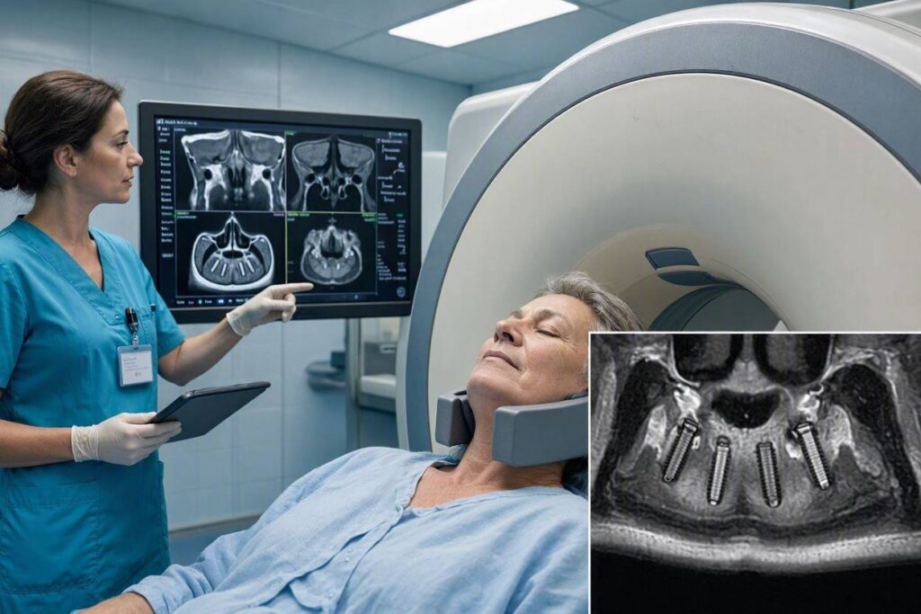
While the dental implant itself is safe in the MRI environment, it is not invisible. The titanium post creates a localized distortion of the magnetic field homogeneity. This distortion, called a susceptibility artifact, appears on the MRI images as a dark void around the implant, often with geometric distortion and bright signal pile-up at the margins. The artifact is larger than the implant itself, and it can obscure the anatomy in the immediate vicinity.
The size of the artifact depends on several factors. The magnetic field strength is the most significant: a 3 Tesla scanner produces a larger artifact than a 1.5 Tesla scanner for the same implant. The pulse sequence parameters chosen by the radiologist also influence artifact size. Certain sequences, such as fast spin-echo sequences, are less sensitive to susceptibility artifacts than gradient-echo sequences. The radiologist can optimize the scan parameters to minimize the artifact when imaging the area near a known dental implant. The size and orientation of the implant also matter: a larger implant produces a larger artifact, and an implant oriented perpendicular to the main magnetic field produces a larger artifact than one oriented parallel.
For MRI scans of the brain, particularly the brainstem and the temporal lobes, dental implants can produce artifacts that degrade the diagnostic quality of the images. The artifact from a single implant is usually small and manageable, but multiple implants, especially in the posterior maxilla or mandible, can create a more extensive zone of distortion. The radiologist interpreting the scan notes the presence of dental implants and accounts for the artifact when evaluating the images. If the clinical question involves a brain lesion near the skull base, and the patient has multiple dental implants, the radiologist may recommend a different imaging modality, such as a CT scan, for that specific area.
For MRI scans of the cervical spine, the artifact from dental implants is usually not a significant problem because the area of interest is below the level of the jaw. For scans of the lumbar spine, the knee, the shoulder, or any other body part distant from the mouth, the dental implant artifact is completely irrelevant. The implant does not affect the image quality in these distant regions.
Communicating with the MRI Team
Your most important responsibility as a patient with a dental implant is to disclose it during the MRI safety screening process. The screening questionnaire will ask about implants. Answer honestly and specifically: “I have a titanium dental implant in my upper left jaw, placed in 2023.” The MRI technologist will note this in your record and may ask additional questions to confirm the material and the date of placement.
If you have any documentation of your implant, such as the implant passport or identification card provided by many implant manufacturers, bring it to the MRI appointment. This documentation confirms the brand, model, and material of the implant, providing the technologist with definitive information. The implant passport is a small card, similar to a credit card, that the surgeon gives you after implant placement. It lists the implant system, the dimensions, and the lot number. Keep this card in your wallet; it is a valuable medical record.
The MRI technologist is trained to evaluate metallic implants for safety. They have access to reference databases and can consult with a radiologist or a medical physicist if there is any uncertainty. Trust their judgment. If they clear you for the scan, proceed with confidence. If they ask for additional information or documentation, provide it promptly. The safety screening process exists to protect you, and cooperation is the most efficient path to a safe and successful scan.<div style=”border-left: 4px solid #2a9d8f; padding: 15px; margin: 25px 0; background-color: #f0fdfa;”> <p style=”font-weight: bold; margin-bottom: 5px;”>💡 Practical Advice for MRI with Dental Implants</p> <p style=”margin: 0;”>Remove any removable dental prostheses—dentures, partial dentures, retainers—before entering the MRI suite. These may contain metal components that are ferromagnetic or of unknown composition. A fixed dental implant crown or bridge cannot be removed and is safe to leave in place. Inform the technologist about all fixed dental work, including implants, crowns, bridges, and fillings, even if you are sure they are non-metallic. The technologist makes the final safety determination.</p> </div>
Special Considerations: Recent Implant Placement and MRI Timing
A question that occasionally arises is whether there is a waiting period after dental implant surgery before an MRI can be safely performed. The concern is that the recently placed implant might not yet be firmly integrated, and the MRI could somehow disrupt the healing process. There is no evidence to support this concern. The forces exerted on a non-ferromagnetic titanium implant by the MRI magnetic field are negligible, far below the threshold that would disturb a healing implant site. A dental implant can be imaged with MRI immediately after placement, though clinically there would rarely be a reason to do so.
The only timing consideration is the presence of postoperative swelling, hematoma, or surgical dressings that might create discomfort or artifact unrelated to the implant itself. A patient who just had implant surgery is unlikely to need an MRI that same week for unrelated reasons. If an MRI is medically necessary during the immediate postoperative period, the dental implant is not a contraindication.
Conclusion
Dental implants made of titanium or zirconia are non-ferromagnetic and completely safe in the MRI environment at standard clinical field strengths of 1.5 and 3 Tesla, experiencing no dangerous magnetic attraction, torque, or heating. The only clinical consideration is a localized image artifact that can obscure anatomy immediately adjacent to the implant, which is relevant for brain and skull base imaging but inconsequential for scans of distant body regions. Full disclosure of your dental implant during MRI safety screening, supported by your implant passport if available, ensures a smooth and safe imaging experience.
Frequently Asked Questions
Q: Will the MRI pull my dental implant out of my jaw?
A: No. Titanium is non-ferromagnetic. It is not attracted to the MRI magnet. The implant experiences negligible magnetic force, far below anything that could disturb an osseointegrated implant. Your implant is physically more secure than many natural teeth.
Q: Can I have an MRI if I have multiple dental implants?
A: Yes. Multiple titanium dental implants are just as safe as a single implant. They will not interact with each other in the magnetic field in any dangerous way. The image artifact will be more extensive, which may be relevant for scans of the head and neck, but the safety profile is unchanged.
Q: What about zirconia ceramic implants? Are they even safer in MRI?
A: Zirconia is a ceramic, not a metal. It produces virtually no magnetic susceptibility artifact. A zirconia implant is essentially invisible on MRI. From a safety perspective, both titanium and zirconia are MRI-safe. Zirconia has the advantage of producing no image distortion, which can be beneficial if future brain or head and neck MRI scans are anticipated.
Q: Should I keep my implant passport card?
A: Absolutely. The implant passport is a permanent medical record. Store it with your other important medical documents. If you ever need an MRI, the passport provides immediate, definitive information about your implant material, eliminating any uncertainty or delay in the screening process.
Additional Resource:
For authoritative information on MRI safety and medical implants, visit the American College of Radiology: https://www.acr.org/