What Computer To Plan Dental Implants?
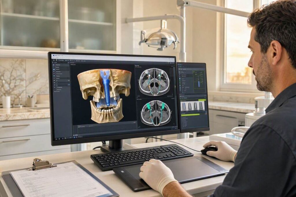
You sit in the dental chair while the surgeon rotates a three-dimensional model of your skull on a high-resolution monitor, zooming into the edentulous space, measuring bone density in Hounsfield units, and virtually placing an implant with sub-millimeter precision. The scene feels like something from a science fiction film, but it is the daily reality of modern implant dentistry. The question that forms in your technically curious mind is: what kind of computer system makes this possible? What hardware and software powers the digital planning that guides a titanium post into the exact right spot in your jawbone?
The answer is that implant planning does not rely on a single “implant planning computer” that you can buy off the shelf at an electronics store. It relies on a digital ecosystem composed of specialized hardware—high-performance workstations with dedicated graphics capabilities—and sophisticated software platforms that integrate Cone Beam CT data, intraoral optical scans, and computer-aided design tools. This technology stack is the invisible backbone of guided implant surgery, the digital bridge between the diagnostic image and the surgical guide that directs the drill.
This guide opens the black box of digital implant planning. We will explore the hardware specifications required to run implant planning software smoothly, the leading software platforms and what they do, the integration of CBCT and intraoral scan data, and the workflow that transforms a digital plan into a physical surgical guide. Understanding this technology not only satisfies curiosity; it gives you the vocabulary to ask your implant surgeon informed questions about how your case is being planned and executed.
The Hardware Foundation: Workstations for Digital Dentistry
The computers used for dental implant planning are not the standard office desktops used for scheduling appointments and processing insurance claims. They are high-performance workstations configured to handle the demanding computational tasks of 3D rendering, volumetric data manipulation, and real-time surgical simulation. A practice investing in digital implant planning invests in hardware capable of keeping pace with the software.
The central processing unit is the engine of the workstation. Implant planning software relies heavily on the CPU for tasks like rendering 3D volumes from CBCT data, segmenting anatomical structures, and performing the complex geometric calculations required for virtual implant placement. A modern multi-core processor—typically an Intel Core i7 or i9, or an AMD Ryzen 7 or 9—is the baseline. More cores allow the software to process multiple tasks simultaneously, such as rendering a 3D model while the surgeon manipulates the view.
The graphics processing unit, the GPU, is equally critical. A dedicated, discrete graphics card with substantial video memory is non-negotiable. The GPU renders the high-resolution, textured 3D models that the surgeon manipulates on screen. Smooth rotation, zooming, and cross-sectional slicing of a CBCT volume depend on the GPU’s ability to refresh the display at high frame rates. Graphics cards designed for professional visualization, such as the NVIDIA RTX series or AMD Radeon Pro series, are common in dental workstations. The GPU also accelerates specific computational tasks in the implant planning software, particularly ray-traced rendering and the simulation of surgical guide sleeves.
Random access memory, RAM, must be generous. A typical implant planning workstation is configured with 16 to 32 gigabytes of RAM, with some high-end systems equipped with 64 gigabytes or more. The RAM holds the large CBCT datasets, the intraoral scan files, and the software’s operating environment simultaneously. Insufficient RAM causes the software to swap data to the slower hard drive, resulting in lag, stuttering, and a frustrating user experience during surgery planning.
Storage is increasingly solid-state. A fast NVMe solid-state drive serves as the primary drive for the operating system and the software. Patient data, including the voluminous CBCT files, is stored on a secondary solid-state drive or on a network-attached storage system with redundancy for data protection. Cloud-based storage solutions are becoming more common, allowing the surgeon to access planning data from multiple locations and facilitating collaboration with remote laboratories and restorative dentists.
The monitor is the surgeon’s window into the 3D patient anatomy. A large, high-resolution display—27 inches or larger, with at least 2560 by 1440 pixel resolution—is standard. Color accuracy is important for evaluating soft tissue and bone density. Some practices use dual-monitor setups, with the planning software on one screen and patient records or reference images on the other. Medical-grade monitors with higher luminance and contrast ratios offer superior visualization of subtle anatomical details.
The Software Ecosystem: From CBCT to Surgical Guide
The hardware runs the software, and the software is where the intellectual work of implant planning occurs. The implant planning software market is dominated by several major platforms, each with its own strengths, supported implant systems, and workflows. These platforms are not interchangeable, and a practice typically standardizes on one or two systems based on the implant brands they use and their preferred clinical workflow.
The leading implant planning software platforms include Simplant, coDiagnostiX, Blue Sky Bio, DTX Studio Implant, and the planning modules integrated into larger dental CAD suites such as exocad and 3Shape Dental System. Simplant, one of the earliest entrants into the market, offers a comprehensive suite of tools for CBCT-based implant planning, virtual tooth setup, and surgical guide design. coDiagnostiX, developed by Dental Wings, is tightly integrated with the Straumann implant system but supports multiple implant brands. Blue Sky Bio offers a free planning software that is widely used, generating revenue through the sale of surgical guides rather than software licenses. DTX Studio Implant, from Envista, integrates with the Nobel Biocare and other Envista implant systems.
The software imports the DICOM dataset from the patient’s CBCT scan. DICOM, or Digital Imaging and Communications in Medicine, is the universal file format for medical imaging. The software reconstructs the 2D projection images from the CBCT into a 3D volumetric dataset. The surgeon can scroll through the slices in axial, coronal, and sagittal planes, and can generate oblique cross-sectional slices that run perpendicular to the curve of the dental arch, providing the ideal view for evaluating the available bone at each potential implant site.
The software allows the surgeon to segment anatomical structures of interest. The mandibular canal, containing the inferior alveolar nerve, is traced on the cross-sectional slices, and the software renders it as a colored tube within the 3D volume, providing a constant visual warning of the no-go zone. The maxillary sinus is segmented, and the sinus floor is mapped. The roots of adjacent teeth are visualized. The bone density is mapped in Hounsfield units, with color-coding that highlights areas of dense cortical bone and areas of soft cancellous bone.
The Integration of CBCT and Intraoral Scan Data
The most powerful feature of modern implant planning software is the ability to merge the CBCT dataset, which shows the hard tissue anatomy, with the intraoral optical scan, which shows the soft tissue and the teeth with exquisite surface detail. The CBCT is excellent at visualizing bone, nerves, and sinuses, but it does not capture the precise contours of the gum tissue or the biting surfaces of the teeth. The intraoral scan captures the teeth and gums in full color and with micron-level accuracy, but it does not show the underlying bone. Merging the two datasets creates a complete virtual patient model that allows the surgeon to plan the implant position relative to both the hidden anatomy and the visible restorative goal.
The merging process, called registration or alignment, uses common reference points visible in both datasets. The software identifies the same tooth cusps, restoration margins, or radiographic markers in both the CBCT and the intraoral scan and mathematically aligns the two datasets into a single coordinate system. The result is a composite 3D model where the surgeon can simultaneously see the bone volume, the nerve position, the planned implant, and the planned crown. This is the “restoratively driven” implant planning philosophy: the implant position is determined by the ideal position of the final crown, not simply by where the most bone is available.
The surgeon uses the software to virtually place implants from a digital library. The library contains accurate 3D models of every implant in the manufacturer’s catalog, including the different diameters, lengths, and platform designs. The surgeon selects an implant, positions it in the bone, and adjusts the angulation and depth. The software provides real-time feedback: the distance from the implant to the nerve, the thickness of the buccal bone plate, the emergence profile of the planned crown. The surgeon can simulate different scenarios, comparing a standard-diameter implant to a narrow-diameter implant, or a straight abutment to an angled abutment.
From Digital Plan to Physical Guide
Once the virtual implant plan is finalized, the software can export the data to fabricate a surgical guide. The surgical guide is a 3D-printed or milled template that fits over the patient’s teeth or directly on the edentulous ridge. It contains metal sleeves at the planned implant positions that direct the surgeon’s drills, controlling the angle, depth, and position of the osteotomy and the implant placement.
The guide design module within the planning software allows the surgeon or a laboratory technician to design the guide. The guide is modeled to fit the teeth or the ridge precisely, using the intraoral scan data. The sleeves are positioned according to the virtual implant plan. The guide is exported as an STL file, the universal format for 3D printing, and sent to a 3D printer. The printer fabricates the guide from a biocompatible resin. The metal sleeves are pressed into the guide after printing. The guide is sterilized and delivered to the surgical operatory.
On the day of surgery, the guide is seated on the patient’s teeth. The surgeon drills through the sleeves, using a sequence of drills with increasing diameters, each guided by a sleeve insert. The depth of the osteotomy is controlled by stops on the drills. The implant is placed through the guide, ensuring that the final implant position matches the virtual plan with a high degree of accuracy. The guided surgery protocol reduces surgical time, minimizes flap reflection, and increases the predictability of the implant placement.
The Role of Artificial Intelligence
Artificial intelligence is beginning to enter the implant planning workflow, not as a replacement for the surgeon’s judgment, but as an assistive tool that automates time-consuming tasks and reduces the risk of oversight. AI algorithms are being trained to automatically segment the mandibular canal, trace the sinus floor, and identify the roots of adjacent teeth. This automated segmentation can be completed in seconds, compared to the minutes or longer required for manual tracing. The surgeon reviews and validates the AI-generated anatomy, making adjustments as needed.
AI is also being applied to suggest optimal implant positions based on the restorative goal and the anatomical constraints. The software analyzes the available bone, the position of the planned crown, and the biomechanical principles of implant support, and it proposes one or more implant configurations. The surgeon evaluates the AI suggestions and modifies them based on clinical judgment and experience. This technology is in its early stages, but it points toward a future where the planning software becomes an increasingly intelligent partner in the treatment planning process.
💡 Questions to Ask Your Surgeon About Digital Planning
You have the right to understand how your implant surgery is being planned. Ask your surgeon: Do you use CBCT-guided planning software? Which software platform do you use? Do you merge an intraoral scan with the CBCT for restoratively driven planning? Will you fabricate a surgical guide for my case? A surgeon who embraces digital planning and can explain the workflow clearly is demonstrating a commitment to precision and modern standards of care.
Conclusion
The computers used to plan dental implants are high-performance workstations with powerful multi-core processors, dedicated graphics cards, ample RAM, and high-resolution monitors, running specialized software platforms like Simplant, coDiagnostiX, or DTX Studio Implant. These platforms integrate CBCT and intraoral scan data into a single virtual patient model, allowing the surgeon to plan the implant position relative to bone, nerves, sinuses, and the final crown restoration. The digital plan is then exported to fabricate a 3D-printed surgical guide that translates the virtual precision into physical accuracy during surgery.
Frequently Asked Questions
Q: Can my implant be planned on a regular laptop?
A: Implant planning software has hardware requirements that exceed the capabilities of most consumer laptops. The software requires a dedicated graphics card with substantial video memory and a multi-core processor. A typical office laptop may struggle to render the 3D CBCT volume smoothly, leading to a slow and frustrating planning experience. Dedicated workstations or high-specification gaming laptops with professional-grade GPUs are the standard.
Q: Is the implant planning software the same for all implant brands?
A: Most planning software platforms support a wide range of implant brands, with digital libraries containing accurate 3D models of the implants and prosthetic components from the major manufacturers. Some software platforms are more tightly integrated with specific implant systems, offering optimized workflows for that brand. Your surgeon selects the software that best supports the implant systems they use in their practice.
Q: How long does the digital planning process take?
A: The computer processing of the CBCT and intraoral scan data, including registration and segmentation, typically takes a skilled operator 15 to 45 minutes per case, depending on the complexity of the anatomy and the number of implants planned. The automated AI-assisted features are reducing this time, but human review and validation remain essential steps.
Q: Is digital implant planning more expensive for the patient?
A: Digital planning and surgical guide fabrication add line-item costs to the treatment plan, typically ranging from $300 to $800 for the guide and the planning service. However, the increased precision and reduced surgical time can translate to fewer complications, faster healing, and a more predictable outcome. Many surgeons bundle the planning and guide cost into their all-inclusive implant fee.
Additional Resource:
For more information on digital dentistry and guided implant surgery, visit the Academy of Osseointegration: https://osseo.org/