Can MRI and Dental Implants? Safety, Artifacts, and What You Must Tell Your Radiologist
The question of whether you can safely undergo a magnetic resonance imaging scan with dental implants is one that arises frequently, often in a moment of unexpected medical need. You may be scheduled for an MRI of your brain, neck, or spine, and you suddenly remember the titanium post in your jaw. This guide provides a definitive, science-based answer to your concerns. We will explain the interaction between the powerful magnetic fields of an MRI machine and the metal in your dental implant, clarify the absolute safety of modern titanium and zirconia implants, and address the practical issue of image quality. Your peace of mind is the goal.
The Core Safety Question: Ferromagnetism vs. Non-Ferromagnetism
The fundamental safety concern with any metal in the body and an MRI scanner is the phenomenon of ferromagnetism. The MRI machine generates a magnetic field that is tens of thousands of times stronger than the Earth’s magnetic field. If a metal object inside your body is ferromagnetic, meaning it contains iron and is strongly attracted to a magnet, this magnetic field could exert a powerful physical force on the object. In a nightmare scenario, a ferromagnetic implant could be pulled, twisted, or dislodged from its position in the bone, causing severe internal injury. Additionally, the rapidly switching radiofrequency fields during the scan can induce electrical currents in conductive metals, potentially causing heating of the implant and the surrounding tissues.
Dental implants are fabricated from commercially pure titanium or a titanium alloy, typically Ti-6Al-4V. Titanium is a paramagnetic metal. This means it is not ferromagnetic. It has a negligible magnetic susceptibility and does not experience any significant attractive force in an MRI magnetic field. It does not move. It does not heat up to a clinically dangerous degree. The titanium post that is osseointegrated into your jawbone is, for all practical purposes, magnetically inert in the MRI environment. Zirconia implants are a ceramic material and are completely non-metallic and non-magnetic. The universal scientific consensus, supported by decades of clinical experience and formal testing by implant manufacturers, is that dental implants are MRI-safe. They are classified as MR Conditional, meaning they pose no known hazards in a specified MRI environment, which encompasses all standard clinical MRI scanners up to 3 Tesla, and often up to 7 Tesla.
The MR Conditional Label and Manufacturer Documentation
The major implant manufacturers, including Straumann, Nobel Biocare, Dentsply Sirona, and Zimmer Biomet, have all performed rigorous testing on their implant systems in MRI environments. Their official documentation declares their products to be safe for patients undergoing MRI. This documentation is readily available and can be provided by your implant dentist or the manufacturer’s website. The formal label is “MR Conditional,” which is the correct regulatory designation for a medical device that has been tested and demonstrated to pose no known hazard under specific MRI conditions of static magnetic field strength, spatial gradient field, and specific absorption rate of radiofrequency energy.
You should never rely on verbal assurances alone. If you are scheduled for an MRI and you know you have dental implants, you should inform your referring physician and the MRI technologist. You can provide them with the implant manufacturer’s name and, ideally, the implant reference card, a small card you should have received at the time your implant was placed, similar to a credit card, that identifies the specific implant system. The radiology team will cross-reference this with their database of tested, safe devices. The likelihood of anyone encountering a ferromagnetic dental implant today is vanishingly small. The era of stainless steel blade implants, which were ferromagnetic, ended decades ago. All modern root-form dental implants are titanium or ceramic.
The Issue of Artifacts: Image Distortion and Diagnostic Quality
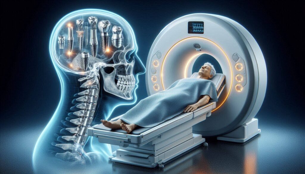
While safety is unequivocally established, the interaction between a dental implant and an MRI scanner does produce an unwanted effect: artifacts. An artifact is a distortion or a signal void in the MRI image. It appears as a dark zone, a warped area, or a bright halo around the implant. This artifact is caused by the difference in magnetic susceptibility between the titanium implant and the surrounding biological tissue. The metal disrupts the homogeneity of the local magnetic field, which is the foundational requirement for creating a clear, accurate MRI image.
The clinical significance of this artifact depends entirely on what body part is being imaged. If you are having an MRI of your knee, your dental implants are of zero consequence. They are outside the field of view. If you are having an MRI of your brain, the implants in your jaw are at the very edge of the field of view and will not obscure any intracranial anatomy. The situation is different if the MRI is specifically focused on the maxillofacial region: the jaws, the temporomandibular joints, the pharynx, or the upper neck. In these cases, the implant can create a significant artifact that may obscure the anatomy immediately adjacent to it. The artifact may hide a small, subtle lesion in the jawbone or the floor of the mouth. The radiologist, the medical doctor who interprets your MRI, is intimately familiar with this phenomenon. They see dental implant artifacts on scans every day. Their report will state that the area immediately adjacent to the implant is obscured by artifact, and they will make their diagnosis based on the remainder of the scan and any other available imaging, such as a CT scan, which is much less affected by titanium.
How to Minimize Artifacts and Optimize Your Scan
You and your medical team can take proactive steps to minimize the artifact and maximize the diagnostic quality of your MRI. The most important step is full disclosure. Do not simply assume the radiology team knows you have an implant because they can see it on a prior X-ray. Tell the technologist explicitly. “I have a dental implant in my upper left first molar position.” This information allows them to adjust the MRI scanning parameters. Certain specialized sequences, called metal artifact reduction sequences, are specifically designed to reduce the blooming and distortion caused by paramagnetic metals. The technologist can apply these sequences if they know the metal is there.
If the MRI is specifically ordered to evaluate a condition in your jaw or the floor of your mouth, your implant dentist or your physician may recommend a different imaging modality as the primary diagnostic tool. A cone beam computed tomography scan or a conventional medical CT scan is largely unaffected by titanium artifacts. An MRI may still be ordered to evaluate the soft tissues, but the CT will provide the detailed bony anatomy. The presence of a dental implant is not a contraindication to MRI. It is a piece of information that the radiology team uses to customize your scan for the best possible result. You should never avoid a medically necessary MRI because you are worried about your implant. The scan’s diagnostic benefit far outweighs the localized, manageable, and well-understood artifact issue.
Conclusion
Dental implants fabricated from titanium or zirconia are unequivocally safe for MRI scans, as these materials are non-ferromagnetic and pose no risk of movement, heating, or injury in the magnetic field, a fact confirmed by decades of clinical experience and manufacturer testing. The only clinical consideration is the localized image artifact, a dark distortion around the implant caused by magnetic susceptibility, which is only relevant when the scan targets the maxillofacial region and is a well-known, manageable phenomenon for radiologists. Your essential action is to proactively inform your medical team and MRI technologist of your implant, providing the manufacturer details to ensure they can apply metal artifact reduction techniques and accurately interpret your scan.
Frequently Asked Questions
Will the MRI machine rip out my dental implant?
No. This is an absolute impossibility with a modern titanium or zirconia dental implant. Titanium is non-ferromagnetic. The magnetic field exerts no meaningful force on the implant. It is as safe in the MRI scanner as a natural tooth.
Do I need to tell the MRI technologist about my implant?
Yes, always. While the implant is safe, the technologist needs to know so they can note it in your record, set up the appropriate artifact reduction sequences if the implant is in the field of view, and interpret the image correctly.
Can I have a brain MRI with dental implants?
Absolutely. Your dental implants are in the jaw, which is at the very bottom edge of the field of view for a brain MRI. They will not obscure the brain tissue and are completely safe. Inform your doctor, but proceed with the scan without any concern.
What about an MRI of my jaw or TMJ?
The implant will create a localized artifact on the scan, which may obscure the anatomy immediately around the implant. Your doctor is aware of this and will take it into account. A CT scan may be ordered to complement the MRI for the bony detail.
Is a metal-free zirconia implant better for MRI scans?
Zirconia implants produce even less artifact than titanium, making them an excellent choice for patients who require frequent, high-resolution MRI scans of the head and neck for ongoing medical conditions. They are completely non-metallic and, from an imaging standpoint, ideal.
Additional Resource
The American College of Radiology provides comprehensive guidelines on MRI safety and the management of patients with implanted medical devices. For detailed information on MR Conditional devices and artifact reduction techniques, you can explore their resources at: https://www.acr.org/