Does A Dental Implant Have Roots?
The language we use shapes our understanding. In dentistry, we routinely say a dental implant “replaces the tooth root.” This phrase, while helpful for patient comprehension, creates a biological misunderstanding. A dental implant does not have roots in any anatomical or physiological sense. It has no periodontal ligament, no cementum, no nerve supply, and no living connection to the bone like a natural tooth root does. The implant is a biocompatible anchor, a threaded post, typically made of titanium or zirconia, surgically placed into the jawbone. It fuses with the bone through a process called osseointegration. This fusion is remarkable, but it is fundamentally different from the relationship between a natural tooth root and its surrounding bone.
Why does this distinction matter? Because understanding the difference illuminates everything about how an implant functions, how it feels, how it responds to force, how it resists infection, and why it requires a different kind of maintenance than a natural tooth. Patients who grasp this distinction care for their implants more effectively. They understand why their implant crown feels slightly different when they bite. They comprehend why the dentist probes around the implant with caution. They appreciate the miracle of osseointegration without mistaking it for a natural biological attachment.
We crafted this guide to delve deep into the structural, biological, and functional differences between a natural tooth root and a dental implant fixture. You will learn what truly anchors an implant in your jaw, how that anchorage compares to a natural tooth, and what the clinical implications are for your daily life and long-term oral health. This knowledge empowers you to be a better steward of your implant investment.
The Anatomy of a Natural Tooth Root
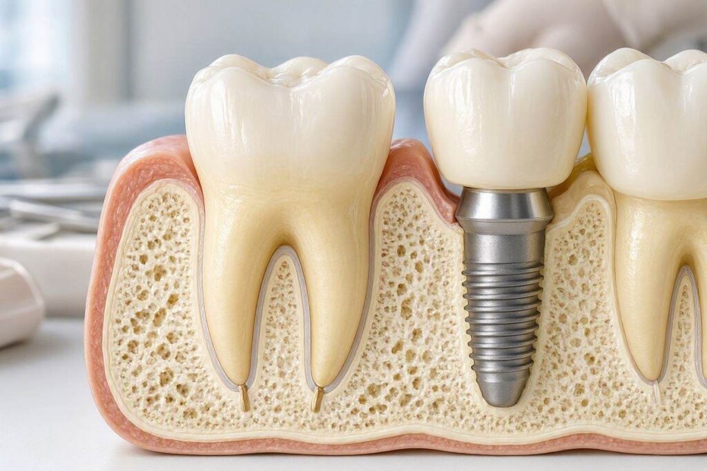
To appreciate what a dental implant is not, we must first understand what a natural tooth root is. The visible part of a tooth, the crown, represents only about one-third of the total tooth structure. The other two-thirds lie beneath the gum line, embedded in the jawbone. This hidden portion is the root. A tooth may have one, two, or three roots, depending on its type and location. Molars, the grinding teeth in the back, typically have two or three roots. Incisors and canines, the front teeth, usually have one.
The root’s outer surface is covered not by enamel, like the crown, but by a thin, bonelike substance called cementum. The cementum is avascular, meaning it has no blood supply. It attaches to the periodontal ligament, a remarkable suspension system composed of hundreds of tiny collagen fiber bundles. These fibers run from the cementum on one side to the alveolar bone socket on the other side. Think of them as thousands of microscopic bungee cords holding the tooth in its bony socket. This ligament is living tissue, richly supplied with blood vessels and nerves. It constantly remodels and adapts.
Inside the root, a canal system houses the dental pulp. The pulp is the living heart of the tooth, containing nerves, blood vessels, and connective tissue. It enters the tooth through tiny openings at the root tip called apical foramina. The pulp provides sensory function—the ability to feel hot, cold, and pressure—and nutritive function, delivering oxygen and nutrients to the tooth’s internal cells. It also has a defensive function, laying down reparative dentin in response to decay or trauma.
The bone surrounding the natural tooth root is not static either. The periodontal ligament transmits the forces of chewing to the bone as tension. This tension signal tells the bone-forming cells (osteoblasts), “This bone is needed. Keep it here.” The bone constantly remodels in response to this functional tension. This is why orthodontists can move teeth through bone with controlled pressure. The periodontal ligament mediates the entire process.
The Periodontal Ligament: Nature’s Shock Absorber
The periodontal ligament deserves special emphasis because it represents the single most important functional difference between a tooth root and an implant. This ligament is roughly 0.2 to 0.4 millimeters wide. Within that tiny space, an intricate network of collagen fibers suspends the tooth like a hammock. When you bite down on a natural tooth, the periodontal ligament compresses slightly, allowing the tooth to move microscopically within its socket. The tooth moves about 25 to 100 microns vertically and up to 50 microns laterally under normal chewing forces. This movement provides a natural shock absorption system.
The periodontal ligament is also densely innervated. Proprioceptive nerve endings constantly send feedback to your brain about the position of your teeth, the force of your bite, and the texture of the food you are chewing. This sensory input is called proprioception. Close your eyes and bite gently on a single sesame seed placed on a back molar. You can feel it. That astonishing sensitivity comes from the periodontal ligament. It makes your teeth feel “alive” and integrated into your nervous system.
Finally, the periodontal ligament serves a defensive role. Its rich blood supply delivers immune cells and antibodies that help ward off bacteria invading from the gum crevice. It acts as a biological barrier. When periodontitis develops, bacterial toxins and the host inflammatory response destroy this ligament and the surrounding bone, leading to tooth mobility and eventual tooth loss.
What Anchors a Dental Implant: The Reality of Osseointegration
Now, place the natural tooth anatomy aside. A dental implant fixture enters the picture. It is a screw-shaped device, typically made of commercially pure titanium or a titanium alloy. Zirconia implants, a metal-free alternative, also exist. The implant’s surface is not smooth. It is microscopically roughened through processes like sandblasting, acid-etching, or anodization. This surface micro-topography is the key to osseointegration.
The implant surgeon drills a precisely sized and shaped hole into the jawbone, called an osteotomy. The implant fixture is inserted into this osteotomy with controlled torque. Over the following weeks and months, the body’s healing response kicks in. Bone cells, specifically osteoblasts, migrate to the implant surface. They attach to the microscopic roughness and begin laying down new bone matrix directly onto the titanium or zirconia surface. There is no intervening soft tissue layer. The bone grows right up to the implant, forming a direct structural and functional connection.
This is osseointegration. The implant becomes mechanically locked into the bone. It is an extraordinarily strong connection. Removing a well-integrated implant requires a significant surgical effort, often involving cutting it out with a trephine bur. The implant does not have the microscopic mobility of a natural tooth. It is ankylosed, meaning it is rigidly fused to the bone with no give.
There is no cementum layer on the implant surface. There is no periodontal ligament. There are no nerve endings inside the implant fixture; it is a solid piece of metal or ceramic. There is no pulp, no blood supply within the implant itself, and no connection to the body’s sensory nervous system. The implant is biologically inert. The body tolerates it, encapsulates it in bone, but does not integrate it into its living sensory or defensive networks in the way a natural tooth root is integrated.
The Connective Tissue Seal: A Fragile Barrier
While the implant has no periodontal ligament, the soft tissue around it does form a biological seal. The gum epithelium grows down and attaches to the implant surface or abutment surface via a hemidesmosomal attachment, similar to how it attaches to a natural tooth. Below this epithelial attachment, a zone of connective tissue forms between the implant/abutment and the surrounding bone. This connective tissue contains collagen fibers, but crucially, these fibers run parallel to the implant surface, not perpendicularly inserting into it as they do with cementum on a natural root.
This parallel fiber orientation creates a much weaker biological seal than the perpendicular insertion into cementum. The implant is more vulnerable to bacterial penetration. If plaque accumulates at the implant-gum interface, bacteria can more easily migrate down into the bone, triggering peri-implantitis. The defensive blood supply from a periodontal ligament is absent. The connective tissue around an implant has fewer blood vessels and fewer fibroblasts. This is why peri-implantitis can progress rapidly and aggressively once established, often without the early warning signs of bleeding and pocketing that characterize periodontitis around natural teeth.
Functional Differences: Feel, Force, and Feedback
The anatomical differences between a tooth root and an implant translate into daily, tangible differences in function. These differences are not necessarily bad. They simply are. A patient with an implant must understand them to use their new tooth properly and protect it over a lifetime.
Bite sensation is profoundly different. Remember the sesame seed test. A patient with a dental implant cannot feel fine texture or light pressure through the implant itself. The implant has no periodontal ligament nerve endings. Any sensation of pressure when biting on the implant crown comes from transference of vibration through the implant fixture into the surrounding bone. The bone itself has some sensory innervation. Additionally, the adjacent natural teeth, if present, provide proprioceptive feedback. But the fine-tuned, delicate sensory information that a natural tooth provides is absent.
This lack of proprioception creates a risk. A patient with a natural tooth feels when they are biting too hard on a hard object and reflexively reduces force. An implant patient may not receive that warning. They can generate enormous biting forces without discomfort, potentially fracturing the porcelain crown, loosening the abutment screw, or even damaging the bone around the implant. Patients with implant-supported restorations must learn to be consciously aware of their biting forces and avoid hard, crunchy foods like ice, hard candies, and unpopped popcorn kernels.
Mobility under force also differs completely. A natural tooth moves slightly, absorbing some of the shock of chewing. An osseointegrated implant does not move. It is rigid. All the force of the bite is transmitted directly and undampened into the bone. This is called an ankylosed state. While the implant fixture itself is strong, the bone supporting it and the prosthesis above it must withstand this rigid load distribution. This is why proper implant treatment planning and fabrication of the crown with precise occlusion are so critical. The bite must be adjusted so that the implant crown is protected from excessive lateral forces.
The way orthodontic forces affect each system highlights the difference perfectly. Orthodontists can move natural teeth through bone by applying controlled pressure. The periodontal ligament mediates bone resorption on the pressure side and bone deposition on the tension side. An osseointegrated dental implant cannot be moved orthodontically. It is fused to the bone. Applying orthodontic force to an implant does not move it through bone; it risks fracturing the bone or failing the implant. This has major implications for patients who have a mix of natural teeth and implants and need orthodontic treatment later.
Comparative Table: Natural Tooth Root vs. Dental Implant
To crystallize these distinctions, we present a side-by-side comparison of the key structural and functional characteristics.
| Feature | Natural Tooth Root | Dental Implant Fixture |
|---|---|---|
| Connection to Bone | Suspended by periodontal ligament (collagen fibers). | Direct bone-to-implant contact (osseointegration). No ligament. |
| Shock Absorption | Yes, provided by the periodontal ligament (25-100 microns of movement). | No. Rigid, ankylosed connection transmits all forces directly to bone. |
| Proprioception (Fine Sensation) | Yes. Rich nerve supply in the periodontal ligament provides exquisite tactile feedback. | No. Sensation is indirect, through bone vibration. Vastly reduced tactile sensitivity. |
| Biological Seal | Strong. Collagen fibers insert perpendicularly into cementum. Rich vascular supply. | Weaker. Collagen fibers run parallel to the implant surface. Reduced vascularity. |
| Response to Bacterial Challenge | Inflammation (gingivitis/periodontitis) mediated by immune system; often progresses slowly. | Inflammation (peri-implant mucositis/peri-implantitis) progresses more rapidly; bone loss often crater-like. |
| Orthodontic Movement | Possible. Tooth moves through bone via controlled pressure and tension. | Not possible. Implant is ankylosed; force will not move it. |
| Internal Structure | Contains living pulp tissue (nerves and blood vessels). | Solid titanium or zirconia. No internal living tissue. |
This table makes clear that a dental implant is an excellent prosthetic replacement for the function of a tooth root but is in no way a biological replica of one.
Clinical Implications: Why the Distinction Matters
Understanding that an implant is a metal anchor, not a natural root, drives every aspect of clinical care, from diagnosis and treatment planning to long-term maintenance and management of complications. Dentists who treat implants as if they were “just like teeth” set their patients up for failure.
Hygiene protocols differ. You cannot use standard stainless steel scalers or ultrasonic tips on a titanium implant or abutment. The metal instruments scratch the implant surface, creating grooves that harbor bacteria and make the surface even harder to clean. Implant maintenance requires specialized plastic, graphite, or titanium-coated scalers that clean without scratching. Dental hygienists receive specific training in peri-implant instrumentation.
Probing around an implant is a nuanced procedure. Dentists probe natural teeth with controlled force (approximately 25 grams) to measure the depth of the gum pocket and detect bleeding, a sign of inflammation. Probing around an implant requires extreme gentleness, often 15 to 20 grams of force. The weaker connective tissue seal around an implant can be inadvertently damaged by aggressive probing, potentially introducing bacteria deeper. Bleeding on probing around an implant is a sign of peri-implant mucositis and must be taken very seriously.
The diagnosis of pathology differs. A natural tooth can develop decay (caries), an infection of the pulp (pulpitis), an abscess at the root tip (periapical abscess), or periodontal disease affecting the supporting bone. An implant cannot get decay. It has no enamel and no pulp. But it is absolutely susceptible to peri-implant disease, an inflammatory condition affecting the soft tissue and bone surrounding the implant. Peri-implant mucositis is the reversible inflammatory condition of the soft tissues alone, analogous to gingivitis. Peri-implantitis involves progressive bone loss around the implant. It is often more challenging to treat than periodontitis because the implant’s surface provides a vast, rough area for bacterial biofilm to colonize once exposed.
Failure modes differ. A natural tooth can fracture at the root level, usually requiring extraction. An implant fixture rarely fractures, though it is possible with massive overload. The more common implant failures are biological (failure of osseointegration leading to mobility, or peri-implantitis leading to bone loss) or mechanical (fracture of the abutment screw, fracture of the crown, loosening of the screw-retained restoration).
Long-Term Maintenance: Caring for Your “Non-Root”
Given these realities, the implant patient must adopt a specific, disciplined maintenance mindset. The implant is a fantastic tooth replacement, but it requires a higher level of daily care than natural teeth because nature provides no backup systems.
Invest in implant-specific hygiene tools. An interdental brush with a plastic-coated wire cleans the curved surfaces of the implant crown and abutment. Implant-specific floss, which has a thicker, fuzzy middle section and a thin threader end, cleans under the crown and around the abutment effectively. A water flosser with a non-metal tip can flush out food debris and disrupt bacterial plaque from hard-to-reach areas around implant-supported bridges.
Attend professional maintenance visits strictly on schedule. For many implant patients, a three-month recall interval is more appropriate than the standard six-month cycle. The dental professional needs to monitor the peri-implant tissues regularly, remove any calculus deposits gently, and assess for early signs of inflammation. Early detection of peri-implant mucositis allows for non-surgical treatment and reversal. Once bone loss from peri-implantitis occurs, the damage is often irreversible without surgical intervention.
Be mindful of parafunctional habits. If you clench or grind your teeth, especially at night, the rigid, non-cushioned implant is at significant risk. The absence of a periodontal ligament means all that grinding force goes directly into the bone and the implant components. A custom-fabricated night guard, properly adjusted to protect the implant prosthesis while allowing a balanced bite, is an essential protective device for any implant patient with bruxism.
Conclusion
A dental implant does not have roots in the biological sense; it is a titanium or zirconia anchor that achieves stability through direct osseointegration with the jawbone, lacking the periodontal ligament, nerve supply, and living pulp tissue that characterize a natural tooth root. This fundamental difference means the implant cannot provide the same sensory feedback or shock absorption, making it vulnerable to overload and requiring a rigorous, tailored hygiene regimen to prevent peri-implant disease. Recognizing the implant as a remarkable prosthetic anchorage device, rather than a replacement root, empowers patients to maintain their investment successfully for decades.
Frequently Asked Questions
Can a dental implant feel pain?
No, the implant fixture itself has no nerves and cannot feel pain. However, the surrounding gum tissue and bone can feel pain if they become inflamed, infected, or traumatized. Pain around an implant is a warning sign that requires immediate professional evaluation.
Why doesn’t my implant feel like my other teeth when I chew?
Your natural teeth have periodontal ligaments filled with nerve endings that provide fine tactile feedback. Your implant is rigidly fused to the bone and lacks these nerves. You feel pressure indirectly through bone vibration, which is a much duller sensation. Your brain adapts over time, but the sensation is never identical to a natural tooth.
Can I get braces if I have a dental implant?
Yes, but the implant itself cannot be moved. It serves as an immovable anchor point. The orthodontist designs the treatment plan to move the natural teeth around the implant, using the implant as a fixed reference. This requires careful planning and is more complex than standard orthodontics.
How long does osseointegration take before the implant feels secure?
Initial mechanical stability is achieved at the time of surgery. Biological osseointegration, where the bone has firmly bonded to the implant surface, typically takes three to four months in the lower jaw and four to six months in the upper jaw. Your dentist confirms integration before attaching the final crown.
Additional Resource
For a deeper scientific understanding of osseointegration, visit the Academy of Osseointegration’s patient education library: Academy of Osseointegration – Patient Info