How Long Do Dental Implants Take to Fuse with Bone?
A dental implant is a masterpiece of bioengineering, but it does not come with a guarantee of instant function. The titanium post placed in your jaw is a foundation, and a foundation must cure. The process of the implant fusing with your bone, known as osseointegration, is the single most important biological event in the entire implant journey. It determines whether your implant will be a stable, lifelong anchor for a tooth or a failure that must be removed. The most common question patients ask is a deceptively simple one: “How long does this fusion take?” The answer is not a single number on a calendar. It is a range, influenced by the location in your mouth, the quality of your bone, your health, and the technology of the implant surface. This article provides a comprehensive, clear, and realistic timeline. You will understand the cellular journey of osseointegration, the factors that speed it up or slow it down, and exactly what to expect during the months between surgery and your final crown.
The Biological Clock of Osseointegration
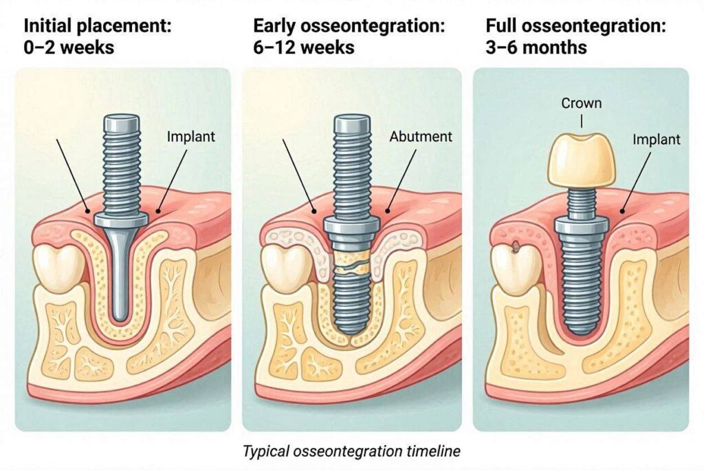
Osseointegration is a living, dynamic process. It is not a chemical glue that sets overnight. It is the slow, orderly growth of living bone cells onto the titanium dioxide surface of your implant. Immediately after the implant is placed, a blood clot forms around the threads. Platelets release growth factors that summon bone-forming cells called osteoblasts. These osteoblasts migrate to the implant surface and begin laying down a soft, unmineralized matrix called osteoid. Over weeks, calcium phosphate crystals are deposited in this matrix, forming a primitive, disorganized type of bone called woven bone. This woven bone provides the first biological grip, but it is weak. The final, crucial step is remodeling. The weak woven bone is gradually resorbed by cells called osteoclasts and replaced by mature, highly organized lamellar bone. This lamellar bone is strong and can bear load. The entire process, from clot to mature lamellar bone, is what constitutes osseointegration.
Primary vs. Secondary Stability: The Crossover Point
To understand the timeline, you must understand two types of implant stability. Primary stability is the mechanical grip the implant gets the moment it is screwed into the bone. It’s like a wood screw biting into a plank. This stability is high right after surgery. Over the next few weeks, the bone that was compressed and slightly damaged during drilling remodels. The primary stability dips. Simultaneously, new bone formation—secondary stability—is increasing. There is a critical period, typically around 3 to 6 weeks, where total stability is at its lowest. This is called the stability dip. If an implant is loaded with a tooth during this vulnerable window, the micro-motion can disrupt osteoblast activity. Fibroblasts then populate the interface, creating a fibrous scar capsule instead of bone. This is why a healing period is non-negotiable for most protocols. The goal is to allow the secondary stability curve to rise and plateau, providing a solid, living anchorage.
The General Timeline for Implant-Bone Fusion
While individual variation exists, the dental community works with a well-established, evidence-based timeline. The implant site in the lower jaw (mandible) and upper jaw (maxilla) heal at different rates due to fundamental differences in bone structure.
Lower Jaw (Mandible) Healing: 3 to 4 Months
The mandible, particularly the anterior region between the mental foramina, is composed of dense cortical bone with a rich blood supply from the inferior alveolar artery and periosteum. This type of bone, classified as Type I or Type II, provides excellent primary stability and heals faster. After implant placement in the anterior mandible, osseointegration is generally sufficiently advanced to support a tooth at the 3-month mark. In the posterior mandible, where bone is slightly less dense, a healing period of 4 months is standard. The dentist will confirm this with clinical tests before loading.
Upper Jaw (Maxilla) Healing: 4 to 6 Months
The maxilla, especially the posterior region, is characterized by a thinner cortical plate and a core of spongy, cancellous bone. This is Type III or Type IV bone. The blood supply is less robust, and the initial mechanical interlock of the implant is lower. The healing process is intrinsically slower. A routine implant in the posterior maxilla requires a minimum of 4 months of healing, and very commonly, 6 months is prescribed to allow the secondary stability to fully mature. The bone here is more forgiving in one sense (it is vascular), but its low density means the stability dip is more pronounced and the remodeling phase takes longer. An anterior maxillary implant, in denser bone, may be ready at 4 months.
Factors That Influence the Fusing Time
The 3-6 month range is not arbitrary. Several powerful variables can shorten the wait or necessitate a longer, more cautious healing phase.
Bone Quality and Quantity
This is the single most important local factor. Dense, thick bone fuses faster and provides more stable anchorage during the critical remodeling phase. Soft, thin bone requires more time because the interface is physically smaller and the initial woven bone is less robust. If a patient has osteoporosis or a history of bisphosphonate use, the bone metabolism is altered, and the surgeon may extend the healing period. The volume of bone also matters. An implant placed into a site that required a simultaneous bone graft is not just fusing to the prepared osteotomy wall; it is also waiting for the graft particles to be revascularized and replaced by new, viable bone. In these cases, the healing clock effectively resets to the graft’s timeline, often extending the wait to 6-9 months before the implant is loaded.
Implant Surface Technology
Modern implant surfaces are not the machined, smooth surfaces of the 1980s. They are moderately roughened by sandblasting and acid etching (SLA). This roughness increases the surface area for bone cell attachment dramatically. More advanced surfaces are hydrophilic, meaning they attract water and proteins to the surface instantly upon insertion, accelerating the initial clotting and osteoblast migration. Implants with hydrophilic surfaces, such as Straumann’s SLActive, have been shown in clinical studies to reduce the healing time from 6-8 weeks to 3-4 weeks in stable bone. This is a genuine biological acceleration. If your dentist uses a premium, active-surface implant, your timeline might be shorter, potentially moving into the early loading window of 6-8 weeks if all other parameters are ideal.
Patient Systemic Health and Lifestyle
Your body is the healing machine. If the machine is compromised, healing slows down.
- Smoking: Nicotine is a potent vasoconstrictor. It starves the healing bone of oxygen and nutrients. Smokers have a significantly higher implant failure rate, and many surgeons will double the healing time for a heavy smoker, or refuse to load the implant until a full 6 months have passed, regardless of jaw location.
- Uncontrolled Diabetes: Elevated blood glucose levels impair white blood cell function and collagen synthesis. A patient with an HbA1c above 7% is a compromised healer and needs a longer, more closely monitored healing period.
- Vitamin D Deficiency: Vitamin D is essential for calcium absorption and bone mineralization. A deficient patient may have a poor healing response.
- Age: Healthy older patients heal similarly to younger patients. Biological age, not chronological age, is the predictor.
The Role of Immediate Loading: Fusion Under Controlled Function
Immediate loading, the “teeth in a day” concept, seems to defy the healing timeline. It does not. The implant is still fusing to the bone over the same 3-6 month period. The difference is that a rigid, temporary prosthesis is splinted to the implant at the time of surgery, with very specific conditions. The implant must have exceptionally high primary stability (insertion torque of 35-45 Ncm or higher). The temporary tooth is meticulously adjusted to be completely out of functional occlusion—it does not touch the opposing teeth during chewing. The forces on the implant are minimal. The temporary splinting of multiple implants in an All-on-4 case uses cross-arch stabilization to nullify micro-motion. The bone is still healing; the patient is simply provided an aesthetic, non-functional tooth during the healing phase. This is an advanced protocol, not a shortcut around biology.
How Dentists Confirm That Fusion Is Complete
You do not guess. The dentist uses objective, documented methods to confirm the implant is ready before taking an impression for the crown. The timeline is a guide, but the final decision is based on diagnostic evidence. This includes a clinical test of mobility (the implant must be absolutely rigid), a percussion test (a high-pitched, ringing sound indicates solid bone contact), a periapical radiograph showing intimate bone-to-implant contact with no radiolucent line, and ideally, a quantitative measurement using Resonance Frequency Analysis. An RFA device gives an Implant Stability Quotient (ISQ) value. A rising or stable ISQ trend above 65-70 is a strong indicator of successful fusion. This process has been detailed in a previous article, “How Dentists Confirm Bone Fusion to Implant,” which you can refer to for an in-depth explanation of these verification methods.
What Happens If You Load an Implant Too Early?
If an implant is loaded before adequate secondary stability has developed, the sequence of events is predictable. The micro-motion exceeds the threshold (around 100-150 microns). Osteoblasts stop laying down bone matrix. The mesenchymal cells instead differentiate into fibroblasts. A soft, non-adherent fibrous capsule forms around the implant. Clinically, the implant becomes mobile. The patient may feel a dull ache or tenderness on biting. Radiographically, a dark radiolucent line appears around the implant. This is a failed osseointegration. The implant must be removed, the site must heal, and after a waiting period of 3-6 months, a new implant can be placed. The cost is time, money, and emotional frustration. This is why the dentist’s discipline to wait for the biological clock is an act of conservative wisdom, not unnecessary delay.
Managing Your Time and Expectations
The waiting period can be the most psychologically challenging part of the implant journey. The surgery is done. The patient feels healed within a week or two. The gum looks normal. The temptation to “just put the tooth on now” is powerful, especially if it’s a visible front tooth. This is where patient education is critical. You must internalize that the visible healing of the gum is not the same as the invisible integration of the bone. The bone needs its own clock, and that clock cannot be hurried by wishful thinking. To manage this period, your dentist will provide a temporary tooth or bridge. An Essix retainer with a denture tooth, a small bonded Maryland bridge, or a partial flipper denture—these temporaries are designed to give you a smile while the implant heals, without applying damaging force to the implant.
Important Note: The 3-6 month healing period is a passive time for you, the patient. You eat a soft diet, avoid the implant site, and maintain meticulous oral hygiene. But for your jawbone, it is a period of intense, silent, microscopic construction. Respect this biological process. A patient who demands a crown early because they are “tired of waiting” is demanding an early failure. Trust the timeline your biology dictates.
Conclusion
Dental implants generally require 3 to 4 months to fuse with the dense bone of the lower jaw and 4 to 6 months for the softer bone of the upper jaw, a timeline governed by the complex biological process of osseointegration. This period can be shortened by advanced implant surface technologies and ideal patient health, or extended by the need for bone grafts or compromised systemic conditions. The final confirmation of fusion is always based on clinical and radiographic evidence before the tooth is attached.
FAQ
1. Can I feel the implant fusing to my bone?
No. Osseointegration is a silent, painless cellular process. Any discomfort you feel after the first week is from the soft tissue healing, not the bone fusion. A lack of sensation is a positive sign.
2. Will antibiotics speed up the fusion?
No. Routine antibiotics are prescribed to prevent post-operative bacterial infection, not to accelerate bone formation. Bone healing has its own genetically determined pace, which antibiotics do not influence.
3. Is the implant securely fused after 3 months, or is it still getting stronger?
At 3 months, the implant is clinically ready to be loaded—meaning the bone-implant interface can safely withstand controlled chewing forces. However, the bone continues to remodel and densify around the implant in response to load for the first year of function, becoming increasingly stronger.
Additional Resource
For further reading on the science of osseointegration, visit the Academy of Osseointegration: https://osseo.org/