Lump Around Healing Cap of Implant: Causes & Solutions
You are in the middle of your dental implant journey. The post is in your jawbone, and your dentist placed a small metal dome on top of it. This is the healing cap, also called a healing abutment. Its job is to shape your gum tissue while the implant fuses to the bone below. Then one day, you run your tongue over the area or look in the mirror and notice something alarming. There is a lump, a bump, or a fleshy mass around that healing cap. Your mind races. Is it an infection? Is the implant failing? Is this normal? A lump around a healing cap is a relatively common occurrence during the implant healing phase. It can mean several different things, ranging from completely benign and normal to a sign of a problem needing immediate attention. This article will guide you through every possible cause, teach you how to distinguish between them, and explain exactly what solutions are available.
What Is a Healing Cap and Its Purpose?
Before we dissect the lump, we must understand the structure it surrounds. A healing cap, officially called a healing abutment, is a small, temporary metal or plastic cylinder. Your dentist screws it into the internal connection of the dental implant post. It sits on top of the implant, protruding through the gum tissue into the oral cavity. Its primary function is not structural but biological. It guides the healing of the soft tissue. When a surgeon places an implant and leaves it to heal submerged under the gum, they place a flat cover screw. At a second, brief surgery, they make a small incision and exchange that cover screw for a taller healing cap. The healing cap allows the gum to heal in a specific shape—a perfectly round, diameter-matched access channel to the implant platform below. This makes the final step of taking an impression and seating the final crown much easier, cleaner, and more precise. Without it, the gum would just grow back closed.
The Normal Appearance Around a Healing Cap
Knowing what “normal” looks like is essential to spotting “abnormal.” A healthy healing cap site is clean and quiet. The gum immediately surrounding the cap should be smooth, pink, and firm. It should hug the cap tightly, forming a cuff. There might be a small, flat, well-healed collar of tissue encircling the metal. There is normally no bleeding when you brush or rinse gently. You should not feel any throbbing pain. Some tenderness for a day or two after the cap’s placement is expected, but this resolves quickly. The healing cap sits there passively, doing its job, while all the intense biological activity happens deep below in the bone. Any significant deviation from this calm state—a growing lump, a change in color, a discharge—is a signal to investigate.
Common Causes of a Lump Around a Healing Cap
A lump can appear for a variety of reasons. The most common causes can be grouped into categories based on the tissue type involved and the underlying trigger.
1. Overgrowth of Gum Tissue (Granulation Tissue Hyperplasia)
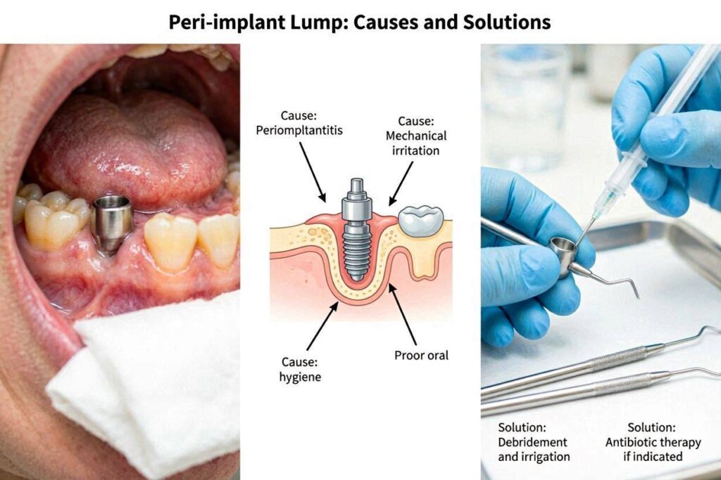
This is the most frequent cause of a fleshy, red bump around a healing cap. The soft tissue of the gum essentially “overreacts.” During the healing phase, the body produces granulation tissue. This is the pinkish-red, velvety, richly vascular tissue that forms in all healing wounds. It is the body’s natural bandage, filling in defects before the outer layer of epithelium grows over it. Sometimes, in a warm, moist, and slightly irritated environment, this granulation tissue receives continued signals to proliferate. It doesn’t stop. It grows exuberantly, building up a soft, bleeding, raspberry-like lump that bulges around and sometimes even over the top of the healing cap. This is called inflammatory fibrous hyperplasia or a hyperplastic tissue response. It is not a true infection, though it looks angry. It is the body’s excessive healing response to a low-grade, persistent stimulus.
2. Loose or Ill-Fitting Healing Cap
The healing cap must be passively stable. It is screwed into the implant with a tiny screw. If that screw loosens, even a microscopic amount, the healing cap develops a micro-motion. Every time you swallow, speak, or chew on the opposite side, the cap wiggles. This constant, tiny mechanical irritation is a powerful trigger for the gum to proliferate. The body recognizes a “foreign body” that is moving and tries to wall it off or cushion it with a reactive soft tissue mass. The lump in this case is a direct response to the physical instability. The tissue may completely engulf a healing cap that has become significantly dislodged, growing right over it and burying it under a dome of pink, firm tissue.
3. Accumulation of Food and Plaque (Peri-Implant Mucositis)
A healing cap is a small cylinder sticking out into a forest of tongue, cheeks, and food. Its metal surface is polished but can still accumulate a biofilm of bacteria if not cleaned. The junction where the healing cap meets the gum is a natural nook for debris. If oral hygiene around the cap is inadequate, plaque buildup triggers an inflammatory response in the surrounding gum. This is peri-implant mucositis at the healing cap stage. The gum becomes red, puffy, and swollen. This inflammatory swelling can present as a soft, boggy, bleeding lump around the cap. It is an infection of the soft tissue only. The bone below is not yet involved. The key sign is that the lump is tender, bleeds profusely with a light touch, and improves dramatically with dedicated cleaning.
4. Bone Spicule or Sequestrum
During implant placement surgery, the bone is drilled. Small, microscopic fragments of non-vital bone can be left behind. Sometimes, a larger piece of the bone crest becomes devitalized. As the healing progresses, the body’s remodeling process pushes these fragments out, much like a splinter. The patient feels a sharp, hard, tiny point emerging through the gum near the healing cap. It can be mistaken for a lump. Often, the overlying gum is slightly irritated or whitish where the bone spicule is pressing from below. This is not related to the healing cap itself but to the surgery that placed the implant. The bone fragment is trying to exfoliate.
5. Suture-Related Reaction
After the healing cap is placed, the gum is often sutured to close the flap snugly around it. If a suture knot is trapped and does not dissolve properly, or if a piece of suture material breaks off, it can act as a foreign body. The tissue reacts by forming a localized inflammatory nodule, sometimes called a suture granuloma. This appears as a localized, pimple-like bump directly at the spot where a suture was tied. It might be reddish-purple and can occasionally drain a tiny amount of fluid.
6. Peri-Implantitis at the Healing Stage
This is the least common but most serious cause of a lump at this stage. If a severe bacterial infection takes hold and progresses beyond the soft tissue, it can cause early bone loss and pus formation around the implant. This presents as an abscess. The lump would be large, soft, very painful, and filled with purulent material (pus). It may have a pointing head, like a gum boil. The patient may have a bad taste, bad breath, and systemic signs like a low-grade fever or malaise. This requires urgent intervention.
Diagnostic Table: What Your Lump Is Telling You
Use this table to correlate the characteristics of your lump with the most likely cause.
| Lump Characteristic | Pain Level | Bleeding | Color | Consistency | Most Likely Cause |
|---|---|---|---|---|---|
| Soft, raspberry-like, growing over cap | Low, mild ache | Very easy, profuse | Bright red/pink | Soft, friable | Inflammatory hyperplasia |
| Circumferential swelling, plaque visible | Mild to moderate | Yes, on brushing | Dusky red/purple | Boggy, swollen | Peri-implant mucositis |
| Cap is mobile, lump is firm | None | None | Normal pink | Firm | Loose healing cap |
| Tiny, hard, sharp point | Sharp when pressed | No | Normal or white | Rock hard | Bone spicule |
| Localized small nodule at suture site | Low | Possible | Red/purple | Firm, nodular | Suture granuloma |
| Large, throbbing, pus-filled | Significant | Pus drainage | Very red, possibly with yellow point | Fluctuant, soft | Abscess/Peri-implantitis |
In-Depth Solutions for Each Cause
Once the cause is identified, the treatment is precise and effective. Ignoring the lump or trying home remedies without a diagnosis can make things worse.
Managing Exuberant Granulation Tissue
For a classic overgrowth of granulation tissue, the solution is removal and control of the stimulus. The dentist will conduct a small, painless procedure. After numbing the area with local anesthesia, they may use a soft tissue laser, electrocautery, or a scalpel to excise the excess tissue. A laser is particularly elegant, as it removes the tissue and seals the blood vessels simultaneously, creating a clean, bloodless field. The healing cap is typically removed, the tissue is contoured back to a healthy anatomy, and the cap is either cleaned and retightened or replaced with a new, sterile one. The tissue almost always heals back normally, but this time, the dentist will ensure there are no loose edges or irritating factors. The removed tissue may be sent for a biopsy just to confirm the diagnosis, which is a standard safety protocol.
Tightening or Replacing a Loose Healing Cap
If the cap is loose, the treatment is simple. The dentist removes the cap, irrigates the area copiously with sterile saline or an antimicrobial rinse like chlorhexidine, and inspects the implant’s internal connection. They will place a new, sterile healing cap or the original one and tighten it to the manufacturer’s specified torque, usually 10-15 Ncm. The firm, reactive lump of tissue will often spontaneously resolve and shrink back to a normal cuff within a week or two once the irritating micro-motion is eliminated. No surgical excision is needed.
Treating Peri-Implant Mucositis
This is treated with non-surgical debridement and improved hygiene. The dentist uses a PEEK plastic-tipped scaler or an ultrasonic instrument with a plastic tip to gently remove the biofilm and any calculus from the healing cap and the surrounding implant collar. The area is irrigated with an antimicrobial agent. The dentist demonstrates how to clean around the healing cap effectively. This often involves using a very soft, single-tufted brush angled at 45 degrees against the cap, or an interproximal brush. Prescription chlorhexidine mouthrinse may be used twice daily for one week. The inflammation resolves rapidly once the bacterial load is reduced. The swollen lump disappears as the gum shrinks back to a firm, healthy state.
Addressing a Bone Spicule
A sharp, hard lump from a bone spicule needs observation or simple removal. If the fragment is small and already pushing through the gum, the dentist can often simply pluck it out with a cotton plier. It comes out with no resistance because it’s a piece of dead bone. This provides instant relief. If it is larger and still partially embedded, a small drop of anesthetic and a tiny incision may be needed to flick it out. The gum heals within days. This is a minor event in the grand scheme of implant healing.
Removing a Suture Granuloma
If a retained suture or a suture granuloma is causing a localized lump, the dentist probes the area. The offending remnant of suture material is identified and removed. The granuloma disappears spontaneously once the foreign body is gone. No other treatment is usually required.
Emergency Management of a Peri-Implant Abscess
An abscess around a healing implant is a dental emergency. The infection must be drained. The dentist will establish drainage through the gum, often through the pocket around the healing cap. The pus is expressed. The area is thoroughly debrided and irrigated with an antiseptic. The healing cap may be removed to access and decontaminate the implant’s internal chamber, which can harbor bacteria. Systemic antibiotics are typically prescribed. The patient is monitored very closely. The bone must be radiographically assessed. If the infection has caused early osseointegration failure and the implant is mobile, the implant must be removed, the site debrided, and healing allowed before a new attempt. If caught extremely early, and the implant is still rigid, the infection can sometimes be resolved, and osseointegration can proceed, though the long-term prognosis must be carefully evaluated.
Prevention: How to Minimize the Risk of Lump Formation
Prevention begins the moment the healing cap is placed. Both the dentist and the patient have roles to play.
During Surgery and Healing Cap Placement
The dentist must follow a meticulous protocol. The healing cap should be sterile and tightened to the correct torque. The gum flap should be closed passively around the healing cap without tension. If the gum is thick, it may need to be thinned so it doesn’t overgrow the cap. Some clinicians prefer a slightly taller healing cap for thick tissue biotypes. A loose, floppy, thick tissue cuff is a setup for hyperplasia. Any rough edges on the healing cap should be polished. The use of a non-submerged (single-stage) technique with a proper healing abutment from the start reduces the need for a second surgery and can simplify soft tissue management.
Patient Home Care Instructions
Your daily routine is your best defense.
- Start gentle cleaning 24 hours after healing cap placement with a prescribed mouth rinse.
- Use a cotton swab dipped in chlorhexidine to gently wipe the top and sides of the healing cap twice daily.
- As the area heals, transition to a very soft, child-sized toothbrush or a single-tufted brush. Place the bristles at the junction of the cap and the gum, and use tiny, gentle, vibrating movements to disrupt the plaque.
- Do not use an electric toothbrush directly on the healing cap at this stage; the vibration can be too intense.
- Do not play with the healing cap with your tongue. Constant tongue pressure is a powerful stimulus for tissue overgrowth.
- Avoid chewing on the side of the healing cap until cleared by your dentist.
Important Note: Never try to cut, burn, or remove a lump around your healing cap yourself at home. Home remedies like applying aspirin, using unsterile needles, or using over-the-counter dental gels for canker sores are dangerous. They can infect the healing site, permanently stain the gum, or cause a severe chemical burn that compromises the entire implant. Leave any tissue removal to a professional with a sterile field and proper instruments.
When to Contact Your Dentist Immediately
While some small bumps can wait for a routine check, certain signs demand a prompt phone call or an emergency visit.
- Rapidly growing mass: A lump that is visibly larger from one day to the next.
- Pus or discharge: Any white, yellow, or greenish fluid oozing from the gum around the cap, especially if accompanied by a foul taste.
- Significant pain: Pain that is not controlled by over-the-counter medication or that wakes you up at night.
- Mobility of the healing cap: If the entire cap feels wobbly or you can push it side to side with your tongue.
- Sudden systemic symptoms: Fever, chills, or swollen lymph nodes under your jaw.
The Psychological Reassurance
Finding a lump is frightening. The mind often jumps to the worst-case scenario: a failed implant, a tumor, a disaster. It is vital to internalize this fact: the vast majority of lumps around healing caps are benign, reactive, and easily treatable. They are a bump in the road, not the end of the road. A loose cap, a bit of food impaction, an exuberant tissue response—these are all problems with straightforward, predictable solutions. True implant loss from an abscess at the healing cap stage is the exception, not the rule. The implant journey is a multistep process, and soft tissue management is a well-known, manageable part of it. Trust your dental professional, describe your symptoms clearly, and allow them to diagnose and treat the issue. There is a clear path to a healthy, healed site and your final crown.
Conclusion
A lump around a healing cap usually results from benign reactive tissue hyperplasia, a loose cap, or soft tissue inflammation due to plaque buildup, rather than a failing implant. Accurate diagnosis, which hinges on the lump’s consistency, color, and accompanying symptoms, guides treatment from simple debridement and cap tightening to minor surgical excision. With prompt professional care and meticulous home hygiene, the site can be returned to health, allowing the implant journey to continue smoothly.
FAQ
1. Is it normal for the healing cap to completely disappear under the gum?
This can happen if the tissue overgrows the cap completely, a process called “submergence.” The cap is not lost, just buried. It is not an emergency, but you should see your dentist so they can gently uncover it. Leaving it buried with a mass over it can complicate the final restoration.
2. Can I use a warm salt water rinse to shrink a lump around my healing cap?
Warm salt water rinses are excellent for soothing mild inflammation and keeping the area clean. They will not shrink an established fleshy lump of granulation tissue or a bone spicule. If the lump persists for more than a few days with saline rinses, you need a clinical evaluation.
3. How long will it take for the gum to heal after a lump is removed?
Healing is rapid. After a minor laser excision or debridement, the gum tissue re-epithelializes in about 7 to 14 days. A tight, healthy cuff reforms within a month, allowing the restorative process to continue.
Additional Resource
For reliable information on peri-implant care, visit the International Congress of Oral Implantologists: https://www.icoi.org/